
Implantable tissue compositions and method

a tissue composition and implantable technology, applied in the field of implantable tissue compositions and methods, can solve the problems of unwanted degradation, control over the degradation process, inability to release bioactive compounds in a controlled manner, etc., and achieve the effect of promoting the localization delivery of a poorly water soluble form

Inactive Publication Date: 2009-05-21
PATHAK HLDG
View PDF66 Cites 94 Cited by
  • Summary
  • Abstract
  • Description
  • Claims
  • Application Information

AI Technical Summary

Benefits of technology

"The present invention provides various methods for modifying and cross-linking biological tissue with unsaturated polymerizable groups. These modifications can lead to the creation of new biological materials with improved properties such as increased strength, flexibility, and biodegradability. The invention also provides compositions of matter comprising biological tissue modified with unsaturated groups and cross-linked biological tissue produced by effective cross-linking conditions. The invention further includes methods for creating a surgical adhesive patch and a non-crosslinked degradable biological tissue. The invention also provides a variety of biological tissue / synthetic biodegradable polymer composites and a method for producing them. Overall, the invention provides new materials and methods for creating stronger, more flexible, and biodegradable biological materials for various applications."

Problems solved by technology

The problems with the animal tissue transplantation include inflammation, unwanted degradation, control over the degradation process, calcification, inability to release bioactive compounds in a controlled manner, and rejection of the transplanted tissue.
Collagen-based biomaterials generally induce a mild inflammatory response, which results in degradation of the protein.
Although glutaraldehyde is the most commonly used chemical fixative for biological tissues, there are a number of drawbacks associated with its use in the production of bioprosthetic devices.
For example, the long term durability of glutaraldehyde-fixed bioprostheses is not well established.
Another drawback to glutaraldehyde fixation of bioprostheses relates to the release of cytotoxic glutaraldehyde on the tissue surface thereby hindering the growth of cells, especially endothelial cells, on the surface of the tissue.
Glutaraldehyde fixed tissue is also susceptible to calcification which leads to device failure.
However, none of alternative chemistries have resulted into a commercial clinical heart valve product.
Tissue crosslinking chemistry is challenging due to a variety of reasons.
This limits the accessibility of tissue functional groups for crosslinking reaction.
The solid state nature of tissue also makes it difficult for large crosslinker molecules such as, by way of example, and not limitation, polymers to penetrate inside the tissue matrix and crosslink the reactive sites.
The denatured tissue / collagen (gelatin) is more susceptible to enzymatic degradation and denatured proteins have inferior mechanical properties as compared to non-denatured tissue.
This often may not be possible due to limited length and mobility of crosslinker.
This results in a number of dangling bonds with incomplete crosslinking.
Therefore, the length of a crosslinker serves as a major limitation in achieving a high degree of crosslinking.
However, polymeric crosslinkers are large molecules which cannot diffuse / penetrate inside the tissue matrix and react with sites present in the bulk of the tissue.
This limits the ability of polymeric crosslinker to surface crosslinking only.
Known techniques generally do not teach the successful use of polymeric crosslinkers in tissue crosslinking.
Known techniques generally do not teach methods and compositions that will affect the degradation behavior of biological tissue.
The useful tissue recovered from one animal is also limited in size.
This size and thickness limitation may limit the use of tissue in making large medical device such as tissue based dialysis catheters.
The tissue size limits also increases production costs due to lower yields.

Method used


Image

  • Implantable tissue compositions and method
  • Implantable tissue compositions and method
  • Implantable tissue compositions and method

Examples


example 1

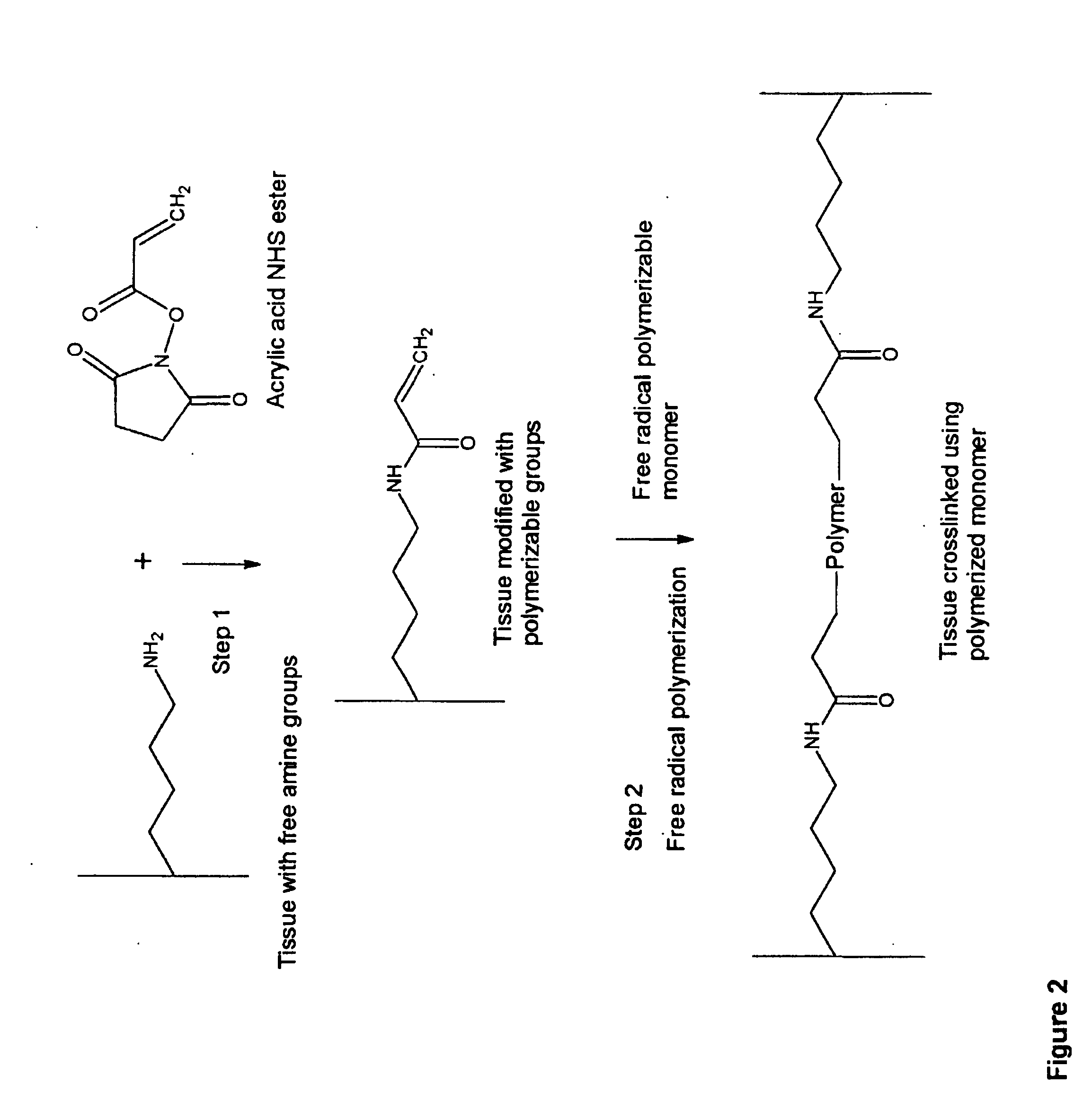
Modification of Tissue Using Unsaturated Succinimide Ester

Acrylic Acid Succinimide Ester

[0287] Modification of Bovine Pericardium Tissue.

[0288] Ten 1 cm by 1 cm bovine pericardium pieces, cut from a freshly obtained bovine pericardial sac, are transferred to 50 ml conical flask containing 10 ml phosphate buffered saline (PBS, pH 7.2). 250 mg acrylic acid succinimide ester (ANHS) (Sigma-Aldrich Product Number: A8060) dissolved in 0.5 ml dimethyl sulfoxide is added to the conical flask and the solution is vortexed for 15 minutes. 0.1 g of ANHS in 0.1 ml DMSO is added to the fixation solution every 2 hours up to six hours. The modification reaction is carried for 6 hours at ambient temperature (25° C.) and then for 12 hours at 4° C. with gentle shaking. The reaction is terminated by washing the tissue with 20 ml PBS 3 times. Finally, the tissue is stored in 30 ml 38% isopropanol and 2% benzyl alcohol solution at 4° C. until further use.

[0289] In another variation of this method, po...

example 2

Modification of Tissue Using Glycidyl Methacrylate

[0296] 10 pieces of 1 cm by 1 cm bovine pericardium tissue are transferred to 100 ml conical flask containing ten ml PBS and a magnetic stir bar. To this solution, 2 ml glycidyl methacrylate, 2 ml triethyl amine and 0.5 g tetrabutylamine hydrobromide are added. The reaction is carried for 16 hours at ambient temperature (25° C.) and then 50° C. for 4 hours with gentle stirring. The solution is cooled and the tissue is isolated from the reaction mixture. The isolated tissue is washed with PBS and 40% ethanol to remove traces of glycidyl methacrylate and other catalysts.

example 3

Modification Tissue Using Acrolein

[0297] Ten 1 cm by 1 cm bovine pericardium pieces are transferred to 50 ml conical flask containing 10 ml PBS and 0.60 ml acrolein. The tissue is incubated for 6 hours at ambient temperature (25° C.). An additional 0.1 g acrolein is added to the mixture at every 1.5 hours up to six hours. Finally, the reaction is continued for 4° C. for 48-120 hours with gentle stirring. The tissue is isolated from the reaction mixture and is washed with PBS and 40% ethanol to remove unreacted acrolein.


PUM


Abstract

Novel implantable tissue fixation methods and compositions are disclosed. Methods and compositions of tissue, fixed using polymeric and / or variable length crosslinks, and di- or polymercapto compounds are described. Also described are the methods and compositions wherein the tissue is fixed using biodegradable crosslinkers. Methods and compositions for making radio-opaque tissue are also described. Methods and compositions to obtain a degradable implantable tissue-synthetic biodegradable polymer composite are also described. Compositions and methods of incorporating substantially water-insoluble bioactive compounds in the implantable tissue are also disclosed. The use of membrane-like implantable tissue to make an implantable drug delivery patch are also disclosed. Also described are the compositions and methods to obtain a coated implantable tissue. Medical applications implantable tissue such as heart valve bioprosthesis, vascular grafts, meniscus implant, drug delivery patch are also disclosed.

Description

CROSS-REFERENCE TO RELATED APPLICATIONS [0001] The present patent application claims priority benefit to PCT Application No. PCT / US2005 / 030187 filed Aug. 24, 2005, which claims priority of the U.S. provisional application for patent No. 60 / 604,737 entitled “Implantable Tissue Composition and Methods” filed on Aug. 26, 2004 under 35 U.S.C. 119(e). The contents of this related provisional application are incorporated herein by reference.FIELD OF THE INVENTION [0002] The present invention relates to methods and compositions for the preparation of biological tissues or extracellular matrices and their medical applications. BACKGROUND OF THE INVENTION [0003] The use of human or animal tissue for medical or surgical use is a rapidly growing therapeutic field. Many uses of processed biological tissues for implantation into humans have been reported. The commercial products or products under development include wound healing dressings, tissue heart valves, ligament substitutes, pericardial ...

Claims


Application Information

Patent Timeline
Patent Type & Authority Applications(United States)
IPC IPC(8): A61F2/02A61K35/12
CPCA61K35/12A61L27/3683A61L2430/40A61L27/38A61L27/18A61L27/24A61L27/3687A61P21/02A61P25/04A61P25/24A61P29/00A61P3/00A61P31/00A61P35/00A61P43/00A61L27/34A61L27/3604A61L27/3608A61L27/3612A61L27/3625A61L27/54A61L27/16A61L27/3633A61L27/3641A61L27/52A61L27/58A61L2300/404A61L2300/406A61L2300/604A61L2300/606A61L2300/608A61L2400/18A61L2420/02A61L2420/08A61L2430/20A61L2430/34
Inventor PATHAK, CHANDRASKEKHARTHIGLE, SANJAY
Owner PATHAK HLDG
Learn More