Wound Closure Devices, Methods of Use, and Kits

Inactive Publication Date: 2009-09-10
INSITU THERAPEUTICS
View PDF41 Cites 113 Cited by
  • Summary
  • Abstract
  • Description
  • Claims
  • Application Information

AI Technical Summary

Benefits of technology

[0011]Provided herein is a wound closure device comprising a plug adaptable to be inserted into an opening formed in two or more tissue layers, one tissue layer transposable relative to a second layer, the plug comprising a material having a first configuration and a second configuration, wherein the plug is adaptable to be inserted into the opening in the first configuration and further adaptable to transition from the first configuration to the second configuration after being inserted into the opening. The wound closure device can be transition between a first configuration and the second configuration after exposure to one or more of an aqueous medium, change in temperature, a chemical environment, pH, ion strength, salt concentration, or light. The wound closure device can be a biocompatible material. The biocompatible material can be selected from at least one of a compressible material, temperature dependent material, shape memory material, a swellable material, and an expandable material. Additionally, the wound closure device can comprise an anchor adaptable to prevent removal of the wound closure device from a wound. The anchor can be a physical feature or a change in the external surface of the device that causes the device to anchor into the wound. The wound closure device

Problems solved by technology

Wound sites generated inside the body or tissue damage are often not accessible and cannot be sufficiently treated or closed.
Open surgery can cause significant additional tissue damage and longer recovery time.
Topical administration of medications often fails to provide therapeutic levels in the vitreous cavity or posterior segment of the eye.
There are significant barriers to solute flux in the corneal epithelium and the topical drops are rapidly lost as the result of drainage and tear fluid turnover.
Drugs can be delivered by frequent injections, but it is not clinically and practically adequate for chronic diseases that can sometimes require multiple weekly administrations over months or years.
In addition, the multiple intraocular injections can lead to an increased likelihood of complications such as vitreous hemorrhages, retinal detachment, and endophthalmitis.
Systemic administration of medication is also very limited to the intraocular diseases due to the presence of blood-ocular barrier (Vel

Method used


Image

  • Wound Closure Devices, Methods of Use, and Kits
  • Wound Closure Devices, Methods of Use, and Kits
  • Wound Closure Devices, Methods of Use, and Kits

Examples


example 1

Preparation of Device

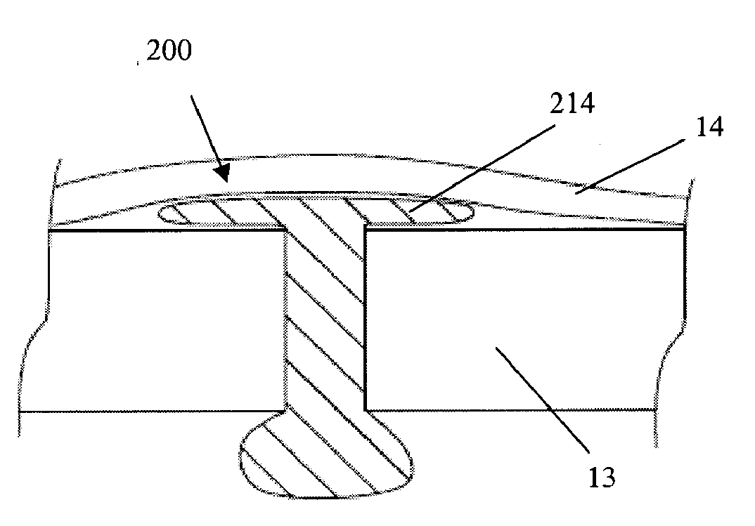
[0082]In some embodiments, a collagen sheet is cut into the size 2 mm by 2 mm by 15 mm, then the cut piece is compressed in two steps such that the final cross-sectional area becomes less than 0.5 mm by 0.5 mm. The compaction is typically done at a room temperature or a temperature between 30 and 37 degree C. Then, the compressed collagen (either rectangular or circular cross-sectional shape) rod is inserted into a tubular mold, preferably Teflon tube, non-adherent polymeric or non-polymeric tubes.

[0083]Polyethylene glycol (PEG, preferably the ones with molecular weight between 1,000 and 10,000) is used as a binder. The PEG is melted at a temperature between 30 and 70 degree C., then the PEG can be sucked into the tube mold containing the compacted collagen rod. The suction can be done by vacuum, wetting by surface tension, or injection. The PEG binder solidified when the temperature drops below its melting temperature. Then, the solid-bound collagen tube is de-...

example 2

Determination of Leakage Rates in Rabbit Eye Using Device

[0088]FIG. 18 illustrates a graph showing the leakage rates of fluid through a wound site in a rabbit eye, at different wound conditions, using one embodiment of the wound closure device described herein. The first 20 minutes just an infusion line was connected to the rabbit eye and was pressured at 35 mmHg. The leakage rate was determined by measuring the flow of the infusion line. At minute 21, a cannula was placed into the wound site and the leakage rate stabilized after about 10 minutes. The leakage rate from the open cannula was measured over 20 min. At minute 60, the cannula was plugged with the wound closure device and the leakage rate declined to base value (the value before the placement of the cannula). After applying the wound closure device, the leakage rate did not increase from the base value, which indicates sealing of the wound site. After removal of the wound closure device, the leakage rate increased rapidly ...


PUM


Abstract

Provided herein is a wound closure device comprising a plug adaptable to be inserted into an opening formed in two or more tissue layers, one tissue layer transposable relative to a second layer, the plug comprising a material having a first configuration and a second configuration, wherein the plug is adaptable to be inserted into the opening in the first configuration and further adaptable to transition from the first configuration to the second configuration after being inserted into the opening. The wound closure device can be used in cases where ocular surgery has been preformed.

Description

CROSS-REFERENCE[0001]This application claims the benefit of U.S. Provisional Application No. 61 / 034,108, filed Mar. 5, 2008, and U.S. Provisional Application No. 61 / 034,110, filed Mar. 5, 2008, and 61 / 040,500, filed March 28, 2008, which applications are incorporated herein by reference in their entireties.BACKGROUND OF THE INVENTION[0002]Surgery is constantly developing less and less invasive surgical techniques such as endoscopic based procedures in order to minimize the trauma inflicted upon a patient during surgery and minimize the recovery time to overcome the trauma of surgery. A wound can include surgical incisions as well as wounds caused by accidental trauma or disease. Wound sites generated inside the body or tissue damage are often not accessible and cannot be sufficiently treated or closed. Often open surgery must be performed to close and repair the wound sites. Open surgery can cause significant additional tissue damage and longer recovery time. Therefore a considerabl...

Claims


Application Information

Patent Timeline
IPC IPC(8): A61F9/007
CPCA61B17/0057A61B17/068A61F9/00736A61B2017/0647A61F9/0017A61B2017/00654A61B17/00234
Inventor FASCHING, RAINERRYU, WONHYOUNGMOSHFEGHI, DARIUS
Owner INSITU THERAPEUTICS
Why Eureka
Try Eureka