

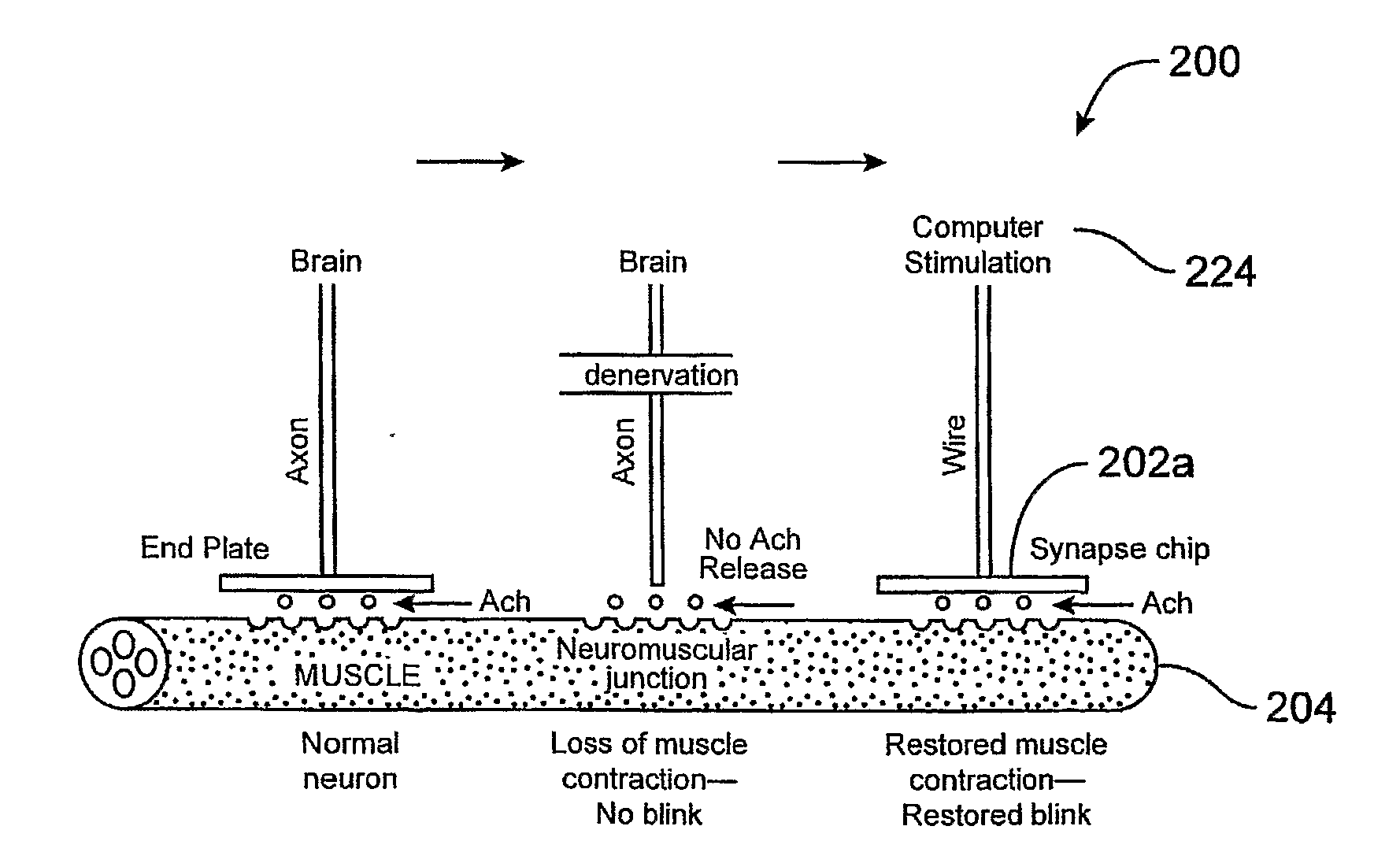
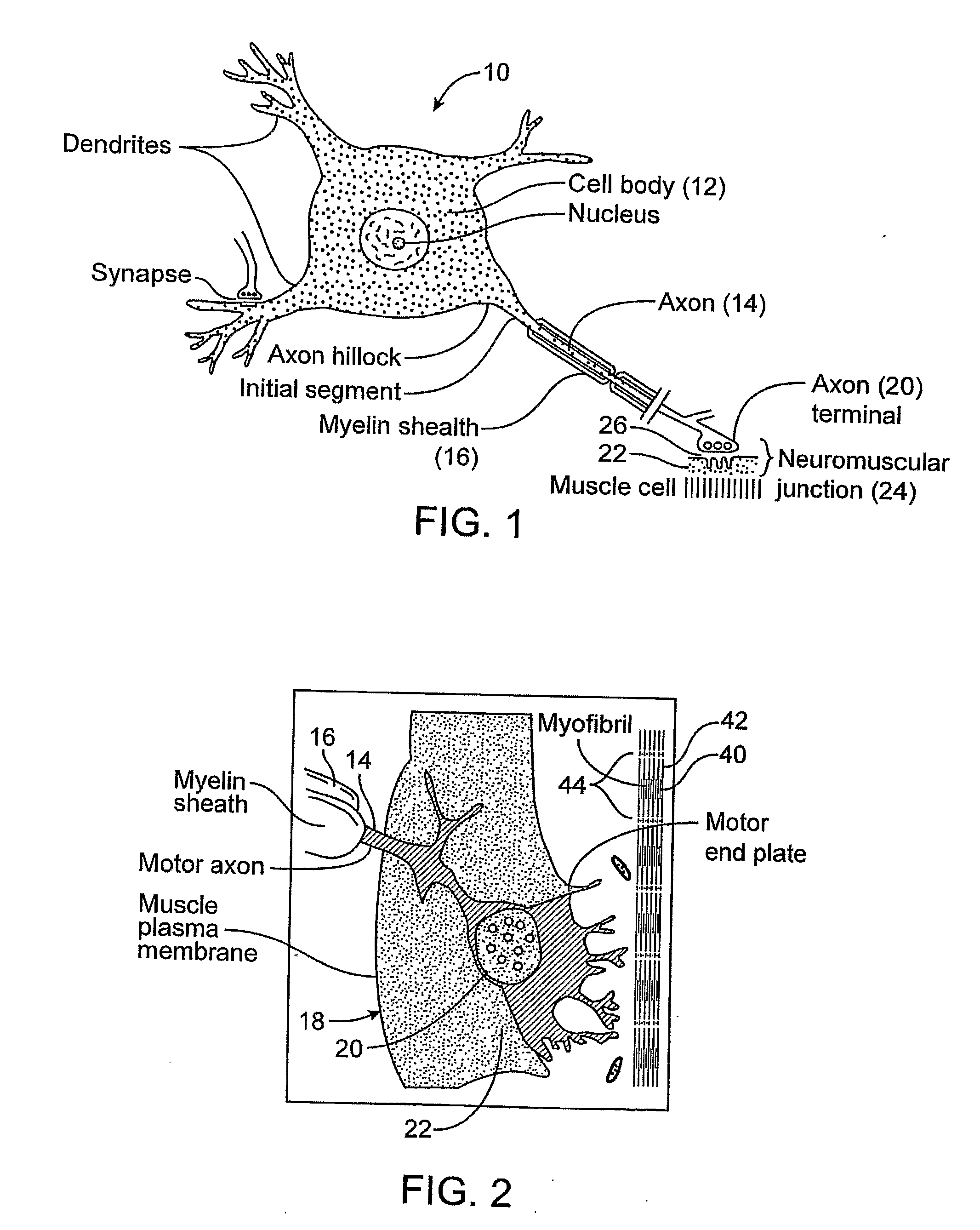
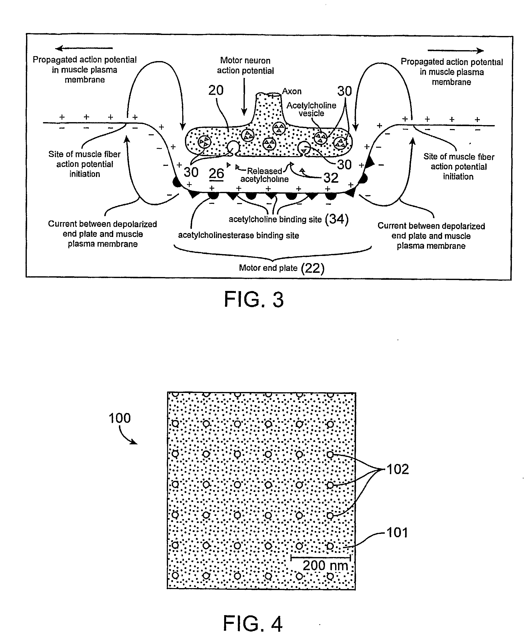
Devices and Methods for Stimulation of Tissue
- Summary
- Abstract
- Description
- Claims
- Application Information
AI Technical Summary
Benefits of technology
Problems solved by technology
Method used
Image

Examples
experiment 1
ts
[0154]4 subjects with denervated orbicularis oculi were tested with electrical-only stimulation at predetermined locations in the preseptal and pretarsal orbicularis oculi, identified by anatomic landmarks. A typical data set is shown below, in this case for a patient denervated on the right side as shown in Table I.
TABLE IPosition of ElectricRight EyelidStimulationPhase DurationCurrentMovementPain5 mm superior to the upper lid0.05 msecUp to 11.8 mANo movement6 / 10punctum0.01 msecUp to 15 mANo movement6 / 105 mm superior the upper lid0.01 msecUp to 26.7 mANo movement6 / 10margin at mid pupil10 mm lateral to the lateral0.01 msecUp to 20.4 mA1 mm twitch5-6 / 10 margin10 mm inferior to lower lid0.01 msecUp to 26.7 mANo movement7 / 10margin at mid pupilPreseptal Surface0.01 msecUp to 36.9 mANo movement7 / 10Electrode, Upper lid
The levels of stimulation in the table were the limits of stimulation tolerable to the patient. Complete functional blinks were not elicited. Notably, a fill body startle ...
experiment 2
timulation In the Denervated Rabbit Model
[0156]To determine if an implantable prototype device capable of delivering electrical stimulation could elicit a complete closure blink of a denervated orbicularis oculi muscle in New Zealand White Rabbits, a rabbit model was used. The rabbit model was selected because of the similarity of the structure and function of their eyelids; specifically the distribution of neuromuscular junctions and muscle fiber type of the orbicularis oculi when compared to humans.
Methods
[0157]a) Two white New Zealand female rabbits were anesthetized by using 3-5% isofluorane inhalation and ketamine / xylazine and monitored by Heska monitor (SP02, heart rate, and rectal temperature). b) A pre-auricular incision was made the facial nerve was surgically sectioned and a five millimeter section was eliminated. The upper eyelid opens when the innervation to the orbicularis oculi is severed creating 6 millimeters of lagophthalmos.
Electrical Stimul...
experiment 2a
[0159]Two weeks post-denervation, one prototype chip with the electrical stimulation delivery facing upwards was placed in the upper and lower lid, with externalized wires to enable stimulation to be controlled by a computer board.
[0160]Results: Stimulation produced a localized muscle contraction of the orbicularis oculi, evidenced by a twitch of the upper and lower eyelids.
[0161]Discussion: Since the pretarsal fibers of the orbicularis oculi only span a third of the length of the muscle, and local electric stimulation can only travel the length of individual fibers, stimulation across a greater portion of the entire length of the muscle may elicit effective contraction. Other possible reasons for limited response to stimulation may include an insufficient size and layout of the gold electrodes, any defect in the connections between the stimulation unit and the chip electrodes, and any localized loss of insulation of the wires causing the wires to short circuit prior to current reac...
PUM
 Abstract
Description
Claims
Application Information