Marker for identification of tissue type of epithelial ovarian cancer, and method for determination of the occurrence of epithelial ovarian cancer based on tissue type by using the marker
- Summary
- Abstract
- Description
- Claims
- Application Information
AI Technical Summary
Benefits of technology
Problems solved by technology
Method used
Image
Examples
example 1
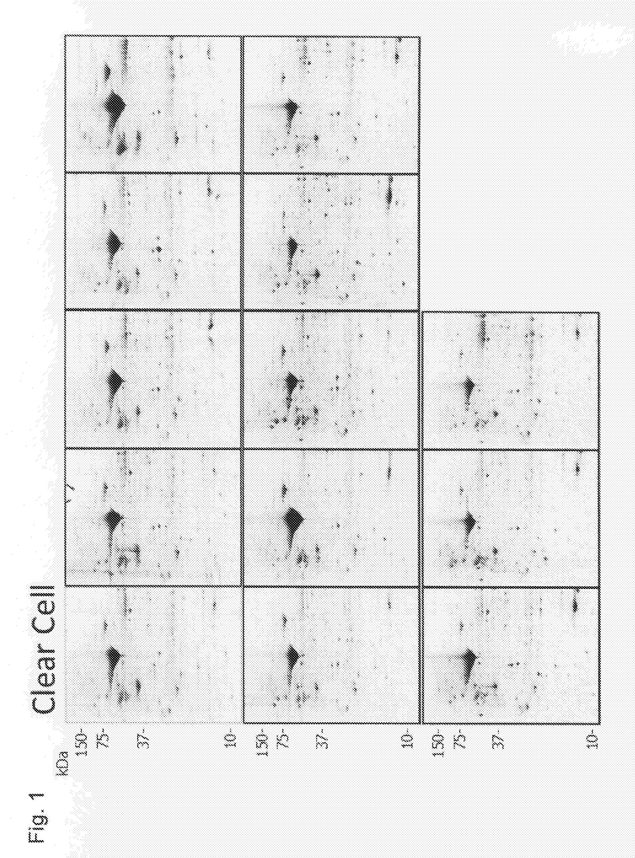
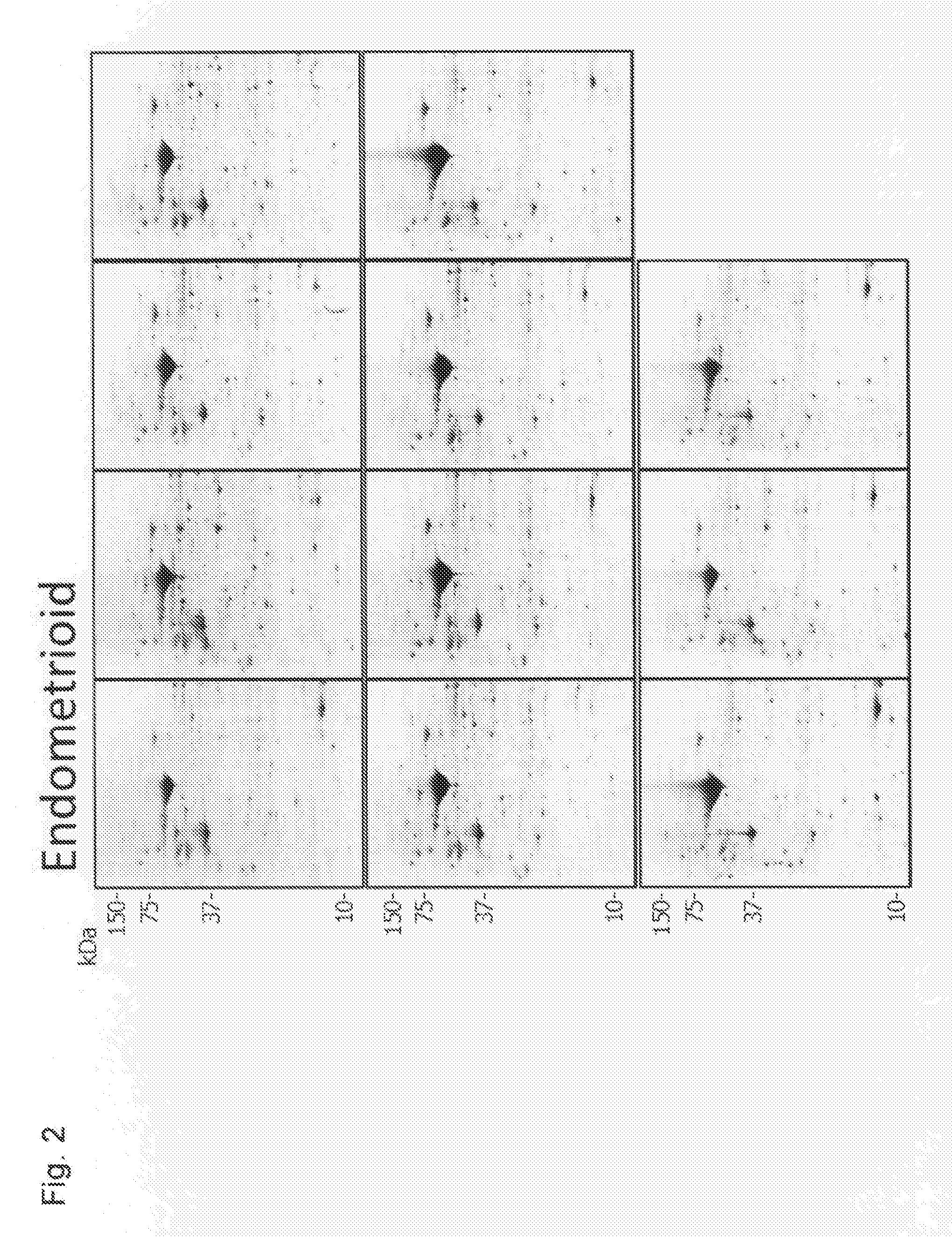
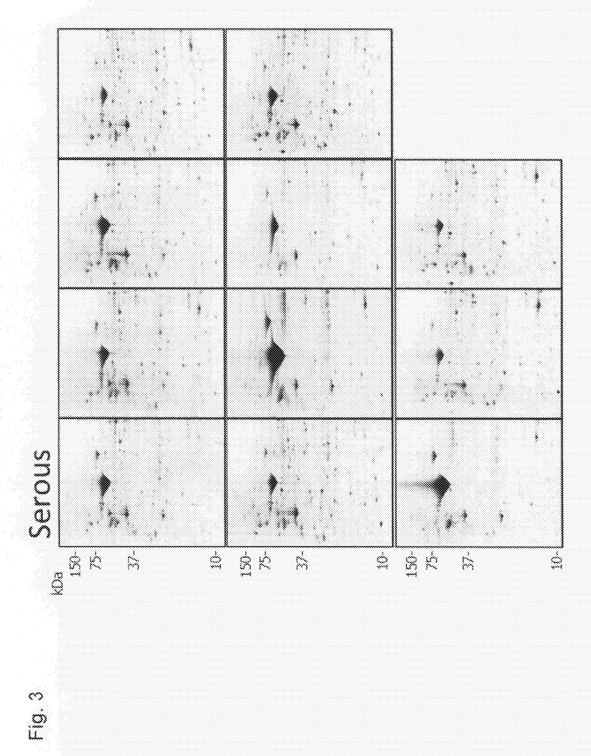
[0407]Profiling was conducted using pathological tissues of epithelial ovarian cancer that had been excised from patients, and for which accurate tissue-type diagnosis had been made from the view point of histopathology (4 tissue-types and 41 specimens: clear cell cancer 13 specimens, endometrioid carcinoma 11 specimens, serous adenocarcinoma 11 specimens, and mucinous adenocarcinoma 6 specimens).
[0408]An excised pathological tissue (stocked in liquid nitrogen) was crushed into fragments of an appropriate size in the presence of liquid nitrogen. The crushed tissue specimen was transferred into a micro tube with a screw cap, and suspended in about 5 times weight of Lysis buffer (50 mM HEPES-NaOH pH 7.5, 100 mM NaCl, 2% CHAPS, 1% Triton X-100). The concentration in the state of a suspension was quantified by the Bradford method, and a specimen corresponding to 300 μg of protein was subjected to desalination by trichloroacetic acid precipitation, to prepare a sample for two-dimensional...
example 2
[0422]For screening the above-described 80 candidate markers into molecules having a determinative role in diagnosing a tissue-type of an unknown ovarian cancer tissue using a molecular technique, a classification prediction model using a decision tree was constructed using statistical analysis software SPSS (SPSS Inc.). Four tissue-types were used as dependent variables, and quantified values of 80 spots of candidate markers scaled into three stages (low expression, high expression, extra-high expression) were used as independent variables. The reference value in scaling was appropriately determined based on the expression pattern and the average quantified value of each candidate marker determined by profiling.
[0423]As one example, FIG. 11 shows a decision tree model for determining whether an unknown specimen is Clear Cell, prepared by using 41 specimens from which the profile was prepared. In FIG. 11, a protein used as a variable of divergence is shown inside the dashed line, an...
example 3
[0426]Using tissue extracts (excluding one specimen of mucinous adenocarcinoma) that are the same with those subjected to two-dimensional electrophoresis in Example 1, quantitative detection by Western blotting was conducted. As antibodies, anti-Annexin-A4 (D-2, Santa Cruz Biotechnology), anti-Maspin precursor (G167-70, BD Transduction Laboratories), anti-Cellular retinoic acid-binding protein 2 (Bethyl Laboratories) and anti-Phosphoserine aminotransferase (Proteintech Group Inc.) were used.
[0427]Results of Western blotting by 40 specimens and 4 antibodies are shown in FIG. 12 (for Clear Cell and Endometrioid) and in FIG. 13 (for Serous and Mucinous). As shown in these drawings, a specific band was detected in a position of respective target molecular weight, and an expression pattern that seems to be tissue-specific was observed similarly to the result obtained by two-dimensional electrophoresis. Each band was quantified based on the band of β-actin (Beta actin) which was a loading...
PUM
| Property | Measurement | Unit |
|---|---|---|
| Current | aaaaa | aaaaa |
| Atomic weight | aaaaa | aaaaa |
| Morbidity rate | aaaaa | aaaaa |
Abstract
Description
Claims
Application Information
 - R&D
- Intellectual Property
- Life Sciences
- Materials
- Tech Scout
- Unparalleled Data Quality
- Higher Quality Content
- 60% Fewer Hallucinations
Browse by: Latest US Patents, China's latest patents, Technical Efficacy Thesaurus, Application Domain, Technology Topic, Popular Technical Reports.
© 2025 PatSnap. All rights reserved.Legal|Privacy policy|Modern Slavery Act Transparency Statement|Sitemap|About US| Contact US: help@patsnap.com


