Method for detecting human parvovirus antigen
a parvovirus and antigen technology, applied in the field of detection of human parvovirus infection, can solve the problems of unclear significance of dna in plasma post-viremia, human parvovirus contamination of human plasma or derived blood products, and is recognised as a significant threat to human health, so as to achieve greater assay sensitivity and improve signal generation
- Summary
- Abstract
- Description
- Claims
- Application Information
AI Technical Summary
Benefits of technology
Problems solved by technology
Method used
Image
Examples
example 1
Screening of the Level of Human Parvovirus Viremia in a Blood Donor Population
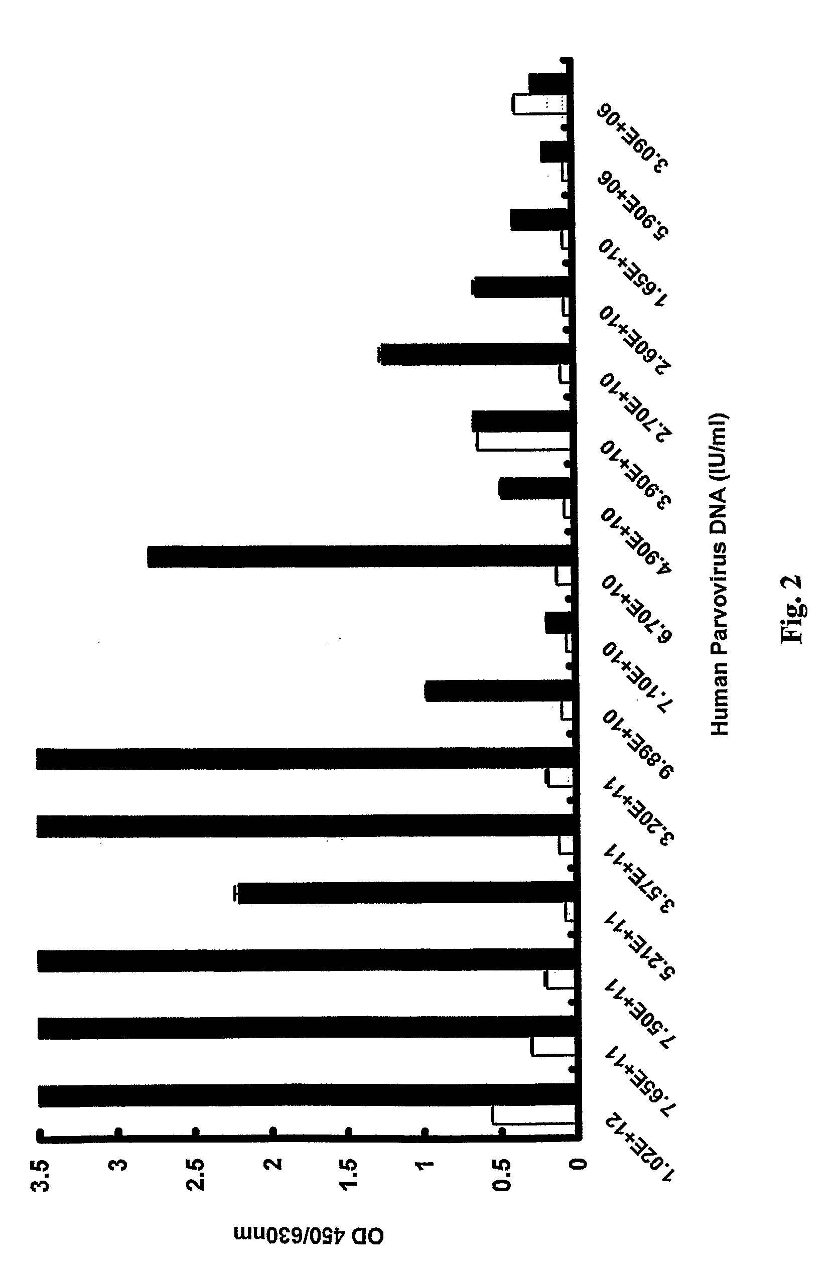
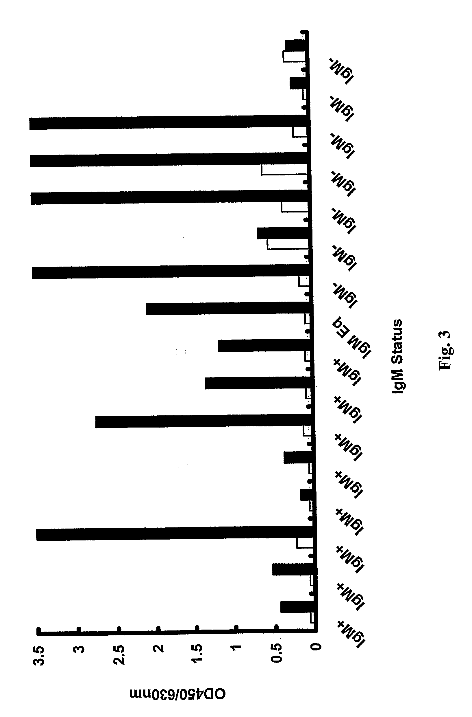
[0036]Dutch blood donors were screened over a 17 month period by the Dutch Blood Bank, which let to the identification of 70 individuals with levels of human parvovirus DNA greater than 106 IU / ml at the time of donation. Sample specimens from these 70 donations were further tested for human parvovirus in accordance with the invention by a virus antigen-capture ETA and also for human parvovirus IgM and IgG antibodies.
Human Parvovirus Antibody Production
[0037]Recombinant human parvovirus VP2 expressed in baculovirus was purified as previously described (Kerr, S. et al (1999) supra) and used for rabbit and sheep immunisations. Once a satisfactory titre was obtained, serum was collected and IgG purified by Protein A affinity chromatography. Purified anti-sheep polyclonal IgG was employed as a human parvovirus-capturing antibody coated on to microtitre plate wells. Rabbit anti-VP2 conjugated to horseradish pero...
example 2
Determination of Assay Sensitivity
[0055]Batches of recombinant VP2 were prepared as follows:
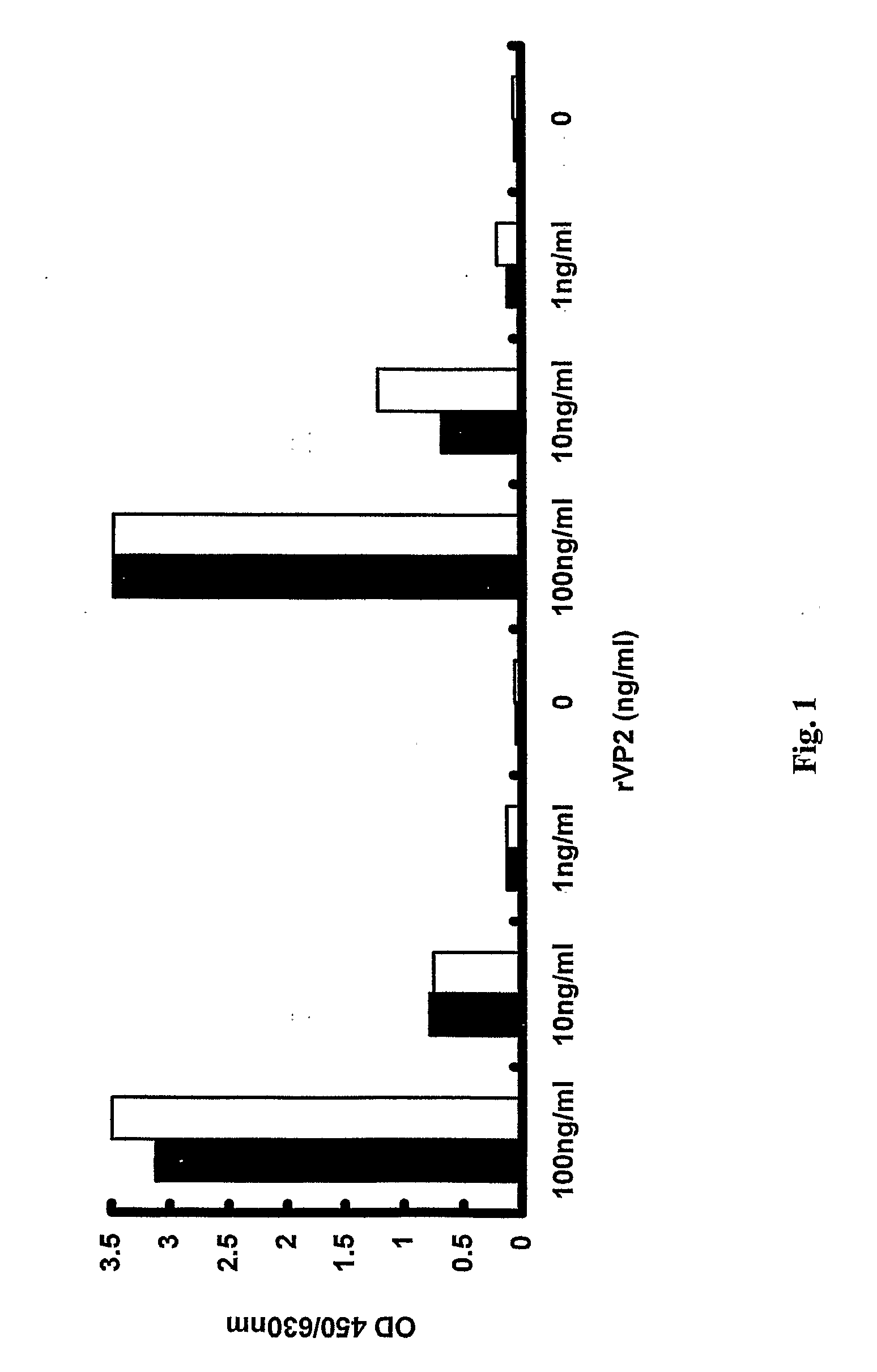
[0056]Infected Sf9 cells were harvested 3 days post-infection and lysed by sonication in PBS. Cell debris was removed by centrifuging at 3000 g for 10 minutes and the resultant supernatant was precipitated with PEG / NaCl overnight. Pellets containing VP2 were then resuspended in PBS and quantified. To estimate the assay sensitivity two separate batches of VP2 were diluted to 1 μg / ml and then decimally diluted to 1 pg / ml to determine the assay cut-off. The virus-capture EIA in accordance with the invention as described in Example 1 was able to detect recombinant VP2 to a concentration of 10 pg / ml. The results are shown in FIG. 6, wherein the cut off of the assay is clearly depicted.
example 3
[0057]The three parvovirus variants were analysed on the virus capture assay according to Example 1. Genotype 3 VP2 was obtained from Jean Pierre Allain, Cambridge Blood Centre, Cambridge, Long Road, CB2 2PT, United Kingdom (reference Parsyan, A., et al (2006) supra). The genotype 2 (A6) sample was received as a gift (Baxter), which had previously been sequenced and quantified (titre of 2.8×10¹¹ IU / ml). Genotype 1 was obtained from the Dutch Blood Bank.
[0058]Genotype 1 VP2 capsid protein was found to exhibit similar reactivity to genotype 3 on the virus capture EIA. The results are shown in FIG. 7.
[0059]As indicated above, FIG. 7 is a comparison of genotype 1 (clear boxes) and genotype 3 (filled boxes) recombinant VP2. Samples were diluted to 1000, 100 and 10 ng / ml of protein in the low pH (3.6) buffer. Error bars represent the standard deviation from the mean. Both genotypes were shown to have very similar reactivity on the virus capture assay. This proves that the virus capture as...
PUM
 Abstract
Description
Claims
Application Information
 - R&D
- Intellectual Property
- Life Sciences
- Materials
- Tech Scout
- Unparalleled Data Quality
- Higher Quality Content
- 60% Fewer Hallucinations
Browse by: Latest US Patents, China's latest patents, Technical Efficacy Thesaurus, Application Domain, Technology Topic, Popular Technical Reports.
© 2025 PatSnap. All rights reserved.Legal|Privacy policy|Modern Slavery Act Transparency Statement|Sitemap|About US| Contact US: help@patsnap.com


