Apparatus for photodynamic therapy and photodetection

a photodynamic therapy and apparatus technology, applied in the field of apparatus for photodynamic therapy and photodetection, can solve the problems of difficult operation, non-coherent white light illumination, and different illuminations of the field of view, so as to achieve effective photodetection and photodynamic therapy

Active Publication Date: 2010-06-10
KOREA ELECTROTECH RES INST
View PDF14 Cites 70 Cited by
  • Summary
  • Abstract
  • Description
  • Claims
  • Application Information

AI Technical Summary

Benefits of technology

[0014]The present invention has been made in an effort to solve the above-described problems associated with prior art. Accordingly, the present invention provides an apparatus for photodynamic therapy and photodetection, which includes a combined light source that can irradiate light having various spectral components to an object tissue, an optical imaging system that can provide direct and three-dimensional images of the object tissue that can be seen with naked eye, and a multispectral imaging system that can obtain images from several spectral portions for these various spectral ranges at the same time, thus performing effective photodetection and photodynamic therapy.

Problems solved by technology

Thus, the above-described U.S. Pat. No. 6,766,184 has problems in that it is impossible to obtain multispectral images by four spectral channels (R, G, B, and NIR) located in the visible and near-infrared spectral ranges at the same time, and further it is impossible to control the spectral configuration of the non-coherent light source in white light illumination.
Moreover, in the case where the apparatus is used as an endoscope, there are problems in that when the illumination is performed through the two difference light guides, one of the two light guides should use the passage of the endoscope so as to deliver two light beams, which makes it difficult to perform the operation and provides different illuminations to the field of view.
Moreover, since the collimating optics is not employed, non-uniform illumination is given to the field of view in a colposcope.
Therefore, there are problems in that it does not provide a color video for monitoring an object tissue in white light illumination and it is impossible to obtain multispectral images in the visible and near-infrared spectral ranges.
Thus, when the apparatus is used as an endoscope, the instrument channel of the endoscope should be used for the light source illumination through two different light guides, and this makes it difficult to perform the endoscope operation and provides non-uniform illumination to the field of view.
However, this system does not include a combined light source that can simultaneously produce wavelengths having two or more differences determined by a user and having a wide spectral range and monochromatic light spectra for photodynamic therapy and / or multispectral fluorescence and reflectance light detection.
Thus, it is impossible to excite two different kinds of fluorescent materials by optimum wavelength combination.
Moreover, the system does not include an optical imaging system for stereoscopic viewing and projection with an additional optical lens on both sensors of a multispectral imaging system, and thus there is no possibility of recording images under white light and fluorescence at the same time.
Furthermore, since the system does not include a movable beam splitter having a dichroic mirror, there is no possibility that the dichroic mirror may be moved out of the optical path.
Thus, light loss occurs under operational conditions in which only a color image sensor is used.
In addition, there is no means for performing the photodynamic therapy.
However, since the camera is disposed in one of stereo paths behind a variable power optical system, only a small amount of illumination light detected by an objective lens of the microscope reaches the camera.
Thus, the stereo microscope disclosed in U.S. Pat. No. 5,571,108 has a problem in that a significant loss of optical signals emerging from the objective lens occurs.

Method used


Image

  • Apparatus for photodynamic therapy and photodetection
  • Apparatus for photodynamic therapy and photodetection
  • Apparatus for photodynamic therapy and photodetection

Examples


example 1-1

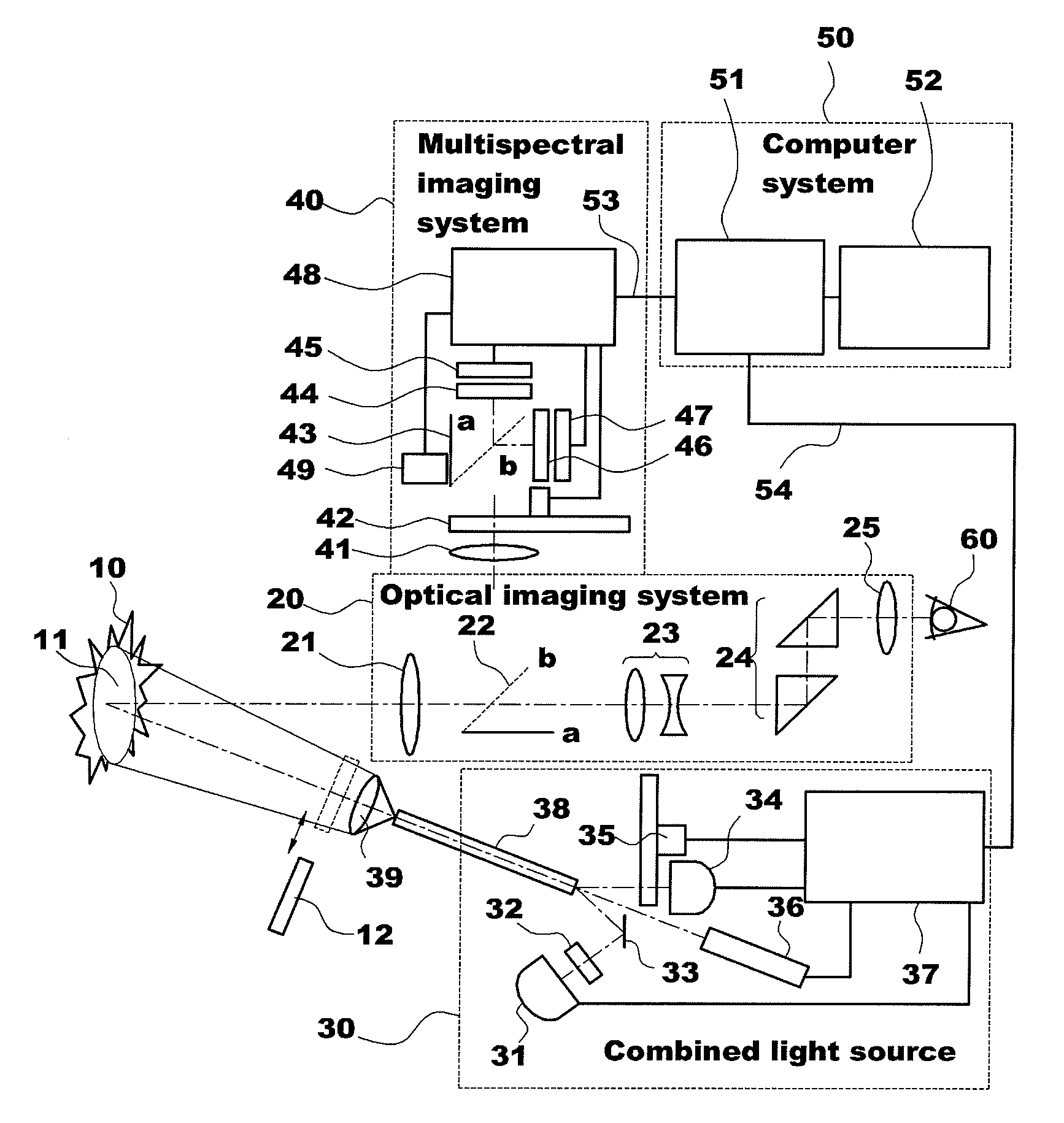
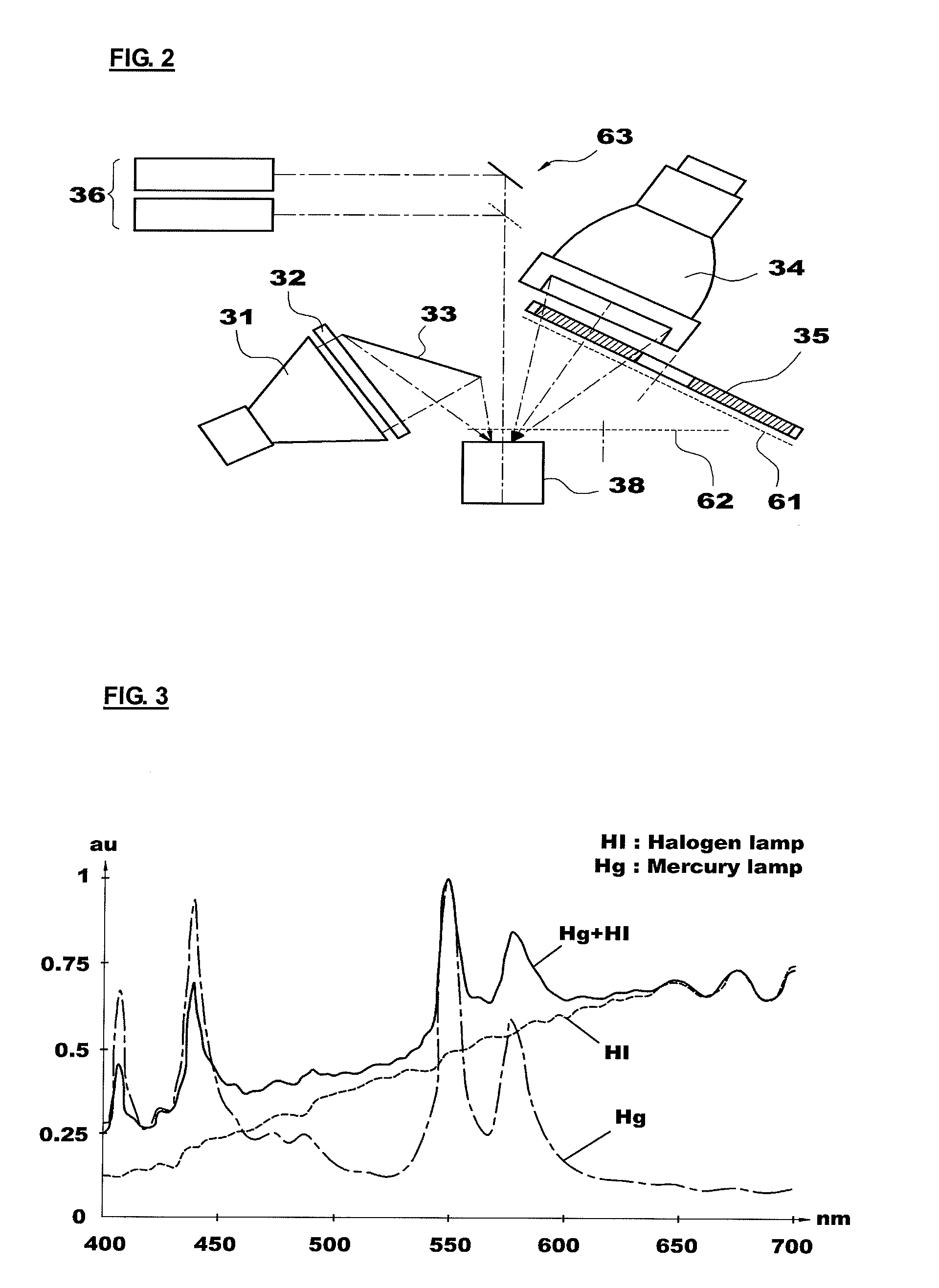
[0085]The combined light source 30 of the present invention employs the halogen lamp 31 and the mercury lamp 34 to irradiate white light to the object tissue and observe the reflectance light and defines a wideband filter having a spectral range of 400 to 700 (or 750) nm as the illumination filter wheel 35. The spectral components of the light irradiated to the object tissue can be flexibly changed by mixing the lights emitted from the mercury lamp 34 in various ratios using the first attenuator and the halogen lamp 31. In this case, the correlated color temperature varies in a range of 3,000 to 6,000 K. Spectral curves obtained from this are shown in FIG. 3. As shown in FIG. 3, when the amounts of lights from two lamps are properly complemented by each other, the spectrum is shown as a gentle and continuous curve.

[0086]The halogen lamp 31 is widely used as a colposcope light source, and a metal halide and an LED lamp may be used as the light source. Meanwhile, the reason that the c...

example 1-2

Excitation of Intrinsic Fluorescence of NADH and Flavin

[0087]In order to optimally excite these fluorescent materials, one of the excitation lights should be located in the ultraviolet region, and the other should be located at the short-wavelength side of the visible spectral region. The mercury lamp 34 with a band-pass filter (Ex1: 340±13 nm) in the illumination filter wheel 35 and the laser 36 (Ex2: 405 nm) satisfy the above conditions as the excitation light sources. The absorption wavelength bands of the two fluorescent materials are significantly different from each other, and the excitation wavelengths of the excitation light sources fall within these absorption wavelength spectral ranges, respectively (FIG. 4). In this case, the fluorescence emission spectra of the two fluorescent materials are different from each other (FIG. 7), and the fluorescence of the two fluorescent materials can be recorded by a single color detector (refer to Example 2-1 to be described below).

example 1-3

Excitation of Intrinsic Fluorescence of Flavin and PpIX

[0088]Since the light absorption wavelength ranges of the above fluorescent materials overlap in the vicinity of 400 nm, it is possible to excite the fluorescent materials at the same wavelength. However, in this case, the penetration depth of the excitation light is not so large. If the fluorescent materials are lo excited at a long-wavelength side of the light absorption spectrum of protoporphyrin IX (PpIX), the penetration depth of the excitation light is increased, which is very important in finding a tumor located in a deep part of the tissue.

[0089]FIG. 5 shows absorption spectra of the above-mentioned fluorescent materials and spectra of the two excitation light sources. In connection with the excitation conditions for flavin and PpIX fluorescence detection, the excitation light sources used in the experiment were the mercury lamp 34 with a band-pass filter (Ex1: 455±15 nm) and the laser 36 (Ex2: 635 nm). Since the outputs...


PUM


Abstract

The present invention provides an apparatus for photodynamic therapy and fluorescence detection, in which a combined light source is provided to illuminate an object body and a multispectral fluorescence-reflectance image is provided to reproduce various and complex spectral images for an object tissue, thus performing effective photodynamic therapy for various diseases both outside and inside of the body.For this purpose, the present invention provides an apparatus for photodynamic therapy and photodetection, which provides illumination with light of various wavelengths and multispectral images, the apparatus including: an optical imaging system producing an image of an object tissue and transmitting the image to a naked eye or an imaging device; a combined light source including a plurality of coherent and non-coherent light sources and a light guide guiding incident light emitted from the light sources; a multispectral imaging system including at least one image sensor; and a computer system outputting an image of the object tissue to the outside. Thus, the apparatus for photodynamic therapy and photodetection of the present invention can effectively perform the photodynamic therapy and photodetection by means of the combined light source capable of irradiating light having various spectral components to an object tissue and the multispectral imaging system capable of obtaining images from several spectral portions for these various spectral ranges at the same time, thus improving the accuracy of diagnosis and efficiency of the photodynamic therapy.

Description

CROSS-REFERENCE TO RELATED APPLICATION[0001]This application claims the benefit of Korean Patent Application No. 10-2008-0124970, filed on Dec. 10, 2008, the entire disclosure of which is hereby incorporated by reference.BACKGROUND OF THE INVENTION[0002]1. Field of the Invention[0003]The present invention relates to an apparatus for photodynamic therapy and photodetection, and more particularly, to an apparatus for photodynamic therapy and fluorescence detection, in which a combined light source is provided to illuminate an object body and a multispectral fluorescence-reflectance image is provided to reproduce various and complex spectral images for an object tissue, thus performing effective photodynamic therapy for various diseases both outside and inside of the body.[0004]2. Description of Related Art[0005]These days, diagnostic and therapeutic methods using light are widely used in the treatment of various skin diseases such as acne, chloasma, dark spots, maculae, scars, wrinkle...

Claims


Application Information

Patent Timeline
Patent Type & Authority Applications(United States)
IPC IPC(8): A61N5/067A61N5/06
CPCA61B5/0059G01N2201/0618A61N5/062G01J3/02G01J3/021G01J3/0248G01J3/10G01J3/12G01J3/36G01J3/4406G01N21/255G01N21/29G01N21/31G01N21/4738G01N21/6456G01N21/6486A61N5/0601A61B6/02G01N21/6447G01N21/64A61N5/06A61B1/00
Inventor KANG, UKV PAPAYAN, GARRI.
Owner KOREA ELECTROTECH RES INST
Why Eureka
Try Eureka