Bioactive Scaffold for Therapeutic and Adhesion Prevention Applications

a bioactive scaffold and adhesion technology, applied in the direction of drugs, prosthesis, peptide/protein ingredients, etc., can solve the problems of reduced tissue oxygenation, inadequate blood supply, and common conditions such as intestinal obstruction, so as to promote contractile cell migration, promote efficient regeneration, and inhibit fibrous adhesion formation

Active Publication Date: 2010-07-01
AXLE INT
View PDF4 Cites 7 Cited by
  • Summary
  • Abstract
  • Description
  • Claims
  • Application Information

AI Technical Summary

Problems solved by technology

Both an inadequate blood supply and reduced tissue oxygenation are common in surgically traumatized tissue.
Adhesion formation is a major complication of serosal repair following surgery, ischemia, or infection, and leads to conditions such as intestinal obstruction, severe abdominal pain, and infertility.
In addition to increased patient morbidity and mortality, adhesions present a significant burden to the health care system.
Postoperative adhesions often require additional surgical procedures to remove obstructions and may increase the risk, cost, and complexity of future operations.
As mentioned already, infection may be an important complication of the wound or injury healing.
These methods of drug delivery are simple but have a disadvantage of very fast clearance of the drug from the treatment area to other areas and therefore suboptimal spatial and temporary drug concentration and distribution patterns in the cavity.
However, these products are usually preparations which are produced already containing a specific drug agent and in addition to that often contain a binding agent to contain and regulate the drug release; they are also often made of materials which do not occur naturally in a human body and have delayed or incomplete clearance from the human body when implanted.
Such a technique, however, has several disadvantages: (1) injection of a collagen form which is not naturally present in a human body; (2) injection of a cross-linking substance which is another unnatural agent; and (3) such technology is suitable for the intramural and subcutaneous delivery, but not convenient for use in surgical procedures involving body cavities and internal surgery.
There has been no report to date of a method that is unequivocally effective at preventing fibrous adhesion formation.
These existing methods are not without drawbacks.
While select therapeutic agents have demonstrated an ability to reduce adhesion formation through their ability to alter various portions of the inflammatory wound healing response, these drugs are rapidly cleared from the wound site, which decreases their overall effectiveness during the approximately week-long fibrous adhesion formation process.
Many of these products have demonstrated a degree of effectiveness but have several common drawbacks with regard to surgical use or clinical complications.
SepraFilm® (carboxymethylcellulose / hyaluronic acid) has documented handling difficulty and may result in intraperitoneal abscesses due to differences in clearance of its polymeric components (i.e. fragmentation of film).
(expanded polytetrafluoroethylene) is difficult to handle during laparoscopy, does not biodegrade and may require additional surgery for removal, introducing the possibility of additional adhesion formation.
While these barriers endeavor to prevent the close, continuous physical contact of adhesiogenic and neighboring serosal surfaces, they do not by their nature address the key cellular aspect of the wound response that is responsible for fibrous adhesion maturation: the migration of fibroblasts into the fibrin gel matrix and its subsequent organization into a fibrous adhesion via collagen synthesis.

Method used


Image

  • Bioactive Scaffold for Therapeutic and Adhesion Prevention Applications
  • Bioactive Scaffold for Therapeutic and Adhesion Prevention Applications
  • Bioactive Scaffold for Therapeutic and Adhesion Prevention Applications

Examples


example

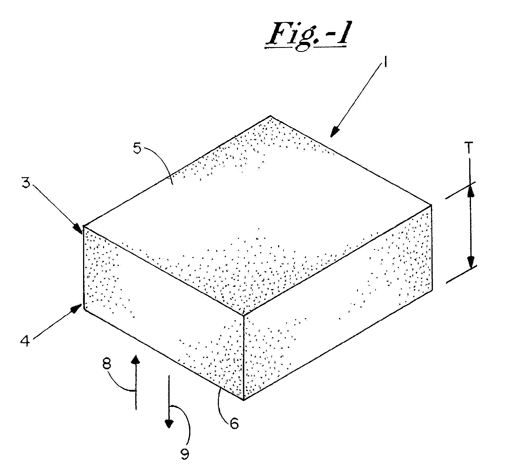
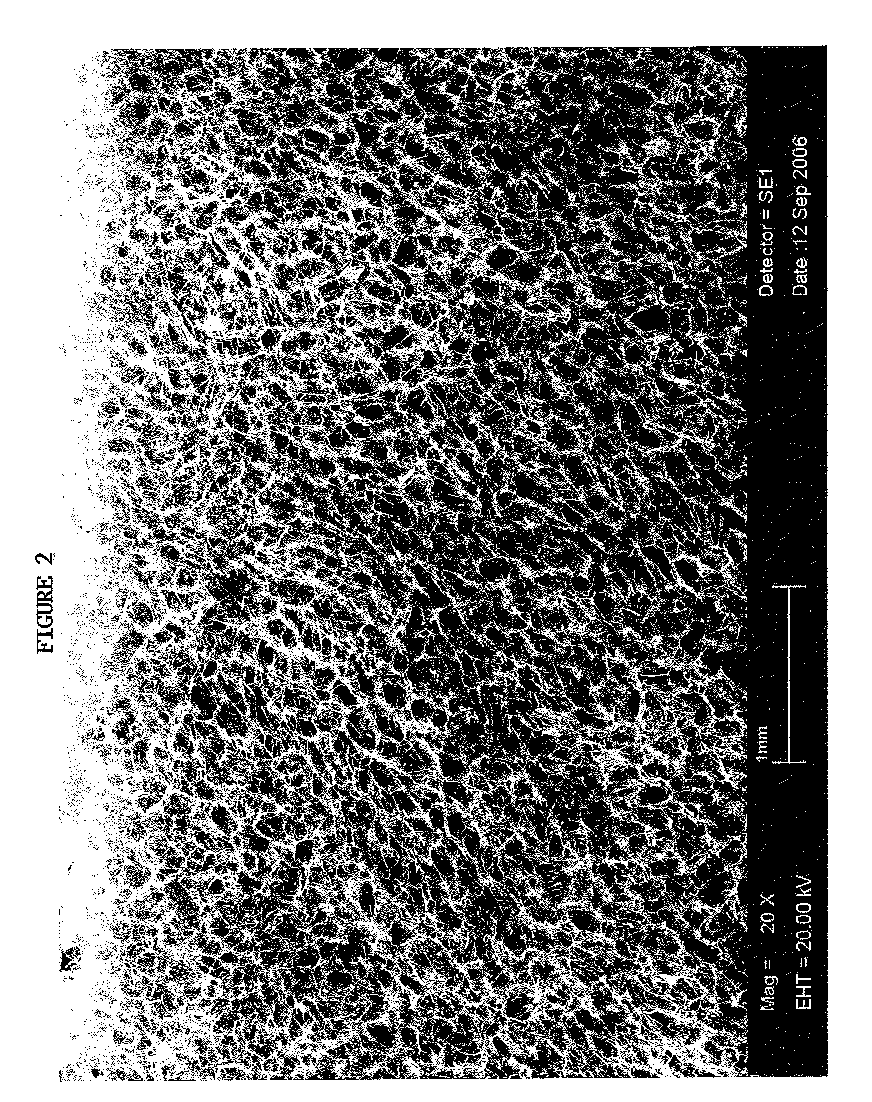
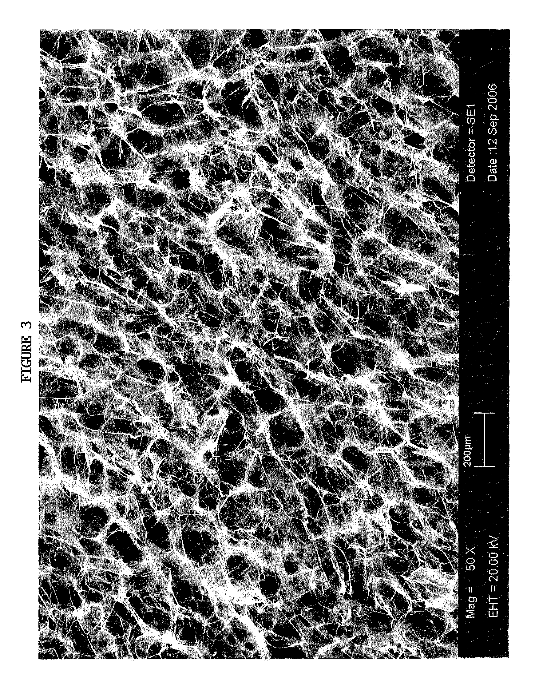
[0036]The TAS is a highly porous, three-dimensional thin sheet or “scaffold” that is prepared by co-precipitation of collagen and a glycosaminoglycan (e.g. chondroitin-6-sulfate), followed by lyophilization. The relevant properties of the TAS (including, but not limited to, its anti-adhesive, bioadhesive, bioresorptive, antithrombogenic and physical properties) can be modified by adjusting the chemical composition, pore size, pore volume fraction, degree of cross-linking, and TAS thickness to yield a scaffold variant that is designed to suit the specific needs of a wide array of wound sites. The initial incarnation of the TAS scaffold is a collagen-glycosaminoglycan (GAG) scaffold with a mean pore size between 5 and 200 μm, preferably between 20 and 150 μm; relative density of 0.5-10%, preferably 0.5-5%; and a degradation half life of 1-6 weeks, preferably 1-4 weeks. Scanning electron microscope photographs taken of a TAS scaffold in accordance with this example are shown inFIGS. 2 ...


PUM

PropertyMeasurementUnit
half lifeaaaaaaaaaa
half lifeaaaaaaaaaa
pore sizeaaaaaaaaaa

Abstract

A device for inhibiting adhesion of apposing human body tissue layers includes a scaffold having a designated mean pore size, relative density, and degradation half-life. The scaffold may be operably positioned between apposing tissue layers, such as proximate adhesiogenic layers at a wound site, so as to permit remesothelialization of the tissue without formation of fibrous adhesions. The scaffold device of the invention inhibits adhesion formation by promoting contractile cell migration away from the wound site for a predetermined period of time. The invention further relates to device and methods for promoting internal tissue regeneration, and for provision and / or dispensation of therapeutic and / or diagnostic agents in vivo.

Description

CROSS-REFERENCE TO RELATED APPLICATION[0001]This application is a divisional of U.S. patent application Ser. No. 11 / 683,393, filed on Mar. 7, 2007, and entitled “BIOACTIVE SCAFFOLD FOR THERAPEUTIC AND ADHESION PREVENTION APPLICATIONS”, which itself claims priority to U.S. Provisional Patent Application Ser. No. 60 / 779,762, filed on Mar. 7, 2006, and entitled “BIOACTIVE SCAFFOLD FOR THERAPEUTIC AND ADHESION PREVENTION APPLICATIONS”, the contents of which are incorporated herein in their entirety.FIELD OF THE INVENTION[0002]The present invention relates to the prevention of undesired tissue adhesion generally, and more particularly to devices for inhibiting tissue adhesion through diversion of fibroblast activities. The present invention further relates to methods and devices for therapeutic agent delivery, diagnostic agent delivery, and for promotion of tissue regeneration.BACKGROUND OF THE INVENTION[0003]The serosa comprises the outermost layer of the visceral structures that lie in...

Claims


Application Information

Patent Timeline
Patent Type & Authority Applications(United States)
IPC IPC(8): A61F2/00
CPCA61L31/129A61L31/146C08L89/06A61K9/0024A61P41/00
Inventor HARLEY, BRENDAN A.SOLLER, ERIC C.AIAZIAN, ERIC
Owner AXLE INT
Why Eureka
Try Eureka