Method for cartilage tissue regeneration via simulated microgravity culture using scaffolds

Inactive Publication Date: 2010-09-02
NAT INST OF ADVANCED IND SCI & TECH
View PDF3 Cites 19 Cited by
  • Summary
  • Abstract
  • Description
  • Claims
  • Application Information

AI Technical Summary

Benefits of technology

[0007]An object of the present invention is to provide a method for engineering of homogeneous cartilage tissue from bone marrow cells in a more rapid manner in a simulated microgravity environment, while allowing control of the shape of the formed cartilage tissue.
[0015]According to an embodiment of the present invention, bone marrow cells isolated from a target (patient) in need of cartilage tissue transplantation are used. Cartilage tissue engineered from bone marrow cells isolated from the target of transplantation is free from the risk of rejection or the like by the patient. Thus, such tissue can be preferably used to regenerate and / or repair cartilage defects of the target.
[0016]According to the present invention, homogeneous cartilage tissue of a desired shape can be more rapidly engineered from bone marrow cells. Accordingly, the present invention is highly applicable in clinical settings, such as regenerative medicine aimed at treating rheumatoid arthritis or osteoarthritis in the orthopedic field or at repairing auricular cartilage in the plastic surgery field.

Problems solved by technology

With conventional techniques, however, considerable mechanical stimuli and damages are imposed on cells.
Accordingly, it is difficult to obtain a large tissue mass.
Even if a large tissue mass were to be obtained, the inner region of the formed tissue would be likely to become necrotic.
Since multiaxial bioreactors cannot minimize shear stress, it is difficult to realize an ideal simulated microgravity environment.
With this technique, however, the shape of the constructed cartilage tissue cannot be controlled.
This disadvantageously limits the clinical applications thereof, where engineering of tissue that is suitable for the damaged area is required.
However, there is no report concerning the engineering of cartilage tissue from bone marrow cells in RWV rotation culture using scaffolds.
A variety of cellular scaffolds for static culture have been known; however, types of scaffolds that are suitable for cell culture in a simulated microgravity environment in a rotary bioreactor cannot be predicted based on the results of static culture.

Method used


Image

  • Method for cartilage tissue regeneration via simulated microgravity culture using scaffolds
  • Method for cartilage tissue regeneration via simulated microgravity culture using scaffolds
  • Method for cartilage tissue regeneration via simulated microgravity culture using scaffolds

Examples


example 1

Cartilage tissue engineering from mesenchymal stem cells derived from rabbit bone marrow using collagen sponge in RWV bioreactor

1. Experimentation

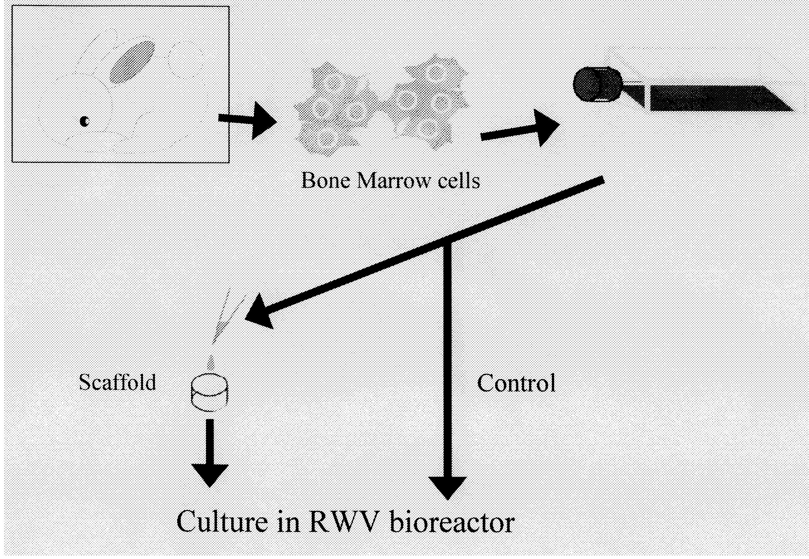
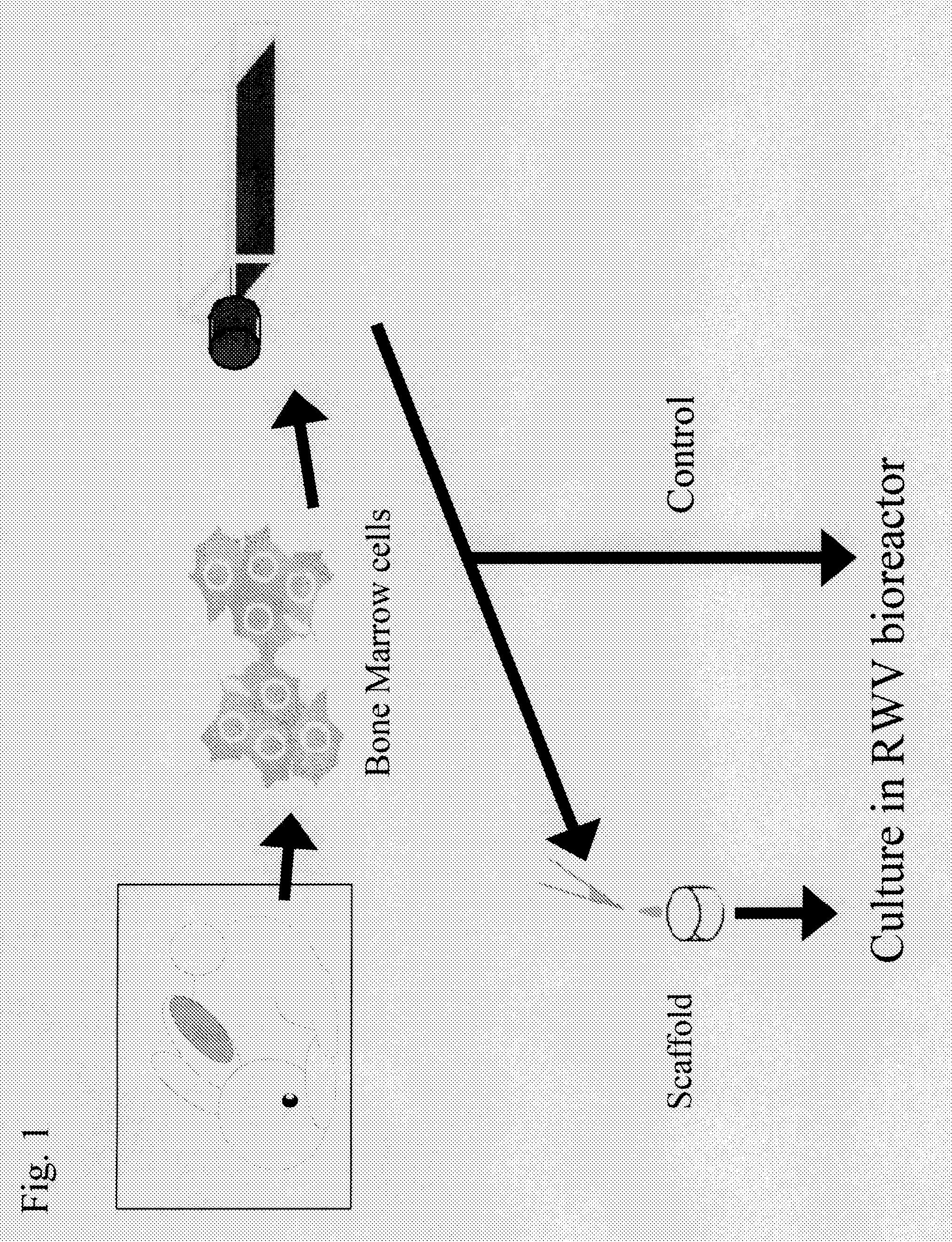
[0060](1) Preparation of Mesenchymal Stem Cells Derived from Rabbit Bone Marrow

[0061]Mesenchymal stem cells derived from rabbit bone marrow were prepared from the femur of a 2-week-old JW rabbit (female) in accordance with the method of Maniatopoulos et al. (Maniatopoulos, C., Sodek, J., and Melcher, A. H., 1988, Cell Tissue Res., 254, pp. 317-330). The harvested cells were cultured in DMEM containing 10% FBS (Sigma) and Antibiotic-Antimycotic (GIBCO BRL) for 3 weeks, and they were allowed to grow.

(2) Culture of Mesenchymal Stem Cells Derived from Rabbit Bone Marrow

[0062]The mesenchymal stem cells derived from rabbit bone marrow thus prepared were seeded on collagen sponges (prepared by extracting and purifying type I collagen from the porcine skin, lyophilizing the same, and crosslinking the same) at a density of 1.5×10⁸ cells / cm3 and sus...

example 2

Comparison of Static Culture and RWV Rotation Culture with the Use of Collagen Sponges

1. Experimentation

[0076]Bovine articular cartilage was harvested and sliced, and the cartilage matrix was removed with the aid of collagenase and cultured in a common cell culture medium (MEM+10% FBS) to prepare bovine articular cartilage-derived chondrocytes. The bovine articular cartilage-derived chondrocytes were seeded on collagen sponges (prepared by extracting and purifying type I collagen from the porcine skin and lyophilizing the same) at a density of 1.5×10⁸ cells / cm3 and suspended in 10 ml of DMEM culture medium (Sigma) containing 10−7 M dexamethasone (Sigma), 10 ng / ml TGF-β (Sigma), 50 μg / ml ascorbic acid (Wako), ITS+Premix (BD), 40 μg / ml L-proline (Sigma), and Antibiotic-Antimycotic (GIBCO BRL). The resultant was subjected to static culture (pellet culture) or rotation culture using an RWV bioreactor (Synthecon) for 3 hours.

[0077]Static culture was conducted by adding 10 ml of the cell ...

example 3

Comparison of Various Cellular Scaffolds in Cartilage Tissue Engineering Using RWV Bioreactor

1. Experimentation

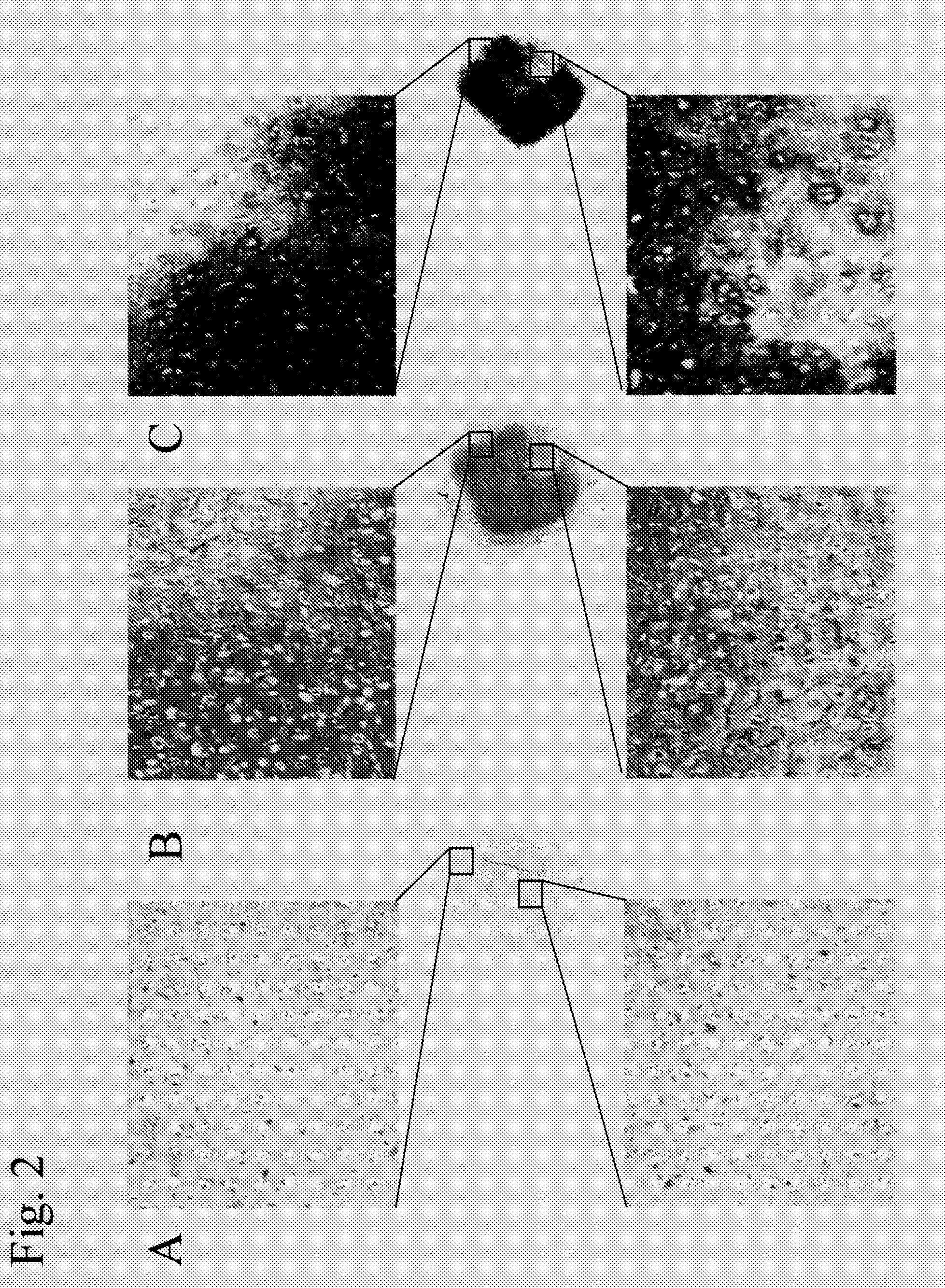
[0079]Using open-cell polylactic acid (OPLA, BD) and a porous composite of hyaluronic acid and hydroxyapatite (hereafter abbreviated as “HAP-HA”) as scaffolds, cartilage tissue regeneration was performed using an RWV bioreactor. OPLA is a synthetic polymer scaffold synthesized from D,D-L,L-polylactic acid (spongy / noncompressive), and the declared pore size thereof is between 100 μm and 200 μm.

[0080]Bovine articular chondrocytes that had been prepared in the same manner as in Example 2 were seeded on OPLA and HAP-HA at a density of 1.5×10⁸ cells / cm3 and suspended in 10 ml of DMEM culture medium (Sigma) containing 10−7 M dexamethasone (Sigma), 10 ng / ml TGF-β (Sigma), 50 μg / ml ascorbic acid (Wako), ITS+Premix (BD), 40 μg / ml L-proline (Sigma), and Antibiotic-Antimycotic (GIBCO BRL). The resultant was subjected to rotation culture using an RWV bioreactor (Synthecon) for 2 weeks....


PUM


Abstract

This invention relates to a method for cartilage tissue engineering using scaffolds in simulated microgravity culture. This invention enables engineering of homogeneous cartilage tissue using bone marrow cells in a more rapid manner in a simulated microgravity environment, while allowing control of the configuration of the resulting cartilage tissue.

Description

TECHNICAL FIELD [0001]The present invention relates to a method for cartilage tissue engineering via simulated microgravity culture using scaffolds. More particularly, the present invention relates to a method for cartilage tissue engineering by seeding bone marrow cells on collagen-based scaffolds or the like and culturing the composite in a simulated microgravity environment.BACKGROUND ART [0002]Three-dimensional tissue regeneration from cells generally requires three-dimensional culture or stirred culture with appropriate scaffolds. With conventional techniques, however, considerable mechanical stimuli and damages are imposed on cells. Accordingly, it is difficult to obtain a large tissue mass. Even if a large tissue mass were to be obtained, the inner region of the formed tissue would be likely to become necrotic.[0003]In order to overcome such drawbacks, there are sets of bioreactors designed to optimize gravity. For example, the rotating wall vessel (RWV) bioreactor is a NASA-...

Claims


Application Information

Patent Timeline
IPC IPC(8): C12N5/077C12N5/0789
CPCA61K35/12A61L27/3834A61L27/3852A61L27/3895A61L2430/06C12N2533/54C12N5/0655C12N2500/25C12N2501/15C12N2501/39C12N2525/00C12N5/00
Inventor TANAKA, JUNZOIKADA, YOSHITOOHYABU, YOSHIMIUEMURA, TOSHIMASA
Owner NAT INST OF ADVANCED IND SCI & TECH
Why Eureka
Try Eureka