Corneal endothelial preparation which enables cells to grow in vivo

Active Publication Date: 2010-09-16
SENJU PHARMA CO LTD
View PDF8 Cites 13 Cited by
  • Summary
  • Abstract
  • Description
  • Claims
  • Application Information

AI Technical Summary

Benefits of technology

[0025]According to the present invention, since a corneal endothelial preparation capable of cell proliferation in vivo is provided, it enables superior transplantation of a corneal endothelial graft, as well as growth of transplanted cells, thus affording a corneal endothelium regenerative effect, with a graft having a smaller area than conventional ones, i.e., a corneal endothelial preparation. The corneal endothelial preparation of the present invention can be utilized as a t

Problems solved by technology

However, corneal donation in Japan is insufficient, and the number of waiting patients for corneal transplantation is about 5500, but the number of corneal transplantation domestically performed per year is

Method used


Image

  • Corneal endothelial preparation which enables cells to grow in vivo
  • Corneal endothelial preparation which enables cells to grow in vivo
  • Corneal endothelial preparation which enables cells to grow in vivo

Examples


Example

Example 1

Preparation of Cultured Corneal Endothelial Cell for Transplantation

[0057]A cynomolgus monkey (3 to 8 years old, male, Keari Co., Ltd.) was euthanized and the eye ball was removed. Corneal endothelial cells were collected together with the Descemet's membrane from the corneal tissue of the removed eye, and treated with DISPASE to separate the corneal endothelial cells. The separated corneal endothelial cells were seeded (1×10⁵ cells / well) on a 12 well culture plate (manufactured by Corning) coated with FNC coating mix (registered trade mark), and cultured in a culture medium (DMEM, manufactured by GIBCO Invitrogen) supplemented with 1% fetal calf serum and 2 mg / ml bFGF (Gibco Invitrogen) under the conditions of 37° C., 5% CO2. When the cells reached confluent, they were detached by a Trypsin-EDTA treatment, and subcultured 3 times at a density of about 1:3. The cultured corneal endothelial cells were finally seeded on a collagen sheet (manufactured by AGC Techno Glass) at a...

Example

Example 2

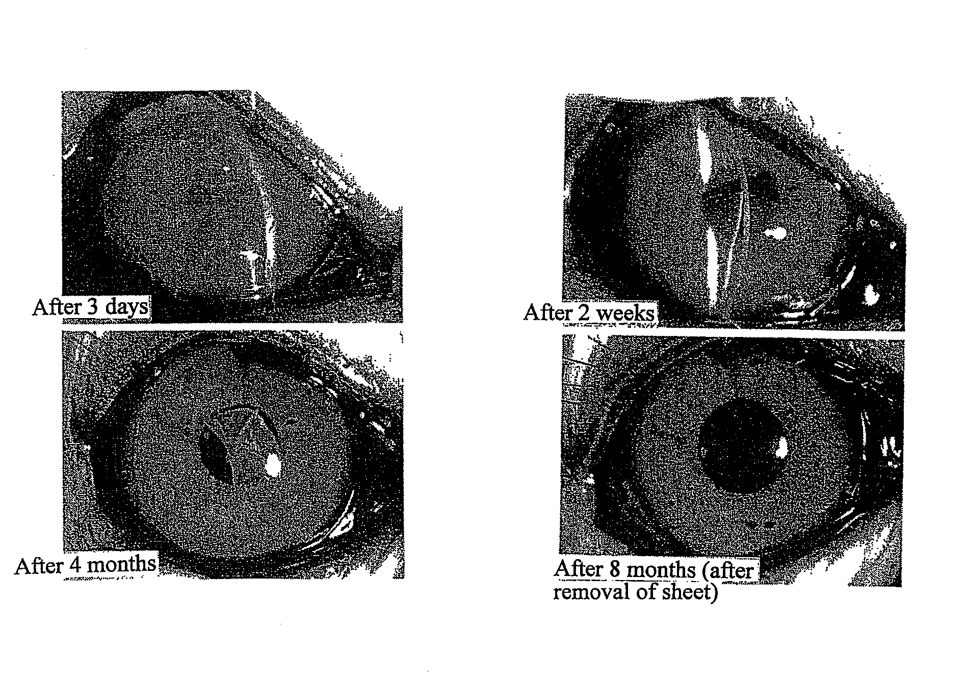
[0058]Transplantation Procedure of Cultured Corneal Endothelium

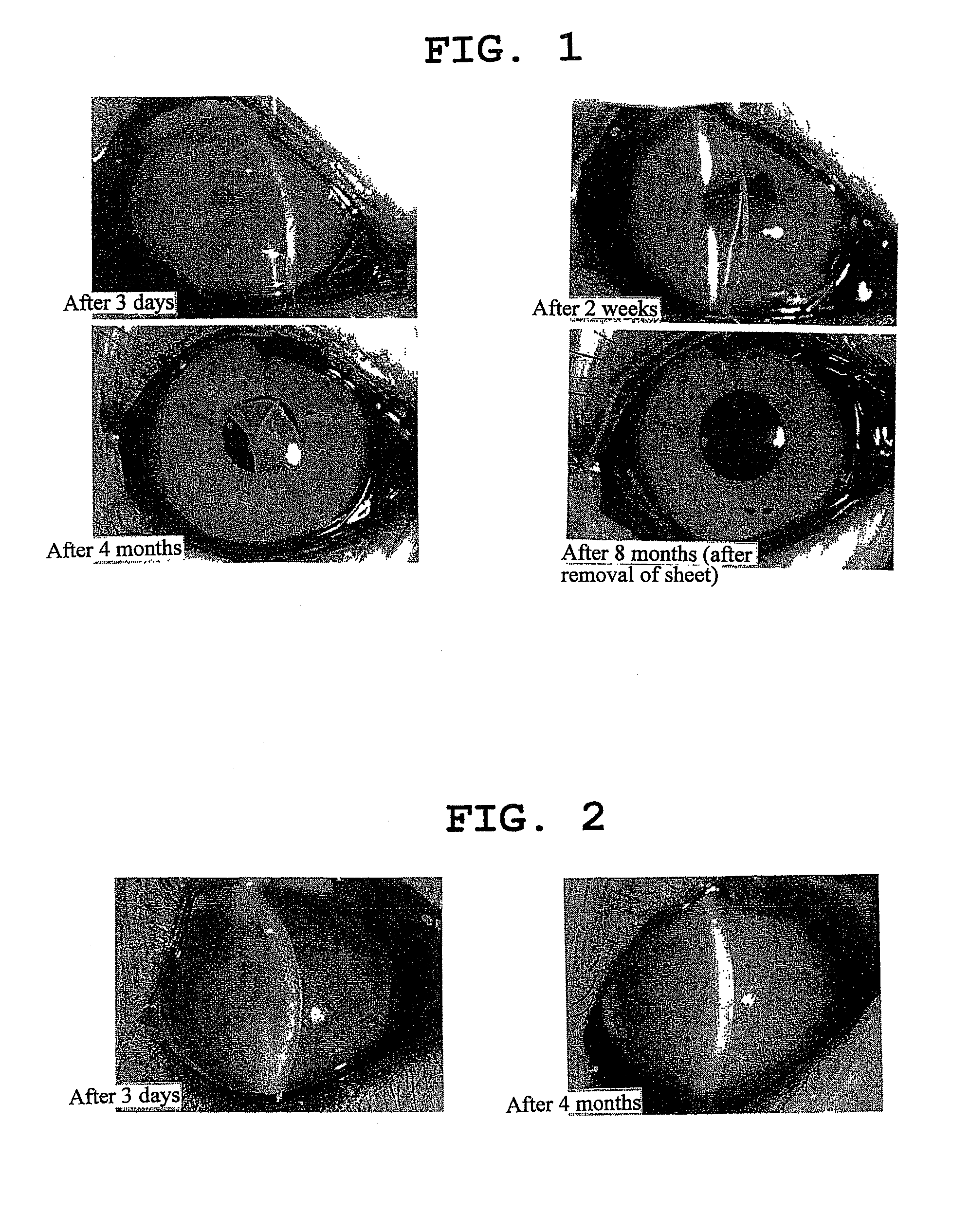
[0059]Four cynomolgus monkeys (3-5 years old, female, KEARI Co., Ltd., CLEA Japan, Inc., LaboProducts Limited) were carried from the cage to a treatment table under systemic anesthesia by intramuscular injection of a mixed anesthesia of ketamine hydrochloride and xylazine hydrochloride. Inhalation anesthesia using a mask was started on the treatment table. After confirmation of the systemic stable conditions, about 5-6 mm of corneoscleral incision was performed in the sclera at 1 mm outside the limb, which is the boundary between cornea and conjunctiva, of 4 eyes (either eye) of the four monkeys, and the corneal endothelial cells were removed from the part as much as possible by physical scraping (diameter about 9 mm). The removed area of corneal endothelial cells was confirmed by Trypan Blue staining. The corneal endothelial sheet obtained in Example 1 was transplanted to the three eyes. As a control, a collage...

Example

Example 3

Histological Evaluation of Corneal Endothelium

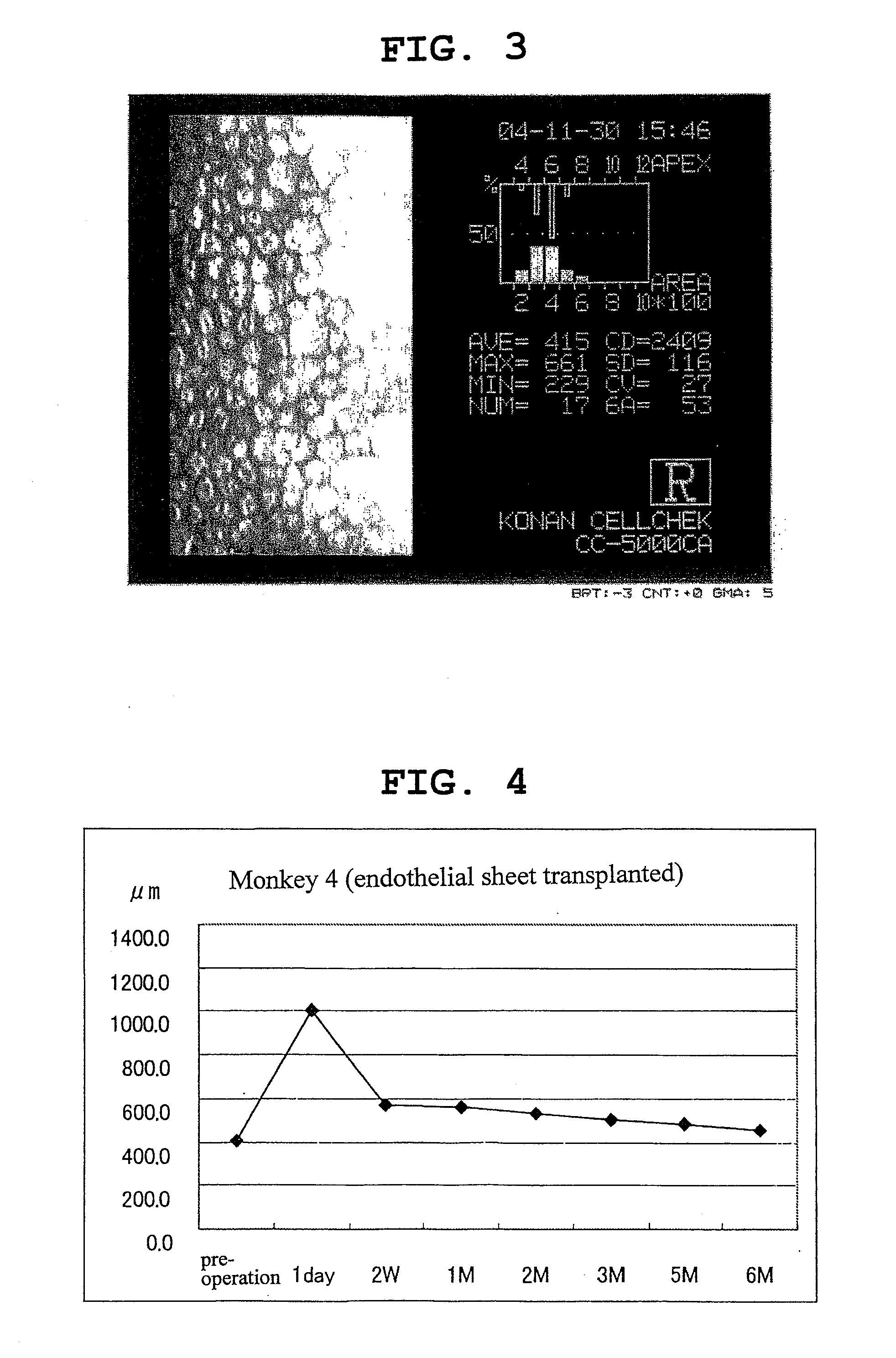
[0062]Among the 4 cynomolgus monkeys that underwent the cultured corneal endothelial transplantation procedure in Example 2, two were euthanized at 6 months post-transplantation, and the isolated corneal tissue was observed with a scanning electron microscope and a transmission electron microscope.

[0063]The both monkeys showed good adhesion between cells since polygonal corneal endothelial cells with a diameter of about 15-30 μm were arranged in one layer inside the cornea (where intrinsic corneal endothelial cells are to be present). In addition, a new basal membrane layer was formed between the corneal endothelial cell and the Descemet's membrane, an intrinsic basal membrane (FIG. 5). It is clear therefrom that the corneal endothelial cell adheres well to the back of the cornea, and constructs a cell biological structure similar to that of intrinsic corneal endothelial cells, thereby exhibiting the normal corneal endothelial f...


PUM

PropertyMeasurementUnit
Fractionaaaaaaaaaa
Timeaaaaaaaaaa
Areaaaaaaaaaaa

Abstract

The present invention provides a graft more suitable for the transplantation of corneal endothelial cells and an application method thereof. Specifically, the present invention provides a corneal endothelial preparation capable of cell proliferation in vivo, which contains a substrate and a corneal endothelial cell layer cultured on the substrate, and a treatment method of a disease selected from the group consisting of bullous keratopathy, corneal edema, corneal leukoma and corneal endothelial inflammation, which includes a step of transplanting the preparation to patients. As the substrate, collagen is used.

Description

TECHNICAL FIELD[0001]The present invention relates to a corneal endothelial preparation capable of cell proliferation in vivo, which is used as a graft for the treatment of corneal endothelial disorders.BACKGROUND ART[0002]Vision information is recognized when the light enters through the cornea, which is a transparent tissue at the front of the eye ball, reaches the retina to excite the retinal nerve cell that generates electric signals, which are transmitted to the cerebral visual area via the optic nerve. Good eyesight requires a transparent cornea. The transparency of the cornea is maintained since the pump function and barrier function of the corneal endothelial cell keep a constant water content.[0003]The corneal endothelial cell density is about 3000 cells / mm2 in human at birth. The cell has no ability to regenerate after damage. In endothelial corneal dystrophy and bullous keratopathy caused by functional disorder of corneal endothelium due to various etiologies, the corneal...

Claims


Application Information

Patent Timeline
IPC IPC(8): A61F2/14A61K35/44A61P27/02A61K35/30A61L27/00A61L27/24A61L27/38A61L27/58
CPCA61L27/24A61L2430/16A61L27/3839A61L27/3808A61P27/02
Inventor KOIZUMI, NORIKOKINOSHITA, SHIGERUSAKAMOTO, YUJI
Owner SENJU PHARMA CO LTD
Why Eureka
Try Eureka