Root canal filler and dental tissue regeneration method

a tissue regeneration and root canal technology, applied in the field of root canal filler and dental tissue regeneration method, can solve the problems of incomplete pulpectomy, loss of teeth, and inability to enlarge root canals and root canal fillings, and achieve the effects of restoring dental pulp function, accelerating blood vessel regeneration, and regenerating dental tissu

Active Publication Date: 2011-01-27
NAT CENT FOR GERIATRICS & GERONTOLOGY
View PDF11 Cites 24 Cited by
  • Summary
  • Abstract
  • Description
  • Claims
  • Application Information

AI Technical Summary

Benefits of technology

[0035]It was possible to regenerate dental tissue by inserting the root canal filler according to the present invention into the root canal after pulpectomy or enlargement and cleaning of the root canal of the infected root canal, even in the case of deep caries with pulpitis or periapical disease with mature teeth with complete apical closure. The root canal filler according to the present invention accelerates regeneration of blood vessel and nerve and regeneration of dental pulp and restoration of dental pulp function, by proliferation of dental pulp cells. In addition, application of a morphogen such as BMP or a growth / differentiation factor on tooth-crown dental pulp resulted in accelerated differentiation of dental pulp cells into odontoblasts and lead to blockage of the tooth crown region with dentin. Further, the apical dental foramen was sealed by addition of tooth cementum.

Problems solved by technology

However, complete pulpectomy, enlargement of root canal and root canal filling are almost impossible.
Thus, pulpectomy often leads to periapical periodontitis, and has high possibility of resultant loss of the tooth.
However when the tissue is irreversible pulpitis with pain, there is no choice but to perform pulpectomy.
In the case of deep caries with pulpitis or periapical periodontitis in mature teeth with complete apical closure, no method nor root canal filler for dental tissue regeneration have been developed yet.
However, in these cases, the root canal filler may be separated from the dentinal wall or ruptured after root canal filling.
In addition, these root canal fillers may cause periapical periodontitis in several years.

Method used


Image

  • Root canal filler and dental tissue regeneration method
  • Root canal filler and dental tissue regeneration method
  • Root canal filler and dental tissue regeneration method

Examples


embodiment 1

[0079]The invention in the present embodiment relates to a dental tissue regeneration method for regeneration of dental tissue in root canal, characterized by injecting an extracellular matrix containing the cells enriched for dental pulp stem cells into the apical side of the root canal after pulpectomy or enlargement and cleaning of an infected root canal. The dental tissues to be regenerated are, for example, blood vessel, nerve, dental pulp, dentin and others in root canal. In the invention in the present embodiment after pulpectomy or enlargement and cleaning of an infected root canal, the dental pulp is removed and disinfected; the apical portion of the root is cut out open (apicoectomized); and a root canal filler is transplanted. In the invention in the present embodiment, a synthetic filler or blood clot is not transplanted into the disinfected hollow root canal, but a scaffold superior in biocompatibility without causing adverse effects and in low immunogenicity with cells...

embodiment 2

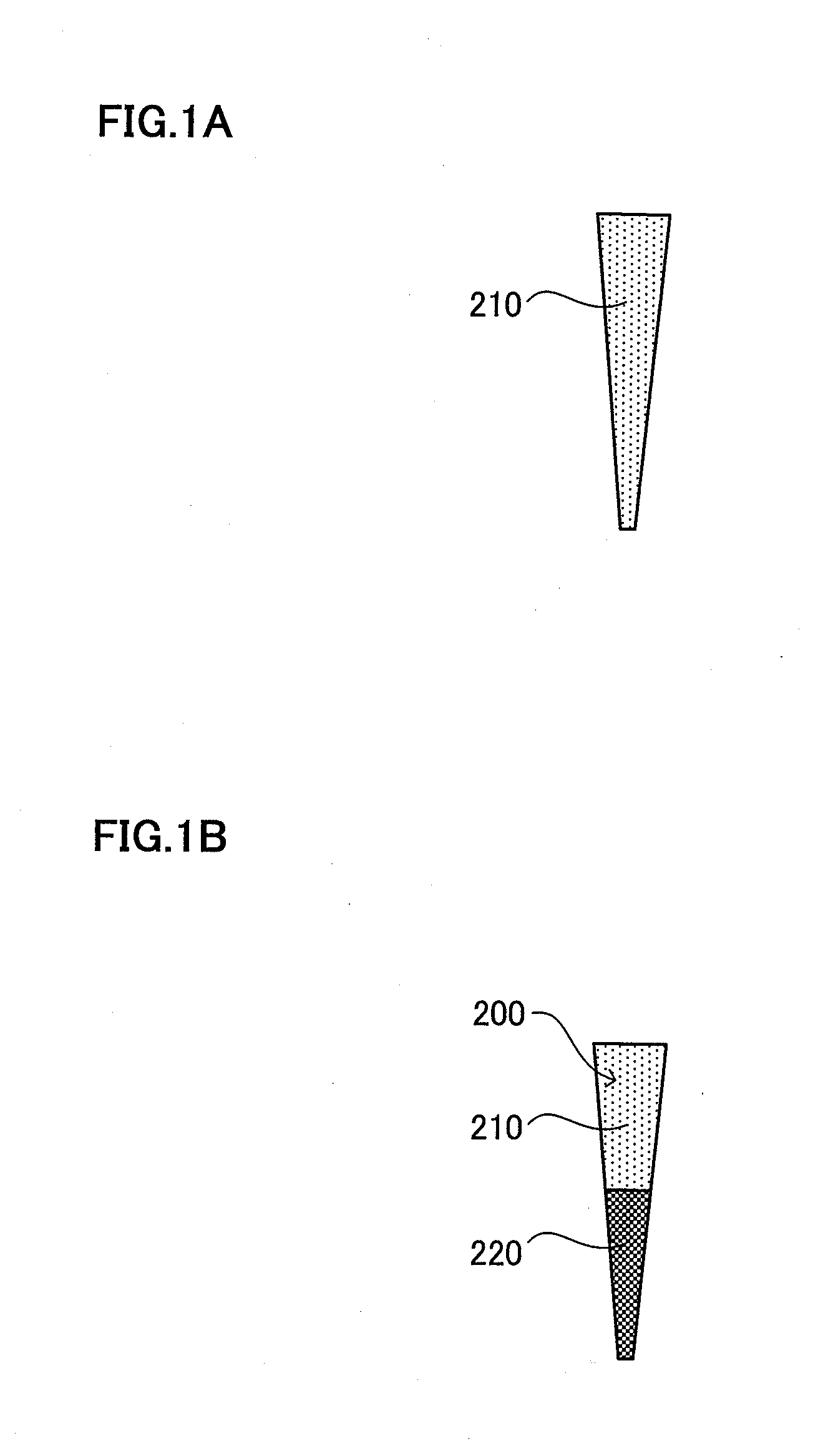
[0099]Hereinafter, the dental tissue regeneration method in embodiment 2 will be described with reference to FIGS. 2A to 2C. In a typical example of the method of producing the root canal filler 200, 30 to 40 μl of collagen mixture (mixing rate of type I and III collagens: 1:1) with a chemotactic factor is first absorbed, and 20 μl to 30 μl of collagen mixture with cells including dental pulp stem cells is then absorbed into the tip of Pipetman, to a total volume of 60 μl. Also in embodiment 2, absorption, for example into the tip of Pipetman is preferable to be sufficiently slow so that no air bubble is generated. The internal diameter of the Pipetman tip is preferably smaller. In this way, the root canal filler 200 shown in FIG. 2A is produced. As described above, in the case of the root canal filler 200, cells including dental pulp stem cells 220 are absorbed in the apical part of the root canal, and a chemotactic factor 230 containing at least one of cell chemotactic factor, cel...

example 1

Fractionation and Characterization of Cells

[0110]Porcine tooth germ was extracted and enzyme-digested with collagenase at 37° C. for 1 hour and a half for separation of dental pulp cells; the cells were dispersed in DMEM containing 2% serum at a concentration of 1×10⁶ cells / ml and labeled with 5 μg / ml Hoechst 33342. The cells were then labeled with CD31 and CD146 antibodies at 4° C. for 30 minutes, before analysis by flow cytometry. FIG. 3 shows the analytical results obtained by flow cytometry of the SP cells. The content of the porcine dental pulp-derived SP cells was 0.2% in the entire cells.

[0111]As shown in FIG. 4, three fractions of dental pulp-derived SP cells: CD31− / CD146− fraction; CD31+ / CD146− fraction and CD31+ / CD146+ fraction were obtained. Further fractionation of the SP cells after labeling with CD31 and CD146 antibodies showed CD31− / CD146− SP cells, CD31+ / CD146− SP cells, and CD31+ / CD146+SP cells, representing 50%, 48% and 2%, respectively. The cells were cultured in ...


PUM


Abstract

Provided is a novel and creative dental tissue regeneration method for regenerating dental tissue after pulpectomy or the enlargement and cleaning of an infected root canal. After pulpectomy or the enlargement and cleaning of an infected root canal, a root canal filler (200) having an extracellular matrix (210) containing the cells (220) enriched for dental pulp stem cells, is inserted into the apical side of the root canal of a target tooth (100). The cells including dental pulp stem cells include at least one of the following: dental pulp SP cells, CD31-negative and CD146-negative cells, CD24-positive cells, CD105-positive cells, and CD150-positive cells. For instance, dental pulp SP cells are CD31− and CD146− negative. Even if pulpitis due to deep caries occurs, appropriate dental pulp regeneration and recovery of dental pulp function are possible.

Description

TECHNICAL FIELD[0001]The present invention relates to a root canal filler and a dental tissue regeneration method by using the root canal filler.BACKGROUND ART[0002]When dental caries is deep enough to reach dental pulp, pulpectomy is usually performed for treatment of the caries. However, the dental pulp not only has a function to block external stimulus by reparative dentin formation, but also functions to inhibit further invasion of bacterial by sense and prevent tooth fracture caused by chewing a hard material with the sense of occlusion. In addition, the dental pulp can maintain protein and water in dentin by metabolism, and additionally keep the tensile strength and other properties of dentin. The dental pulp is also known to have an infection defense mechanism by immune system.[0003]NiTi alloy rotary files are used popularly in endodontics, because of morphological complexity of the root canal. However, complete pulpectomy, enlargement of root canal and root canal filling are...

Claims


Application Information

Patent Timeline
Patent Type & Authority Applications(United States)
IPC IPC(8): A61K35/12A61P1/02
CPCA61K6/0032A61L27/3633A61L27/3834A61L2430/12A61L2400/06A61L27/3865A61K6/50A61P1/02A61P43/00
Inventor NAKASHIMA, MISAKOIOHARA, KOICHIRO
Owner NAT CENT FOR GERIATRICS & GERONTOLOGY
Why Eureka
Try Eureka