

Mammary artery derived cells and methods of use in tissue repair and regeneration
a technology of mammary arteries and derived cells, which is applied in the field of mammalian mammary arteries, can solve the problems of ischemia, congestive heart failure, systolic hypertension, and coronary insufficiency, and achieve the effects of reducing the risk of ischemia, and improving the survival ra
- Summary
- Abstract
- Description
- Claims
- Application Information
AI Technical Summary
Benefits of technology
Problems solved by technology
Method used
Image

Examples
example 1
Cell Isolation Optimization
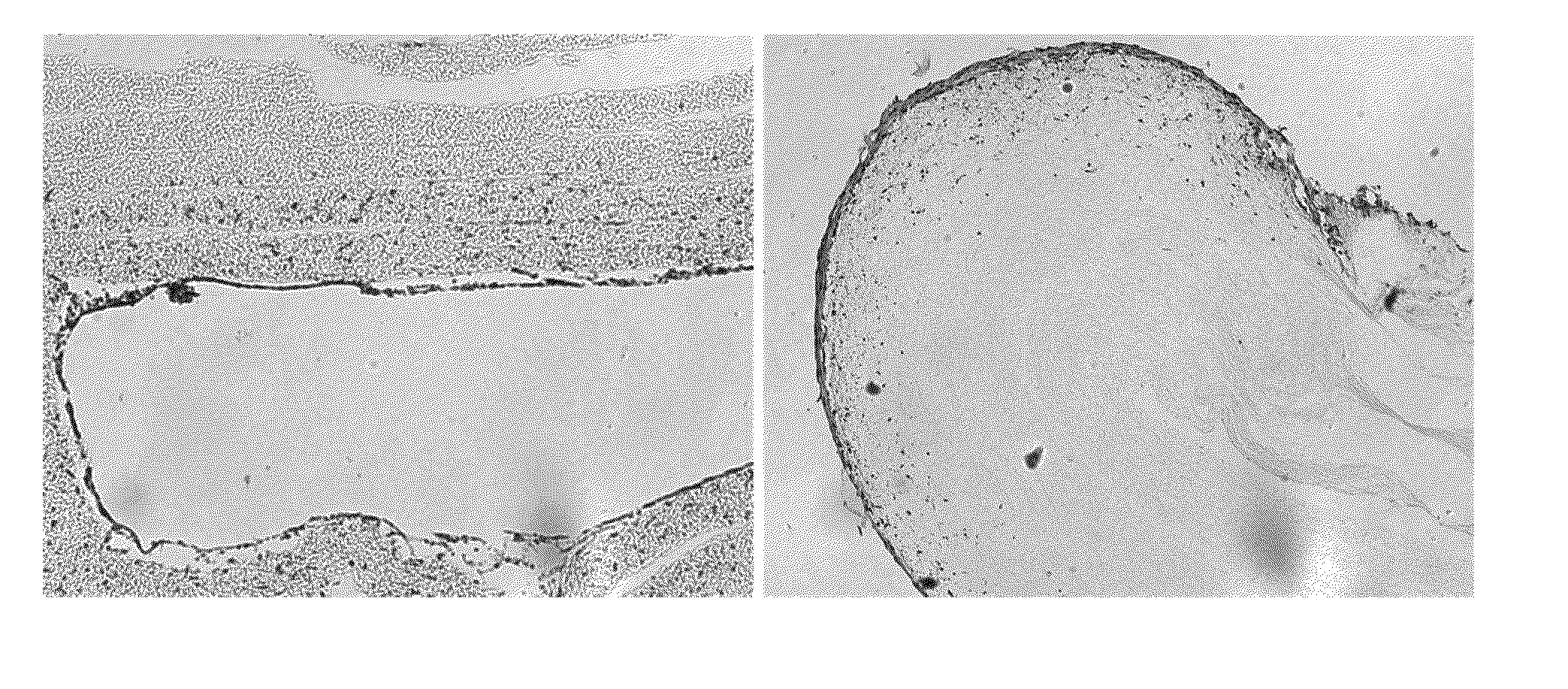
[0043]Initial experiments were conducted to determine the optimal time necessary for tissue digestion. A five centimeter portion of the human internal mammary artery was obtained from the National Disease Research Interchange (NDRI, Philadelphia, Pa.). The artery was trimmed and washed in Dulbecco's modified Eagles medium (DMEM-low glucose; Invitrogen, Carlsbad, Calif.) or phosphate buffered saline (PBS; Invitrogen) to remove blood and debris. The entire artery was then transferred to a 50-milliliter conical tube.
[0044]The tissue was then digested, for varying amounts of time in an enzyme mixture containing 0.25 Units / milliliter collagenase (Serva Electrophoresis, Heidelberg, Germany) and 2.5 Units / milliliter dispase (Roche Diagnostics Corporation, Indianapolis Ind.). The enzyme mixture was then combined with endothelial growth medium-2 (EGM-2) (Lonza, Walkersville, Md.). The conical tube containing the tissue, EGM-2 and digestion enzymes was incubated at ...
example 2
Isolation of Human Internal Mammary Artery-Derived Cells
[0046]The optimal digestion time interval (described in Example 1) was then applied to isolate internal mammary artery-derived cells. A five centimeter portion of the human internal mammary artery was obtained from the National Disease Research Interchange (NDRI, Philadelphia, Pa.). The artery was trimmed and washed in Dulbecco's modified Eagles medium (DMEM-low glucose; Invitrogen, Carlsbad, Calif.) or phosphate buffered saline (PBS; Invitrogen) to remove blood and debris. The entire artery was then transferred to a 50-milliliter conical tube.
[0047]The tissue was then digested in an enzyme mixture containing 0.25 Units / milliliter collagenase (Serva Electrophoresis, Heidelberg, Germany) and 2.5 Units / milliliter dispase (Roche Diagnostics Corporation, Indianapolis Ind.). The enzyme mixture was then combined with iMAC Growth Medium (Advanced DMEM / F12 (Gibco), L-glutamine (Gibco) penicillin (50 Units / milliliter) and streptomycin (...
example 3
Human Internal Mammary Artery-Derived Cell Morphology
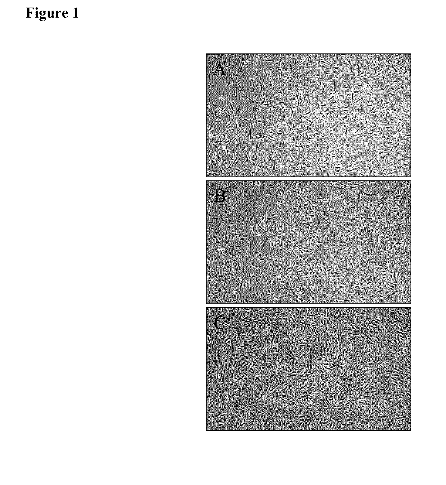
[0048]Fresh iMACs, isolated as described in Example 2, were plated at 5000 cells / cm2 onto type I rat tail collagen coated T75 flasks in iMAC Growth Medium and cultured at 37° C. in 5% carbon dioxide. Cells were passaged every 3-5 days. At each passage, cells were harvested with TypleLE (Gibco), counted and viability was measured using a Guava instrument (Guava Technologies, Hayward, Calif.). For morphological evaluation, iMACs were assessed by light microscopy and morphological characteristics of the cells were observed using a Nikon microscope and LCD digital camera.
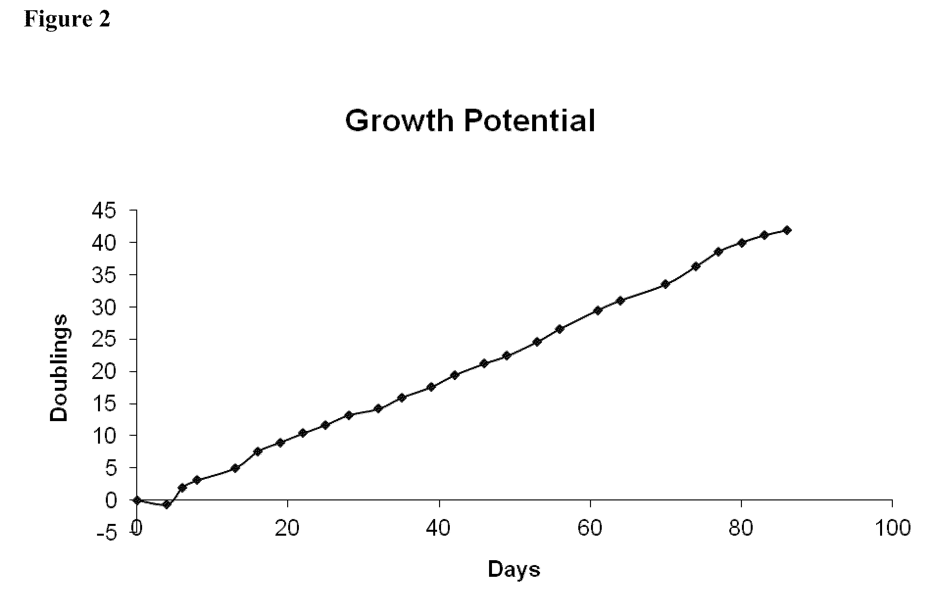
[0049]iMACs were assessed by light microscopy and morphological characteristics of the cells were observed using a Nikon Microscope and LCD digital camera (FIG. 1). Consistently, cultures of iMACs showed a fibroblastic morphology. Morphology was stable at late passage (Passage 23).
PUM
 Abstract
Description
Claims
Application Information