
Method for typing and identification of micro-organisms
a microorganism and typing technology, applied in the field of typing and identification of microorganisms, can solve the problems of high financial burden on the health care system, poor patient experience, and inability to implement programs
- Summary
- Abstract
- Description
- Claims
- Application Information
AI Technical Summary
Benefits of technology
Problems solved by technology
Method used
Image

Examples
example
[0111]Raman Spectroscopy
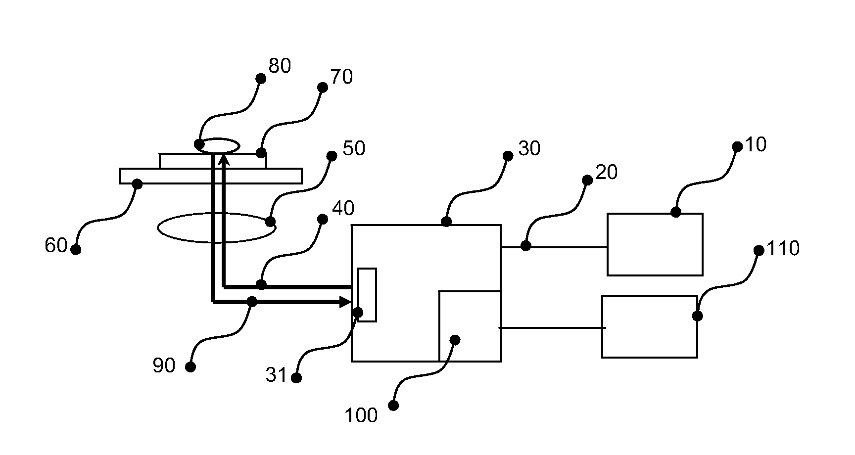
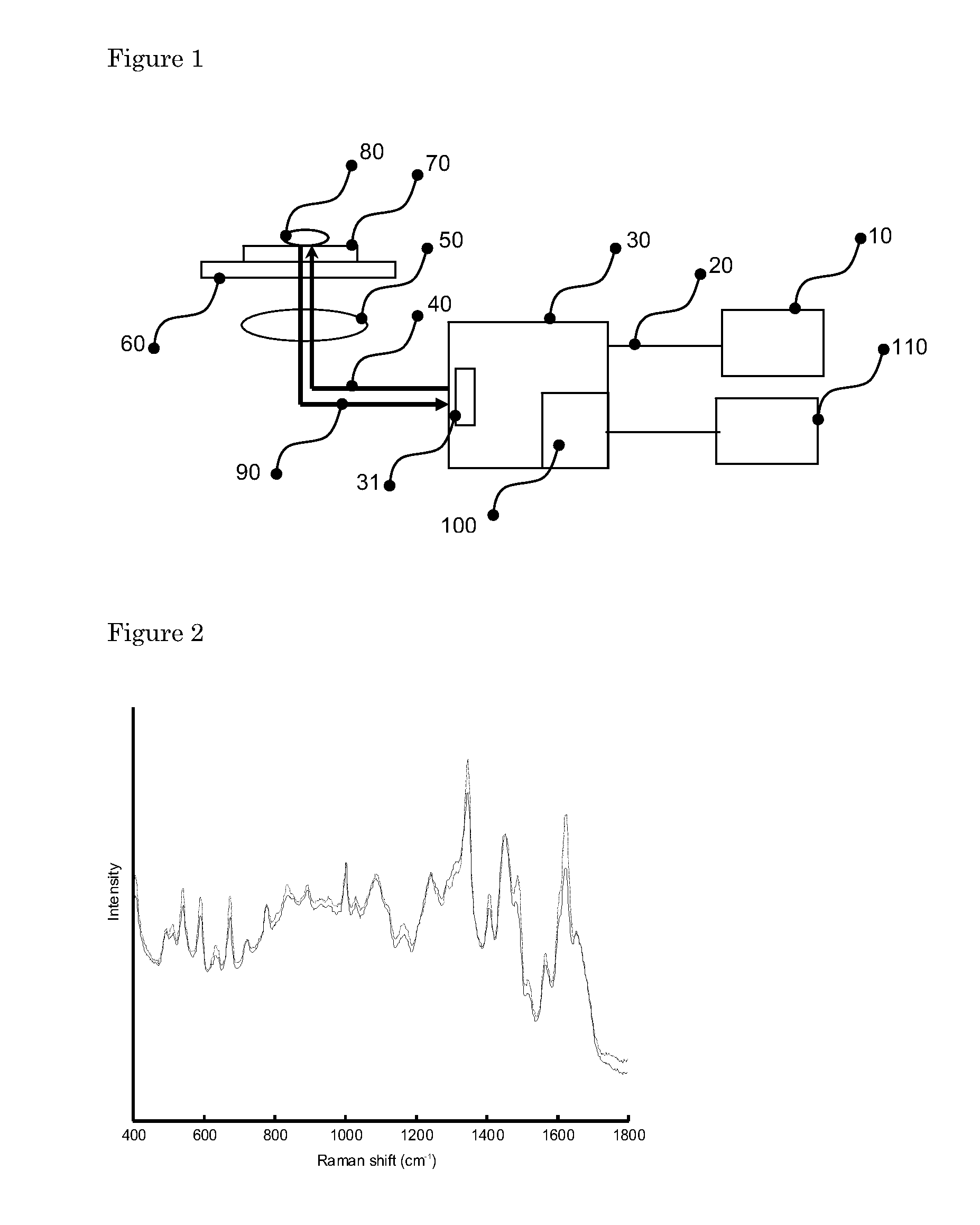
[0112]A schematic of the Raman spectroscopic measurement setup is shown in FIG. 1. Laser light of 785 nm from a diode laser 10 (Tiger Laser System 1000, Sacher LaserTechnik, Marburg, Germany) was guided into a Model 2500 High Performance Raman Module (Model 2500 HPRM) 30 (River Diagnostics B.V., Rotterdam, The Netherlands) by means of a single mode optical fiber 20. The Model 2500 HPRM emitted the laser light 40 through a pinhole 31 of 50 μm in diameter. The laser light was focused by a lens 50 with a numerical aperture of 0.7 in a dried microbial sample 80, deposited on a fused silica slide 70, on a translation stage 60 which could move the fused silica slide 70, in 2 directions perpendicular to the optical axis of lens 50. The laser power on the sample was about 150 mW. Lens 50 collected scattered light 90 from the sample, which followed the same optical path, as laser light 40 but in opposite direction back into the Model 2500 HPRM 30, through pinhole 31, ...
PUM
| Property | Measurement | Unit |
|---|---|---|
| power | aaaaa | aaaaa |
| temperature | aaaaa | aaaaa |
| temperature | aaaaa | aaaaa |
Abstract
Description
Claims
Application Information