Method and kit for detecting antibody to avibacterium paragallinarum

a technology of avibacterium paragallinarum and kit, which is applied in the field of methods and kits for detecting antibodies to avibacterium paragallinarum, can solve the problems of troublesome procedures, troublesome preparation and adding plates, etc., and achieve the effect of higher detection sensitivity

Active Publication Date: 2011-08-18
KM BIOLOGICS CO LTD
View PDF2 Cites 1 Cited by
  • Summary
  • Abstract
  • Description
  • Claims
  • Application Information

AI Technical Summary

Benefits of technology

[0040]In accordance with the present invention, a method and a kit for detecting an antibody to Avibacterium paragallinarum are provided. The peptides from A.pg-A and from A.pg-C as used herein do not include an amino acid sequence homologous to each other and therefore may specifically bind to an antibody induced by a vaccine from A.pg-A and an antibody induced by a vaccine from A.pg-C, respectively. Accordingly, a method and a kit for detection of the present invention enables to distinctly measure an antibody titer for the respective antibodies to vaccines from A.pg-A and A.pg-C in chicken sera not only when the respective vaccines are separately administered to chickens but also when a mixture of the vaccines is administered.
[0041]Furthermore, the present invention, as using a purified recombinant antigen, allows for immobilization of a higher concentration of an antigen than the prior art using cell debris of A.pg and hence allows for construction of a system for measuring an antibody with higher detection sensitivity. The method for detecting an antibody of the present invention, as using the antigenic peptide which is capable of distinguishing a serotype of A.pg, detects an antibody more simply and, in view of antigenic mutation of A.pg, more reliably than B-ELISA using a serotype specific monoclonal antibody.
[0042]Besides, by the method for detection of the present invention, antibodies are distinguished not only in chicken sera after vaccination but also in sera from chickens infected with A.pg.

Problems solved by technology

However, it requires four steps, i.e. addition of sera, addition of monoclonal antibodies, addition of anti-mouse IgG-HRP labeled antibody and addition of a substrate for development, one more step than in ordinary ELISA, rendering troublesome procedures.
For manufacturing a kit, it will take trouble of preparing and adding a plate immobilized with an antigen and reference serum as well as monoclonal antibodies.

Method used


Image

  • Method and kit for detecting antibody to avibacterium paragallinarum
  • Method and kit for detecting antibody to avibacterium paragallinarum
  • Method and kit for detecting antibody to avibacterium paragallinarum

Examples


example 1

Preparation of A.pg-1.3 Gene and DNA Fragments within Region of Said Gene

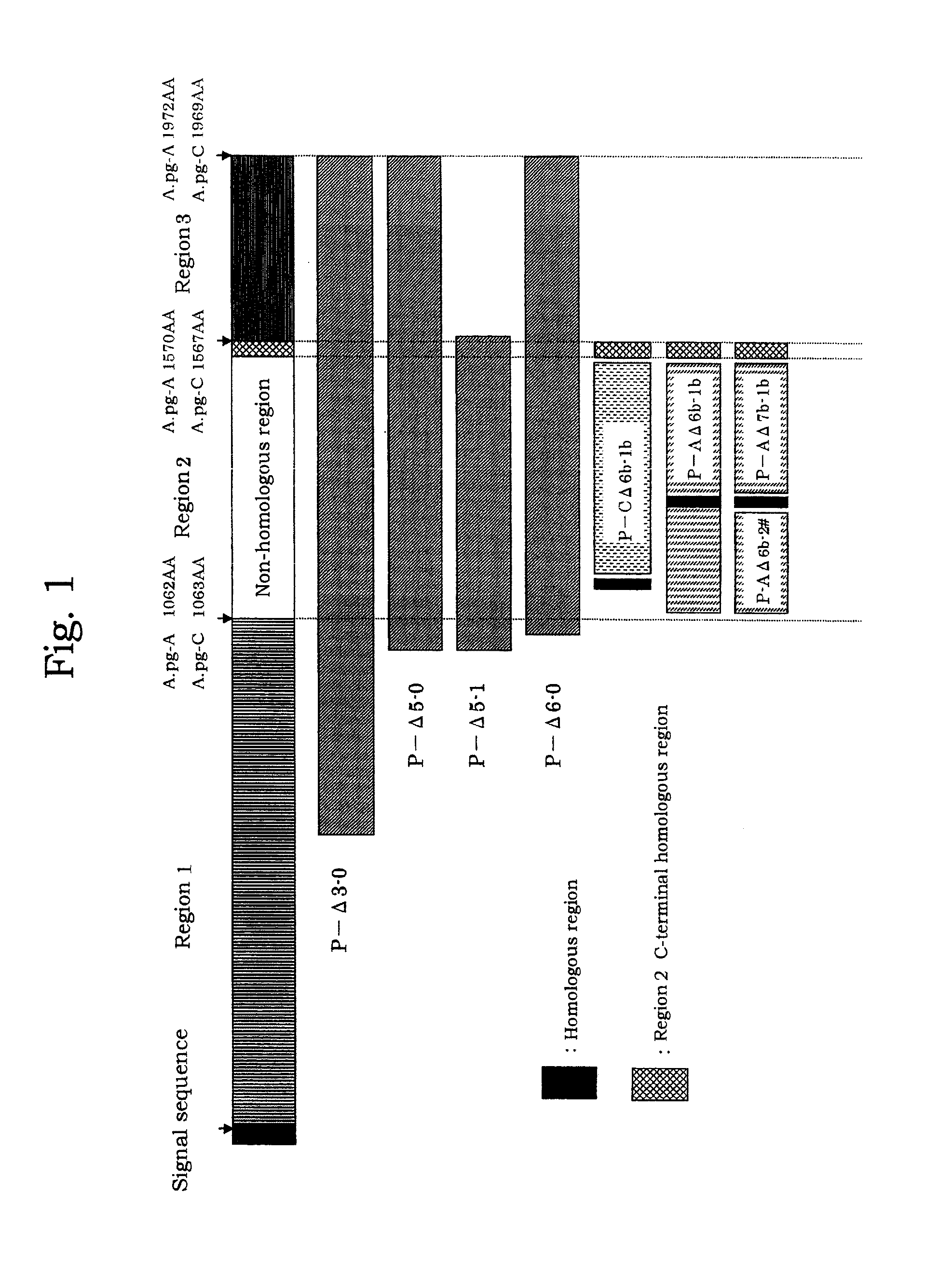
[0070]Genomic DNA libraries were prepared from A.pg-A 221 strain and A.pg-C 53-47 strain in accordance with the method of Tokunaga et al. (Japanese Patent Publication No. 10-84969). Briefly, chromosomal DNAs were extracted from cells collected by centrifuge (8,000 rpm, 20 minutes) using SepaGene kit (Sanko Junyaku Co., Ltd.), and digested with restriction enzyme Sau3AI to prepare DNA libraries. Using DNA fragments from the DNA libraries as a template, PCR was performed with LA Taq (TAKARA BIO Inc.) in accordance with protocol attached thereto to amplify fragments each consisting of a portion of the A.pg-1.3 gene. PCR reaction was carried out with LA PCR kit vert (TAKARA SHUZO CO., LTD.) at 96° C. for 1 minute; then 30 cycles of at 96° C. for 40 seconds, at 56° C. for 60 seconds, and at 72° C. for 120 seconds; and at 72° C. for 10 minutes. FIG. 1 shows positional relationship of each of the DNA fragments. Amplif...

example 2

Expression of Each of DNA Fragments

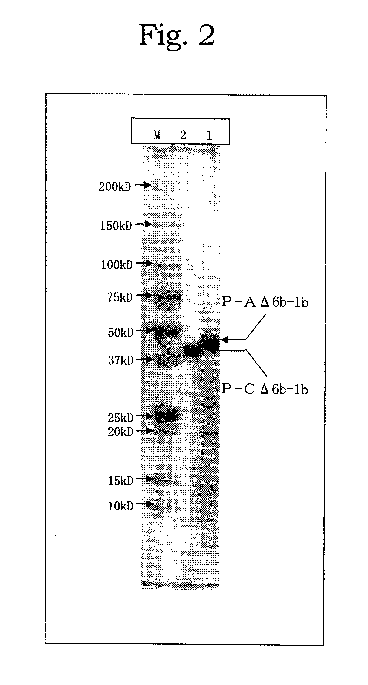
[0071]Each of the DNA fragments obtained in Example 1 was digested with suitable restriction enzymes and after separation on 0.8% agarose electrophoresis the amplified fragments were eluted and recovered with Quantum Prep Freeze N Squeeze DNA Gel Extraction Spin Co kit. The obtained fragments were ligated to plasmid pQE30 (QIAGEN: 6 His-tag sequences are present immediate downstream the initiation codon) or pET22b, which has been digested with the same restriction enzymes as above and the 5′-end of which has been dephosphorylated. The resulting plasmids were used to transform E. coli JM109 strain. E. coli comprising the amplified fragments was cultured overnight in Circle Grow medium (Funakoshi Co., Ltd.) supplemented with ampicillin and on the next day IPTG was added at a final concentration of 1 mM for further culture for 2.5 hours. After centrifuge (10,000 rpm, 5 minutes), supernatant was discarded and precipitate was suspended in an amount of 1...

example 3

Preparation of Anti-Polypeptide Chicken Sera

[0072]The polypeptide P-AΔ5-1 obtained in Example 2 was diluted with PBS to 10 μg / dose and added with aluminum hydroxide at a final concentration of 20%. The resultant mixture was administered subcutaneously to SPF chickens of 5 weeks old at the neck twice at an interval of 3 weeks for immunization. For the other polypeptides P-AΔ3-0, P-AΔ5-0, P-CΔ5-1 and P-CΔ6-0, they were diluted with PBS to 10 μg / dose and emulsified with an oil adjuvant (0.01 g antigen, 0.001 mL or less formalin, 0.01 mL Polysolbate 80, 0.04 mL sorbitan sesquioleate, 0.36 mL light liquid paraffin, the remainder phosphate buffer per dose (0.5 mL)) and the resultant mixture was once administered subcutaneously to SPF chickens of 5 weeks old at the neck. The chickens at 9-11 weeks old were bled to give immunized sera to each of the polypeptides.


PUM

PropertyMeasurementUnit
temperatureaaaaaaaaaa
pHaaaaaaaaaa
temperatureaaaaaaaaaa

Abstract

A method and a kit for detecting an antibody to Avibacterium paragallinarum are provided. A method for detecting an antibody to Avibacterium paragallinarum which comprises detecting an antibody induced by an outer-membrane protein of Avibacterium paragallinarum serotype A and/or serotype C by ELISA with a solid phase to which a peptide consisting of an amino acid sequence of non-homologous region of said outer-membrane protein or a portion thereof is immobilized, and a detection kit used for said method.

Description

TECHNICAL FIELD[0001]The present invention relates to a method and a kit for detecting an antibody to Avibacterium paragallinarum. Specifically, the present invention relates to a method for detecting an antibody to Avibacterium paragallinarum (hereinafter also refer to as “A.pg”; previously referred to as Haemophilus paragallinarum) which comprises detecting an antibody induced by an outer-membrane protein (hereinafter also refer to as “HMT p210 polypeptide”) of Avibacterium paragallinarum serotype A and / or serotype C by ELISA with a solid phase to which a peptide consisting of an amino acid sequence of non-homologous region of said outer-membrane protein or a portion thereof is immobilized, and a detection kit used for said method. Although a peptide is referred to as dipeptide, tripeptide, oligopeptide, polypeptide, and the like depending on the number of amino acid residues constituting of said peptide, a peptide as used herein is defined merely as “peptide” as encompassing any ...

Claims


Application Information

Patent Timeline
Patent Type & Authority Applications(United States)
IPC IPC(8): G01N33/566C07K7/06C07K7/08
CPCC07K14/195C07K16/1242G01N2800/14G01N33/56911G01N2469/20C07K2317/23
Inventor USHIJIMA, TOSHIHIROIMAMURA, TAKASHISAKAMOTO, RYUICHISAKAGUCHI, MASASHI
Owner KM BIOLOGICS CO LTD
Why Eureka
Try Eureka