Diabetes mellitus is a leading cause of vision loss in industrialized and developed countries.
Thus, diseased
retina might have multiple strong back reflections resulting in a relatively high OCT
signal variance.
Segmentation of the object of interest is considered a difficult step in the analysis of medical images.
It is well known that the lines drawn by the detection algorithms in the current commercial Stratus OCT system are frequently and dramatically erroneously drawn, which can lead to inaccurate measurements of
retinal thickness.
These errors are mainly due to both the presence of
high reflectivity regions in the inner
retina and loss of
retinal structure information in local regions along the
retinal cross-section as visualized by the commercial OCT system.
However, sometimes the
boundary detection algorithm fails in such specific cases when localized bright spots of
high intensity appear on some regions of the RNFL layer; and falsely displays the outer boundary of the RNFL layer as a result of the peak
search algorithm which looks for zero crossings in the structure.
These results evidence that the RNFL and GCL+IPL complex are more susceptible to initial damage when comparing MDR with DM eyes.
However, it appears that diabetes also inflicts additional damage to the outer retinal segment demonstrated by the optical changes of the IS / OS and OS / RPE junction.
However, it appears that diabetes also inflicts additional damage to the outer retinal segment demonstrated by the optical changes of the IS / OS and OS / RPE junction.
Recent studies have shown that currently available FD-OCT devices are giving significantly higher RT measurements than Stratus OCT due to different assumptions considered for the detection of the outer retinal boundary, making it difficult to compare data obtained by different devices.
These differences also make it difficult to adequately evaluate the performance of FD-OCT to detect the progression of
disease.
However, no information is provided about which boundary of the RPE layer (i.e. inner or outer) is detected.
Although RTVue allows manual correction of the
automated segmentation, the manual adjustment was not used to change the segmentation results, while differences in axial resolution and calibration might also contribute to thickness measurement differences.
Particularly, minor errors in the segmentation of the outer border of the
retina were observed at the periphery in some RTVue scans.
Thus, it was inferred that RTVue's thinner measurements in R6 and R7 were possibly due to the low quality observed in some OCT scans, which could be a direct result of
image acquisition pitfalls leading to segmentation errors.
Note that visualizing and quantifying microstructural changes within the photoreceptor and RPE,
layers is difficult using Stratus OCT images due to weakened
signal energy after penetrating the neuroretina, RPE, and choriocapillaries.
Discussion: Although a layer-editing tool to manually adjust the retinal layer boundaries for macula and RNFL was recently incorporated in the current Stratus OCT
software, its quantitative analysis does not provide thickness measurements of the various intraretinal
layers.
The
high variability in the thickness measurements of the OS / RPE junction is due to the fact that the outer boundary of this layer is not clearly visualized in Stratus OCT images because of the
low contrast between the OS / RPE junction (outer border) and the RPE inner boundary, which can be attributed to the limitation of the Stratus OCT system to penetrate deeper structures in the retina.
Moreover, the interobserver coefficients of reproducibility calculated for the total retina and intraretinal
layers (except for the RNFL) were higher than corresponding values for intervisit reproducibility, which may possibly be explained by the fact that subject fatigue and normal
drying of the eye during repeated sessions the same day induced more
noise into the overall measurements.
In addition the scans were not aligned between visits because the Stratus OCT did not provide this feature.
Thus, this limitation affected the intervisit variability results.
In addition, computer-aided detection and diagnosis based on automatic / semiautomatic robust algorithms will be essential in clinical studies where large data sets will be impractical for manual grading approaches.
In addition, recent studies have shown that retinal thickness measurements between SDOCT devices are significantly different due to different assumptions considered for the detection of the outer retinal boundary, which makes it difficult to compare data obtained by different devices.
These differences also make it difficult to adequately evaluate the performance of SDOCT to detect the progression of
disease.
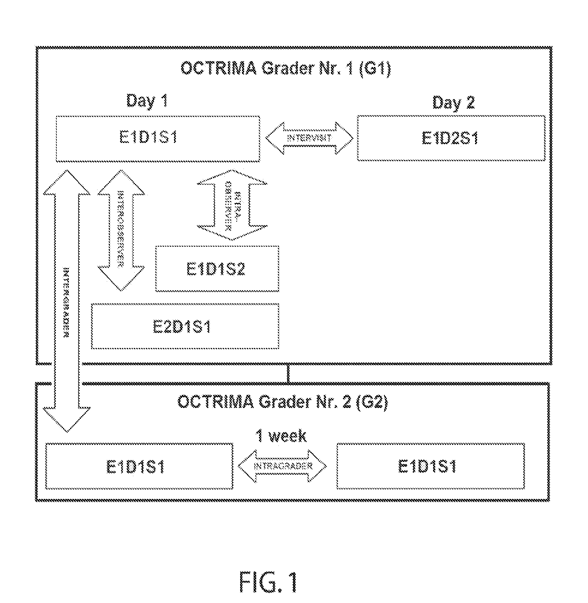
Direct comparison of this study with previous reproducibility and
repeatability studies is difficult because the age group of
healthy subjects along with
image segmentation and experimental and statistical methods vary between studies.
It is well known that it is difficult to visualize and quantify changes within the photoreceptor and RPE layers using Stratus OCT images because of these layers are too close to be resolved and often appear as a single hyperreflective band in TD-OCT images.
The current Stratus OCT
software provides a map of the macula and
nerve fiber layer thickness along with regional averages, but it does not provide thickness measurements of the separate retinal layers.
Conversely, this will increase the
image acquisition time, which could lead to measurement errors due to eye motion.
However, acquisition speed and resolution, which presented early challenges in OCT development, have experienced remarkable improvements with the introduction of a newer generation of OCT known as
spectral domain OCT.
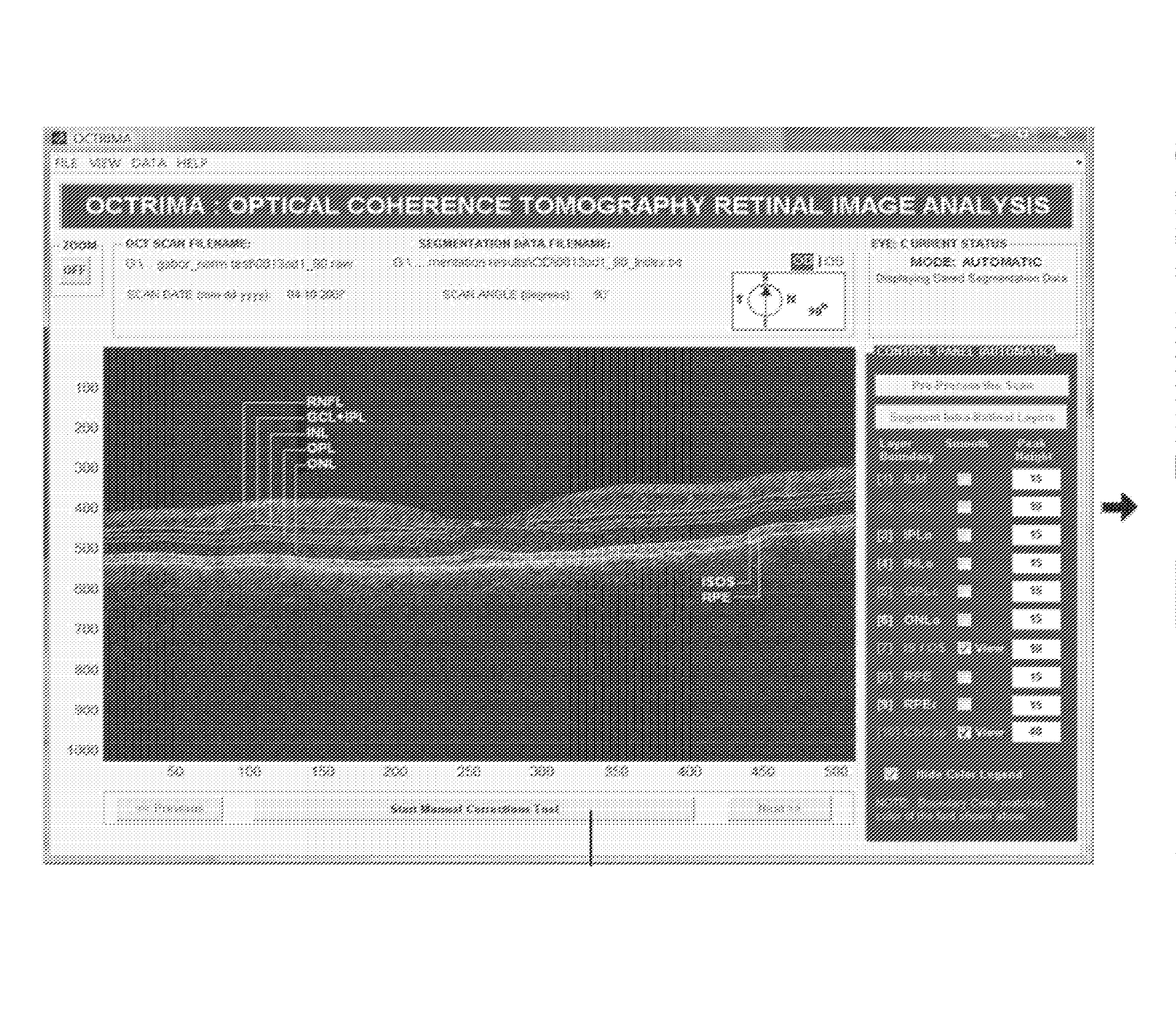
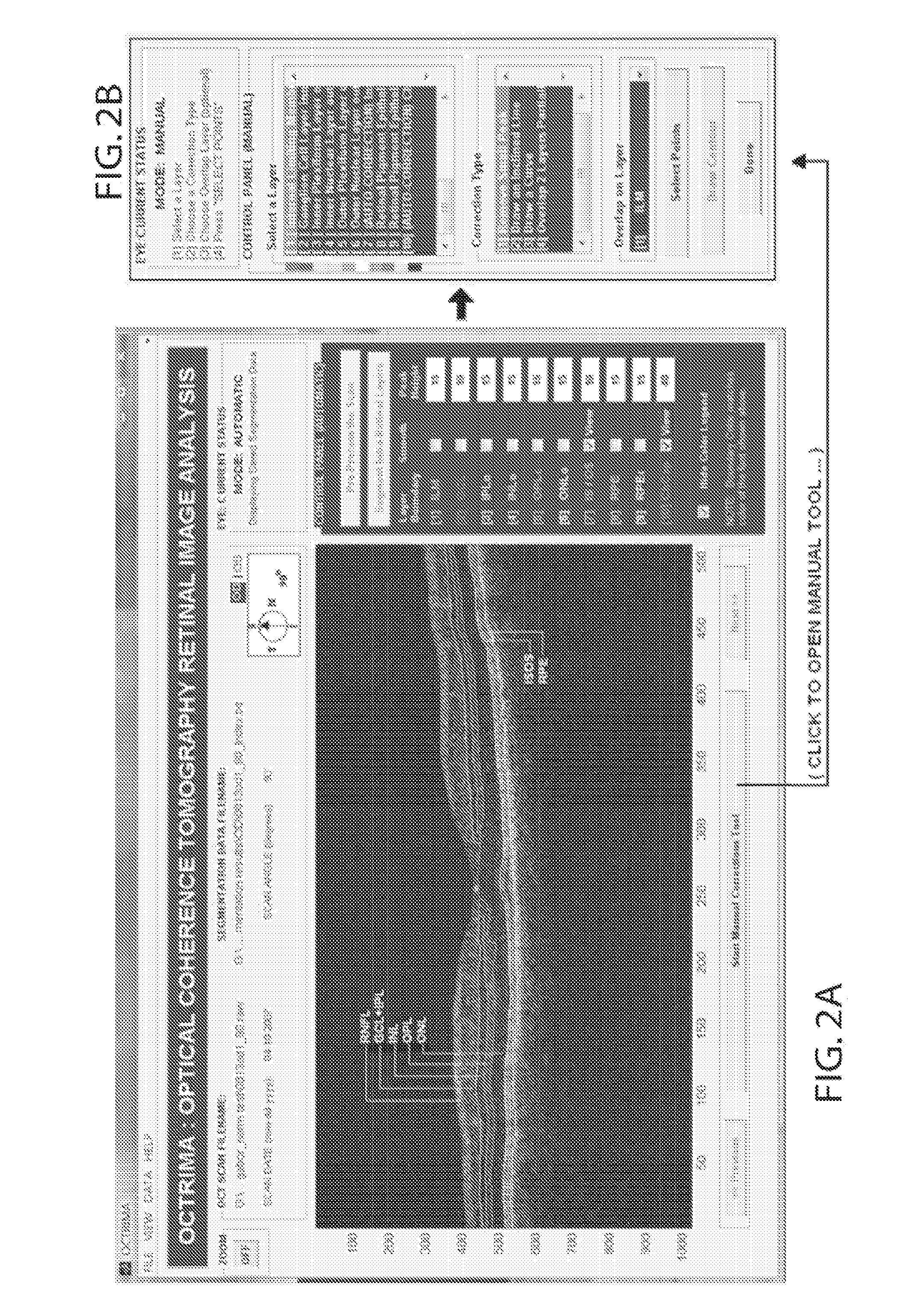
Manual correction module: Segmentation of the object of interest is considered a difficult step in the analysis of medical images.
This is often true in ophthalmic applications such as OCT where
image segmentation is particularly difficult due to restrictions imposed by image acquisition, ocular
pathology and biological variation.
For example, it is well known that detection algorithms fail when the
retinal structure is disrupted by
fluid accumulation which can lead to inaccurate measurements of retinal thickness.
These errors are mainly due to both the presence of
high reflectivity regions in the inner retina and loss of
retinal structure information in local regions along the retinal cross-section as visualized by the commercial OCT system.
However, sometimes the
boundary detection algorithm fails in such specific cases when localized bright spots of
high intensity appear on some regions of the RNFL layer; and falsely displays the outer boundary of the RNFL layer as a result of the peak
search algorithm which looks for zero crossings in the structure.
Thus, the algorithms are unable to independently assess the area and volume of the fluid filled cystic areas within and under the retina that represents leakage from
choroidal neovascularization (CNV).
However,
automatic segmentation algorithms for OCT data have tendency to give erroneous segmentation results especially in
pathological cases, which is actually a result of the algorithm performance independently of how good the OCT image could be reproduced with a high
level of detail.
In addition, computer-aided detection and diagnosis based on automatic / semiautomatic robust algorithms will be essential in clinical studies where large datasets will be impractical for manual grading approaches.
In this study it was found that the scattering coefficients extracted from OCT images were more affected as the
noise level increased.
The commercial time-domain Stratus OCT(Carl Zeiss Meditec, Dublin, CA) has a measurement capability limited to retinal thickness (RT) and cannot give quantitative information on intraretinal layers.

