
Use of gingival fibroblasts for vascular cell therapy

a vascular cell and fibroblast technology, applied in the field of vascular cell therapy, can solve the problems of pathological arterial remodeling, imbalance between degradation and abnormal healing reaction,

Active Publication Date: 2012-07-12
UNIVERSITÉ PARIS CITÉ
View PDF0 Cites 0 Cited by
  • Summary
  • Abstract
  • Description
  • Claims
  • Application Information

AI Technical Summary

Benefits of technology

The patent text describes the use of gingival fibroblasts, a type of mesenchymal cell, for the treatment of various vascular pathologies such as atherosclerosis, vulnerable plaque, restenosis, and arterial aneurysms. These fibroblasts have been found to have inhibitory effects on the main factors responsible for aneurysm formation and to promote the stabilization of aneurysms by producing factors that favor the elastic network. The use of autologous fibroblasts, which are derived from the individual's own gingival tissue and placed in culture, is preferred. The patent also describes the coculturing of gingival fibroblasts with arterial smooth muscle cells and the resulting inhibition of MMP9 activity and potentiation of TGFβ secretion. Overall, the patent presents a novel and promising approach for the treatment of vascular pathologies involving extracellular matrix impairments.

Problems solved by technology

During the repair of these lesions, an abnormal healing reaction can occur, resulting from an imbalance between degradation and synthesis of the extracellular matrix, and inducing pathological arterial remodeling, which can be reflected by a vascular enlargement (aneurism) or, on the other hand, by a constriction (stenosis, occurring secondarily during atherogenesis, or restenosis, subsequent in particular to an angioplasty, evolving in the course of cicatricial remodeling toward the reappearance of a stenosis).
However, these invasive techniques do not treat the cause, but the consequences of the pathology.
However, a major obstacle to the use of SMCs for the treatment of aneurisms by cell therapy comes from the need to obtain these SMCs from stem cells.
The stem cells used come from the marrow or the blood and are very difficult to obtain.

Method used


Image

  • Use of gingival fibroblasts for vascular cell therapy
  • Use of gingival fibroblasts for vascular cell therapy
  • Use of gingival fibroblasts for vascular cell therapy

Examples


example 1

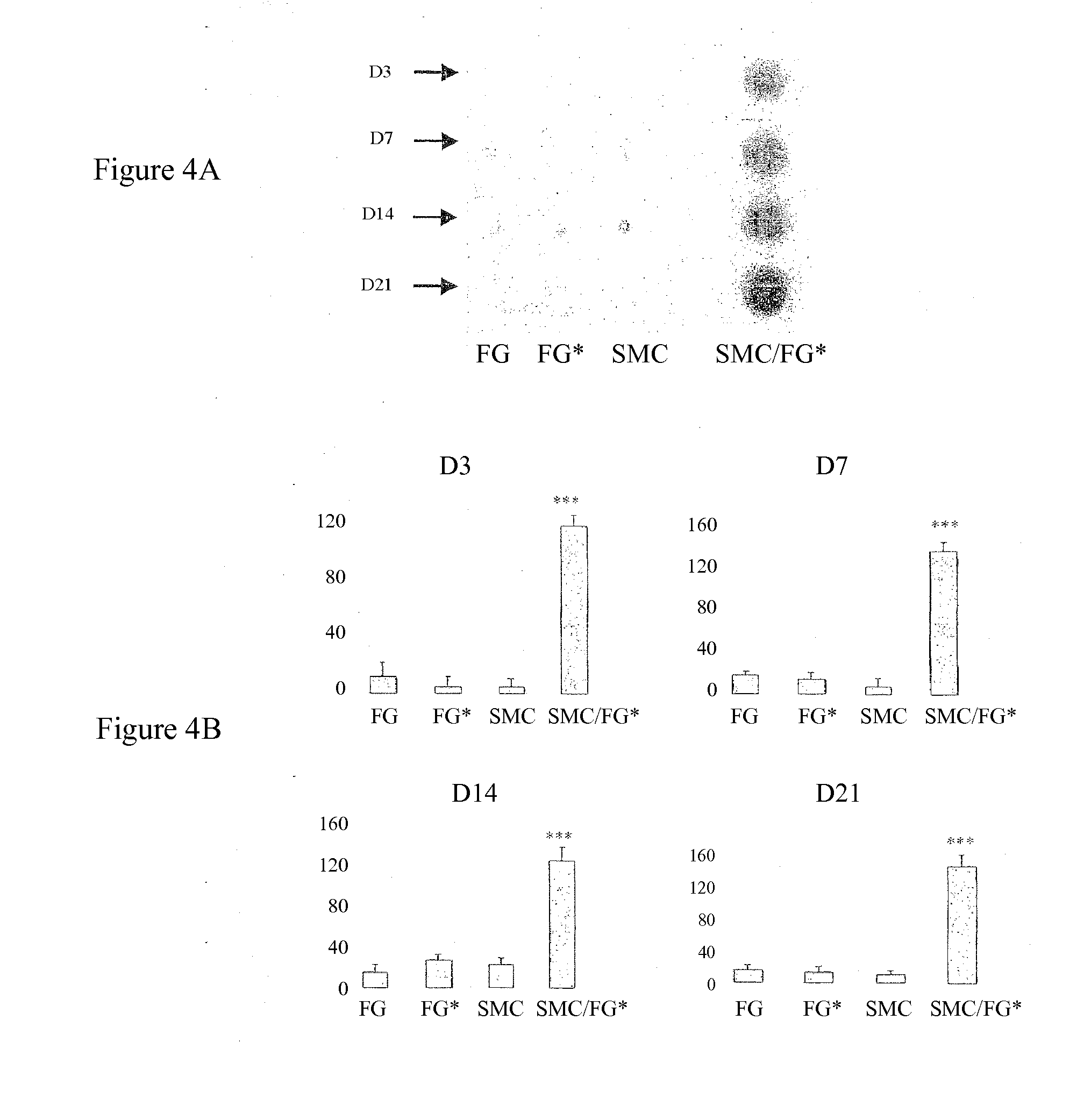
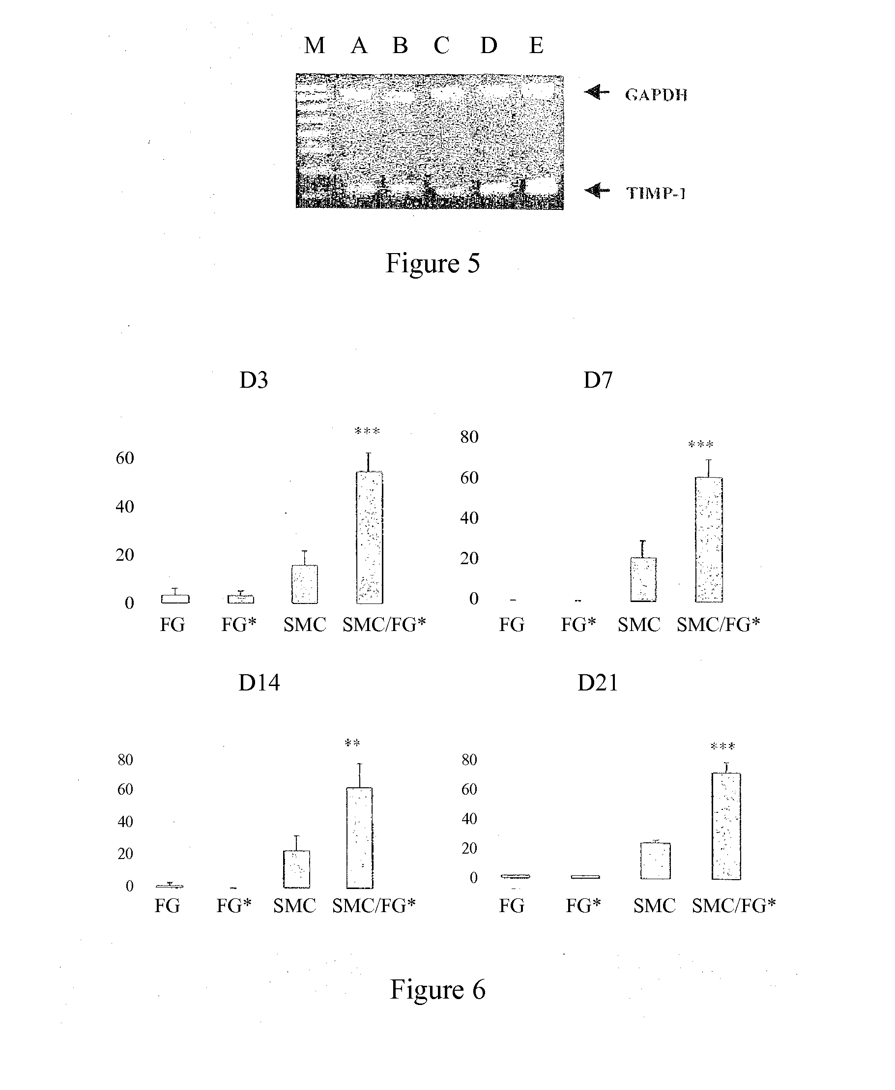
Secretion of MMP2, MMP9, TIMP-1 and TGFβ in Cocultures of Gingival Fibroblasts and Smooth Muscle Cells

Labeling of Fibroblasts

[0051]The gingival fibroblasts are labeled with anionic nanoparticles of maghemite as described by WILHEM et al. (Biomaterials 24: 1001-1011, 2003).

[0052]The gingival fibroblasts (six different cultures) are cultured to confluence in 10% fetal calf serum (FCS); and, after 48 h, the culture supernatant is removed and the cells are cultured in serum-free medium.

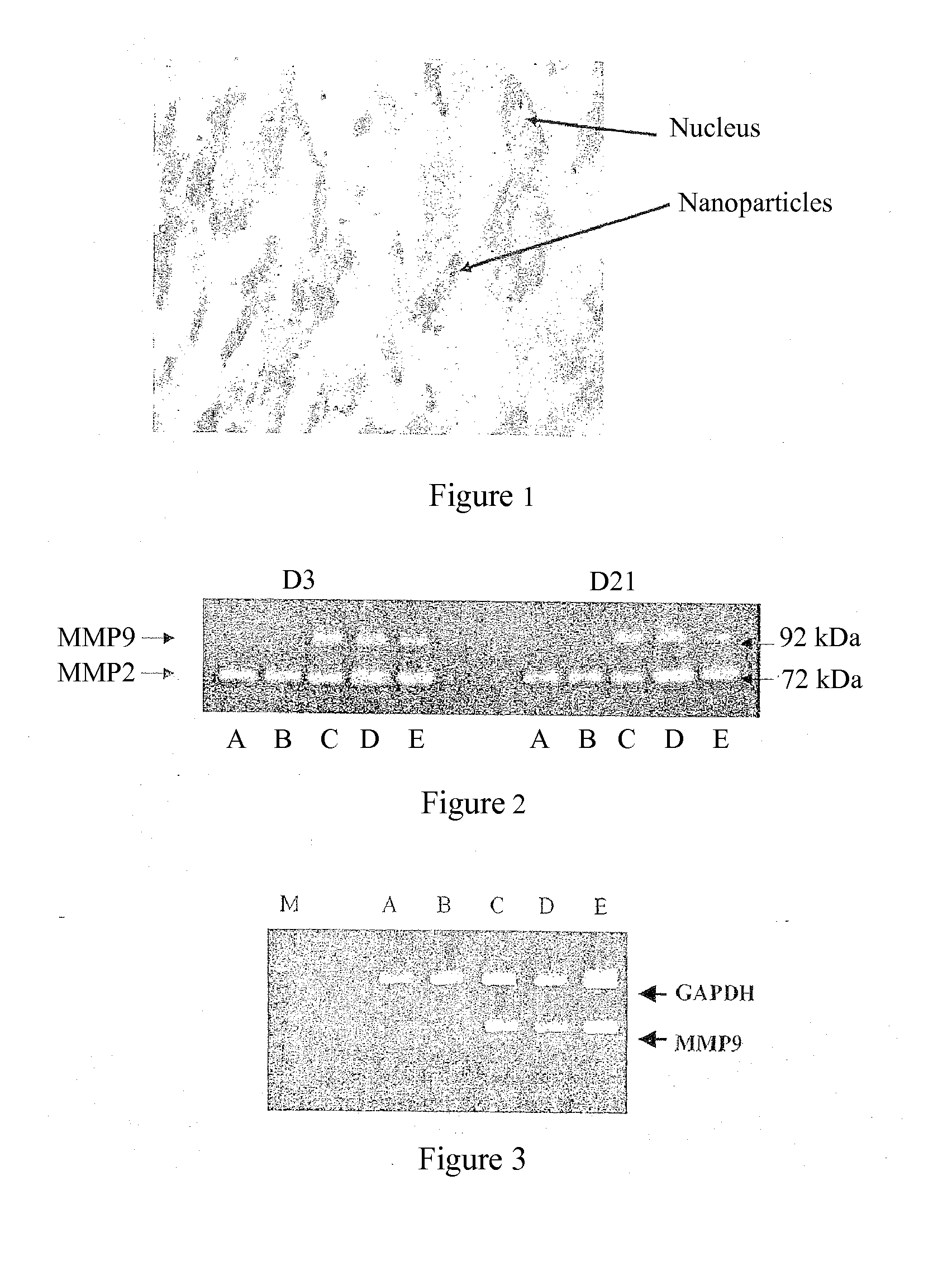
[0053]The quality of gingival fibroblast labeling with these nanoparticles is controlled by Perl staining (Prussian blue). The nanoparticles are absorbed and internalized into the endosome of the gingival fibroblasts, as shown in FIG. 1.

[0054]In order to verify that the labeling does not modify the phenotype of the gingival fibroblasts, the effect of the incorporation of the nanoparticles on the secretion of matrix metalloproteinase-2 (MMP2) and of cytokines IL-1β and TGFβ was evaluated at D1, D3 and D5 (...

example 2

Comparison of the Effects of the Gingival Fibroblasts with Those of Dermal or Adventitial Fibroblasts on the MMP2 or MMP9 activity of SMCs

[0084]Cocultures of dermal fibroblasts (FDs) or adventitial fibroblasts (FAs) and of smooth muscle cells (SMCs) are carried out in a collagen gel, using the same protocol as that described in Example 1 above for the gingival fibroblasts.

[0085]The cells are obtained from skin samples for the dermal fibroblasts, from samples of adventitia for the adventitial fibroblasts, and from samples of arterial media for the smooth muscle cells.

[0086]Dermal or adventitial fibroblast cultures and smooth muscle cell cultures are carried out separately, as a control, under the same conditions.

[0087]The cell interactions are studied at D7 and D14 by determination of the activities of MMP2 and of MMP9, secreted into the culture medium, by zymography as described in Example 1 above. In parallel, 20 μl of recombinant MMP9 (R&D Systems, ref. 911-MP) are subjected to th...

example 3

Organotypic Cultures of Lesioned Arteries

[0089]The three-dimensional culture of lesioned arteries in collagen gel was developed by the inventors in order to provide a model for analyzing arterial remodeling over time.

Obtaining the Cultures

[0090]Atherosclerotic lesions are induced in 5 New Zealand white rabbits by means of the combination of air desiccation and a high-cholesterol diet, as described by LAFONT et al. (Circ. Res. 76(6): 996-1002, 1995; Circulation 100(10): 1109-1115, 1999). After 4 weeks, the presence of atherosclerotic lesions is confirmed by arteriography inside the two femoral arteries. Iatrogenic lesions are then produced on the left femoral arteries using an angioplasty balloon catheter (3 insufflations at 6 atm for 60 sec).

[0091]hours after the angioplasty, the rabbits are sacrificed by intracardiac injection of Phenobarbital. The arteries are collected after dissection and stored in DMEM containing 20% FCS, at 4° C., for 3-10 hours, before placing in culture. In ...


PUM

PropertyMeasurementUnit
pHaaaaaaaaaa
pHaaaaaaaaaa
pHaaaaaaaaaa

Abstract

The invention relates to the use of gingival fibroblasts for obtaining a cellular composition for treating arterial-remodelling pathology, for example an aneurysm, post-cryoplasty stenosis and restenosis, an aortic dissection or atherosclerosis.

Description

[0001]This application is a continuation of U.S. application Ser. No. 11 / 571,542, filed Aug. 9, 2007, which is a national phase of PCT / FR05 / 01690, filed Jul. 1, 2005, which claims the benefit of French application 0407357, filed Jul. 2, 2004.BACKGROUND OF THE INVENTION[0002]The present invention relates to the use of gingival fibroblasts in vascular cell therapy, in particular for the treatment of arterial pathologies.[0003]The arterial wall consists of three superimposed layers called tunicae. The inner tunica (intimae) consists of a layer of endothelial cells. The intermediate tunica (media) consists mainly of smooth muscle fibers and connective tissue rich in elastic fibers. The outer tunica (adventica) is a connective envelope that surrounds the whole assembly.[0004]Arteries are subjected to numerous attacks of various origin (hypoxia, lipid overload, hemodynamic forces, atheroma, hypertension, etc.) that can induce arterial wall lesions. During the repair of these lesions, an a...

Claims


Application Information

Patent Timeline
Patent Type & Authority Applications(United States)
IPC IPC(8): A61K35/12A61P9/10A61K35/33A61K35/37A61L27/38B82Y5/00
CPCA61L27/3804A61K35/33A61L27/3843A61P9/00A61P9/10
Inventor LAFONT, ANTOINEGOGLY, BRUNO
Owner UNIVERSITÉ PARIS CITÉ
Learn More