Laser isolation of viable cells

a technology of viable cells and lasers, applied in cell dissociation methods, instruments, enzymology, etc., can solve the problems of inability to obtain eye donors

Inactive Publication Date: 2012-10-11
ADVANCED CELL TECH INC
View PDF0 Cites 14 Cited by
  • Summary
  • Abstract
  • Description
  • Claims
  • Application Information

AI Technical Summary

Benefits of technology

[0041]In one embodiment, the viable cell may be a RPE cell selected based on pigmentation. In another embodiment, the viable cell may be an RPE cell selected based on at least one detectable characteristic of RPE cells. The detectable characteristic of RPE cells may be at least one of presence of brown pigmentation accumulated within the cytoplasm, a cobblestone, epithelial-like morphology, or expression of at least one RPE cell markers. The RPE cell marker may be selected from the group consisting of bestrophin, RPE65, CRALBP, and PEDF. The RPE marker may be detected by a method selected from the group consisting of binding to an antibody directly or indirectly coupled to a detectable label; incubation with magnetic beads—conjugated antibodies; detecting the expression of a fluorescent protein; detecting an intracellular mRNA, detecting an intracellular protein; and detecting an intracellular small molecule. The viable cell may exhibit at least one detectable characteristics of RPE cells. The detectable characteristics of RPE cells may be morphology or expression of at least one RPE cell markers. The RPE cell marker may be selected from the group consisting of markers identified in Table 1.
[0042]In one embodiment, the differentiated cell may be a RPE cell selected based on pigmentation. In another embodiment, the differentiated cell may be an RPE cell selected based on at least one detectable characteristic of RPE cells. The detectable characteristic of RPE cells may be at least one of presence of brown pigmentation accumulated within the cytoplasm, a cobblestone, epithelial-like morphology, or expression of at least one RPE cell markers. The RPE cell marker may be selected from the group consisting of bestrophin, RPE65, CRALBP, and PEDF. The RPE marker may be detected by a method selected from the group consisting of binding to an antibody directly or indirectly coupled to a detectable label; incubation with magnetic beads-conjugated antibodies; detecting the expression of a fluorescent protein; detecting an intracellular mRNA, detecting an intracellular protein; and detecting an intracellular small molecule. The differentiated cell may exhibit at least one detectable characteristics of RPE cells. The detectable characteristics of RPE cells may be morphology or expression of at least one RPE cell markers. The RPE cell marker may be selected from the group consisting of markers identified in Table 1.
[0043]In one embodiment, the viable cell may be differentiated from one or more pluripotent cells. In another embodiment, the pluripotent cells may be selected from the group consisting of induced pluripotent stem (iPS) cells, embryonic stem (ES) cells, blastomeres, morula cells, embroid bodies, adult stem cells, hematopoietic stem cells, fetal stem cells, mesenchymal stem cells, postpartum stem cells, multipotent stem cells, and embryonic germ cells. In a further embodiment, the pluripotent stem cell may be an embryonic stem cell. In a still further embodiment, the pluripotent stem cell may be a human embryonic stem cell.
[0044]In one embodiment, the differentiated cell may be differentiated from one or more pluripotent cells. In another embodiment, the pluripotent cells may be selected from the group consisting of induced pluripotent stem (iPS) cells, embryonic stem (ES) cells, blastomeres, morula cells, embroid bodies, adult stem cells, hematopoietic stem cells, fetal stem cells, mesenchymal stem cells, postpartum stem cells, multipotent stem cells, and embryonic germ cells. In a further embodiment, the pluripotent stem cell may be an embryonic stem cell. In a still further embodiment, the pluripotent stem cell may be a human embryonic stem cell.

Problems solved by technology

(2007) Br J Opthalmol 91: 349-353 describes the successfully transplantation of autologous RPE-choroid sheet after removal of a subfoveal choroidal neovascularization (CNV) in patients with age related macular degeneration (AMD), but this procedure only resulted in a moderate increase in mean visual acuity.
However, RPE cells sourced from human donors has several intractable problems.
First, is the shortage of eye donors, and the current need is beyond what could be met by donated eye tissue.
For example, RPE cells sourced from human donors are an inherently limited pool of available tissue that prevent it from scaling up for widespread use.
Second, the RPE cells from human donors may be contaminated with pathogens and may have genetic defects.
The cadaver-sourced RPE cells have an additional problem of age where the RPE cells are may be close to senesce (e.g., shorter telomeres) and thus have a limited useful lifespan following transplantation.
Reliance on RPE cells derived from fetal tissue does not solve this problem because these cells have shown a very low proliferative potential.
Any human sourced tissue may also have problems with tissue compatibility leading to immunological response (graft-rejection).
Also, cadaver-sourced RPE cells may not be of sufficient quality as to be useful in transplantation (e.g., the cells may not be stable or functional).
Fourth, sourcing RPE cells from human donors may incur donor consent problems and must pass regulatory obstacles, complicating the harvesting and use of RPE cells for therapy.
Fifth, a fundamental limitation is that the RPE cells transplanted in an autologous transplantation carry the same genetic information that may have lead to the development of AMD.
Thus, a shorter useful lifespan of the RPE cells limits their utility in therapeutic applications (e.g., the RPE cells may not transplant well and are less likely to last long enough for more complete recovery of vision).
However, animal testing alone may be considered insufficient because a human ES cell may be more prone to produce a teratoma in a human host than in the animal model.
Moreover, manual selection of pigmented clusters is very tedious and fully relies on the operator's skills and judgment which may get impaired after several hours of such scrupulous selection and the microscope involving eye and back-straining work.

Method used


Image

  • Laser isolation of viable cells
  • Laser isolation of viable cells
  • Laser isolation of viable cells

Examples


example 1

Protocol for Laser Microdissection of Living In Vitro Cells

Introduction

[0142]Laser capture microdissection (LCM) is a proven technique for the isolation of pure cell populations for downstream molecular analysis. The combined use of UV laser cutting with LCM using an infrared (IR) laser permits rapid and precise isolation of larger numbers of cells while maintaining cellular and nucleic acid integrity necessary for downstream analysis. In this application note, it is shown that these established techniques can also be used for the isolation of living cells, avoiding other more laborious methods of cell selection and enabling a wide range of research applications. This example describes a protocol for the isolation of living adherent cells and the subsequent recultivation of homogeneous subpopulations.

Methods

Specimen Preparation

[0143]PEN membrane slide may be hourly rinsed with 100% ethanol and air-dry prior to use and keep in a sterile environment (e.g., slide should be completely d...

example 2

ES Cell Differentiation to Produce RPE Cells

[0153]Human RPE cells were produced by differentiation of human ES cells essentially as described in U.S. Pat. No. 7,795,025. In brief, hES cell cultures were maintained and expanded on mouse embryo fibroblast (MEF) feeder cells, then trypsinized and cultured on low adherent plates (Costar) until embryoid bodies formed. The embryoid bodies were cultured until regions containing pigmented cells having epithelial morphology were formed therein. The embryoid bodies were then digested with enzymes (trypsin, and / or collagenase, and / or dispase), and pigmented cells were selectively picked, plated, and cultured. After about two weeks in culture at low density, the cultured cells lost their pigmentation, but after another two to three weeks in culture regions of pigmented cells having a cobblestone, epithelial-like morphology again appeared. This pigmentation behavior—temporary loss from cells in proliferating cultures, and restoration in quiescen...

example 3

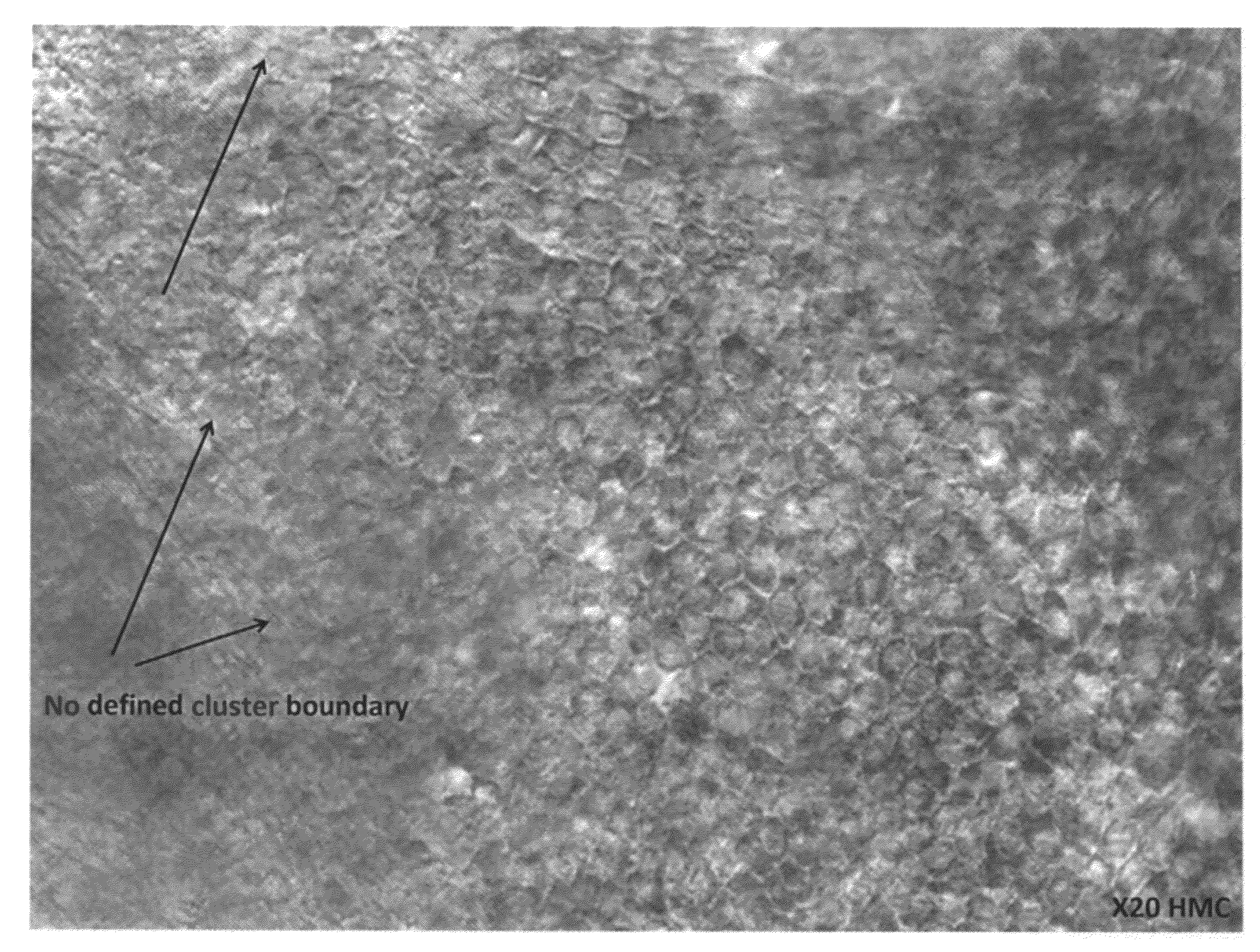
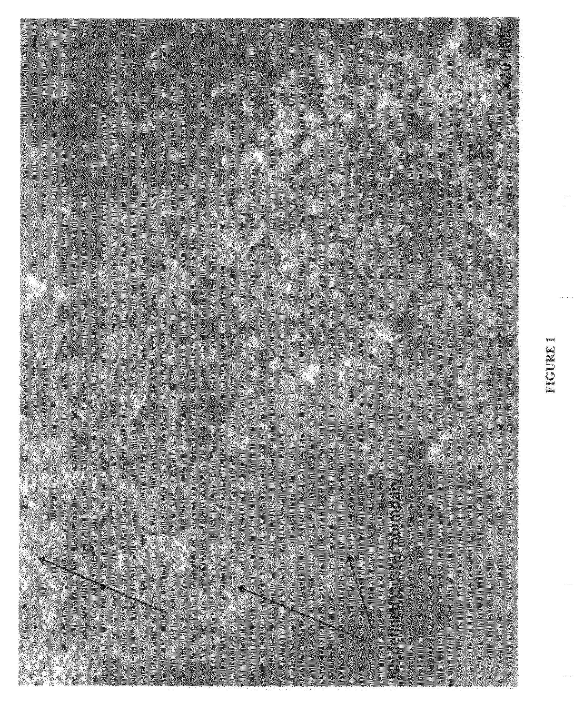
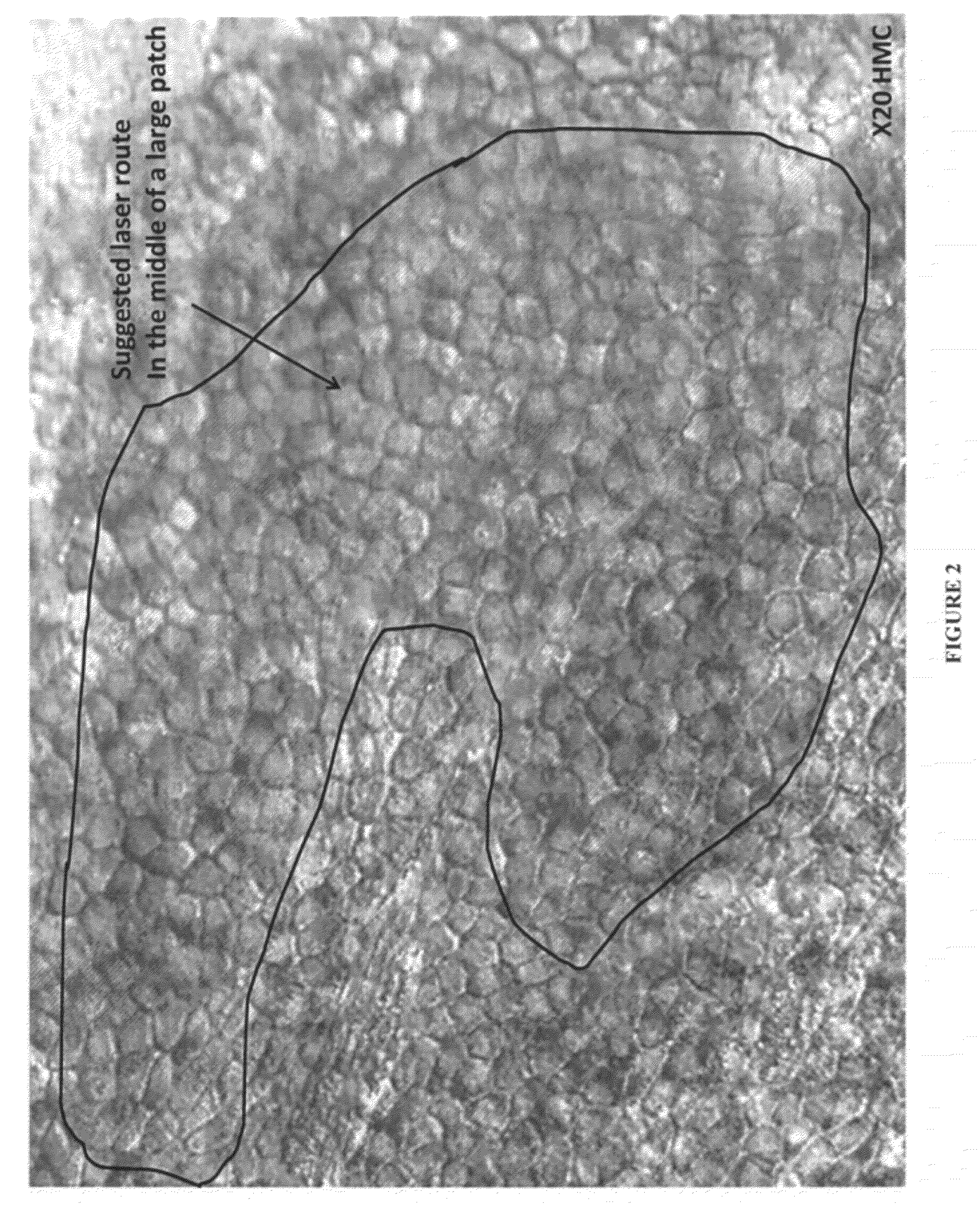
Isolation of Viable RPE Cells Using Laser Microdissection

[0154]Culture containing RPE cells differentiated from human ES cells were produced as described in the preceding example. Laser microdissection was then used to isolate islands of pigmented epithelial cells for further culture. ES-derived RPE cells were grown in multiwell culture plates and maintained as quiescent cultures until pigmented epithelial islands were perceptible (e.g., at least about 7 days). The multiwell plate was then placed on a microscope fitted with the STILETTO® laser system (Hamilton Thorne Ltd., Beverly, Mass.) Islands of pigmented epithelial cells were then visualized, and the provided control software was used to manually draw a target zone circumscribing and immediately outside of each pigmented island. Cells in the target zone were then ablated by laser pulses which were caused to strike the target zone by computer-controlled movement of the microscope stage. After ablation of the target zone, each is...


PUM

PropertyMeasurementUnit
distanceaaaaaaaaaa
distanceaaaaaaaaaa
distanceaaaaaaaaaa

Abstract

Methods for laser microdissection isolation of viable cells are provided. Cells of a desired type may be isolated from a diverse population, optionally with detection and exclusion of undesired cells. Desired cells may be isolated from a population that arose from differentiation of pluripotent cells, preferably embryonic stem cells or induced pluripotent stem cells, and undifferentiated stem cells may be detected and excluded from selection including the isolation of RPE cells sleeted based on morphology (e.g., characteristic mottled appearance) from a population of ES cells. The cells isolated by these methods, including RPE cells, may be essentially free of undifferentiated cells and thus suitable for use in cell-based therapies.

Description

BACKGROUND[0001]1. Field of the Invention[0002]The invention relates to laser microdissection methods for obtaining viable cells. The invention provides methods for the isolation of viable cells differentiated from pluripotent or multipotent cells, preferably embryonic stem cells or induced pluripotent stem cells (iPSCs), including ocular cells such as retinal pigment epithelium cells, iris pigment epithelium cells, vision-associated neural cells, lens cells, rods, cones, or corneal cells. The methods provided by the invention may provide high-purity cell cultures suitable for cell-based therapies.[0003]2. Description of the Related Art[0004]Laser microdissection methods may allow for the isolation of an individual cell to be separated from the surrounding preparation (e.g., a tissue section) by the laser beam, and then released. The released cells may then be moved to a collection device, for example by mechanical means or a laser-induced transport process with the aid of a laser p...

Claims


Application Information

Patent Timeline
Patent Type & Authority Applications(United States)
IPC IPC(8): C12N13/00C12Q1/02C12Q1/68G01N33/53
CPCC12N5/0621G01N2001/2886C12N2509/00
Inventor KLIMANSKAYA, IRINA VITALY
Owner ADVANCED CELL TECH INC
Why Eureka
Try Eureka