
Diagnostic BioMarkers for Fibrotic Disorders

a biomarker and fibrotic disease technology, applied in the direction of biological material analysis, peptides, tissue culture, etc., can solve the problems of affecting tissue or organ function, and affecting the detection of bmp9 and/or bmp10. the effect of facilitating the detection of bmp10

Inactive Publication Date: 2013-08-15
NOVARTIS AG
View PDF2 Cites 11 Cited by
  • Summary
  • Abstract
  • Description
  • Claims
  • Application Information

AI Technical Summary

Benefits of technology

The present invention provides methods for inhibiting fibrosis in a cell, treating fibrotic disorders in a subject, and assessing whether a subject has or is at risk of developing a fibrotic disorder. These methods involve contacting a cell or tissue with a BMP9 or BMP10 antagonist, such as an antibody or nucleic acid, to inhibit the activity of BMP9 and BMP10, which are proteins involved in fibrotic disorders. The methods can be used to treat or prevent fibrotic disorders in various subjects, such as humans, animals, and plants. The invention also provides methods for assessing the efficacy of a treatment regimen for fibrotic disorders by comparing the levels of BMP9 or BMP10 activity before and after treatment.

Problems solved by technology

Although processes related to fibrosis can occur as part of normal tissue formation or repair, dysregulation of these processes can lead to altered cellular composition and excess connective tissue deposition that progressively impairs tissue or organ function.
For example, more than five million people worldwide are afflicted with pulmonary fibrosis, which occurs when the air sacs of the lungs gradually become replaced by fibrotic tissue, causing an irreversible loss of the tissue's ability to transfer oxygen into the bloodstream.
Additionally, hepatic fibrosis represents a major worldwide healthcare burden and results from excessive connective tissue formation in the liver which can lead to portal hypertension or even cirrhosis (see Murphy et al., Expert Opin Investig Drugs.
Although therapeutic agents, such as anti-inflammatory drugs, are often used to treat fibrosis, such treatments can have low efficacy and undesirable side effects.
Moreover, there are currently no wholly effective treatments or cures for fibrotic disorders.

Method used


Image

  • Diagnostic BioMarkers for Fibrotic Disorders
  • Diagnostic BioMarkers for Fibrotic Disorders
  • Diagnostic BioMarkers for Fibrotic Disorders

Examples


example 1

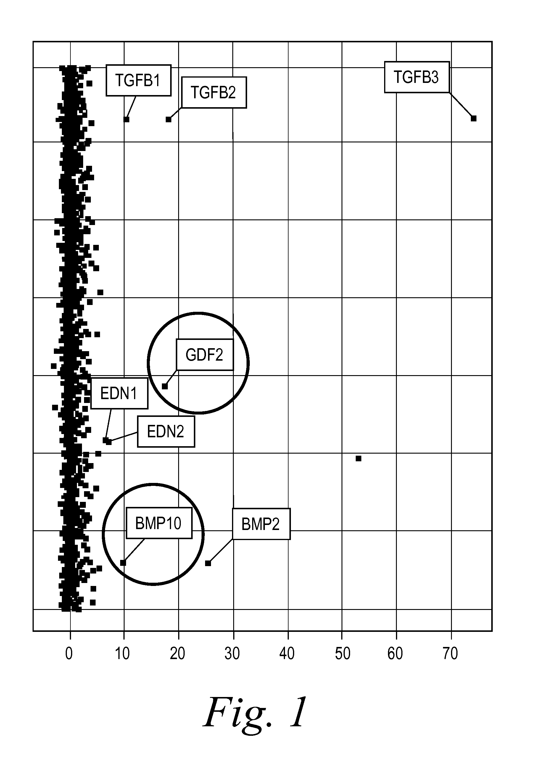
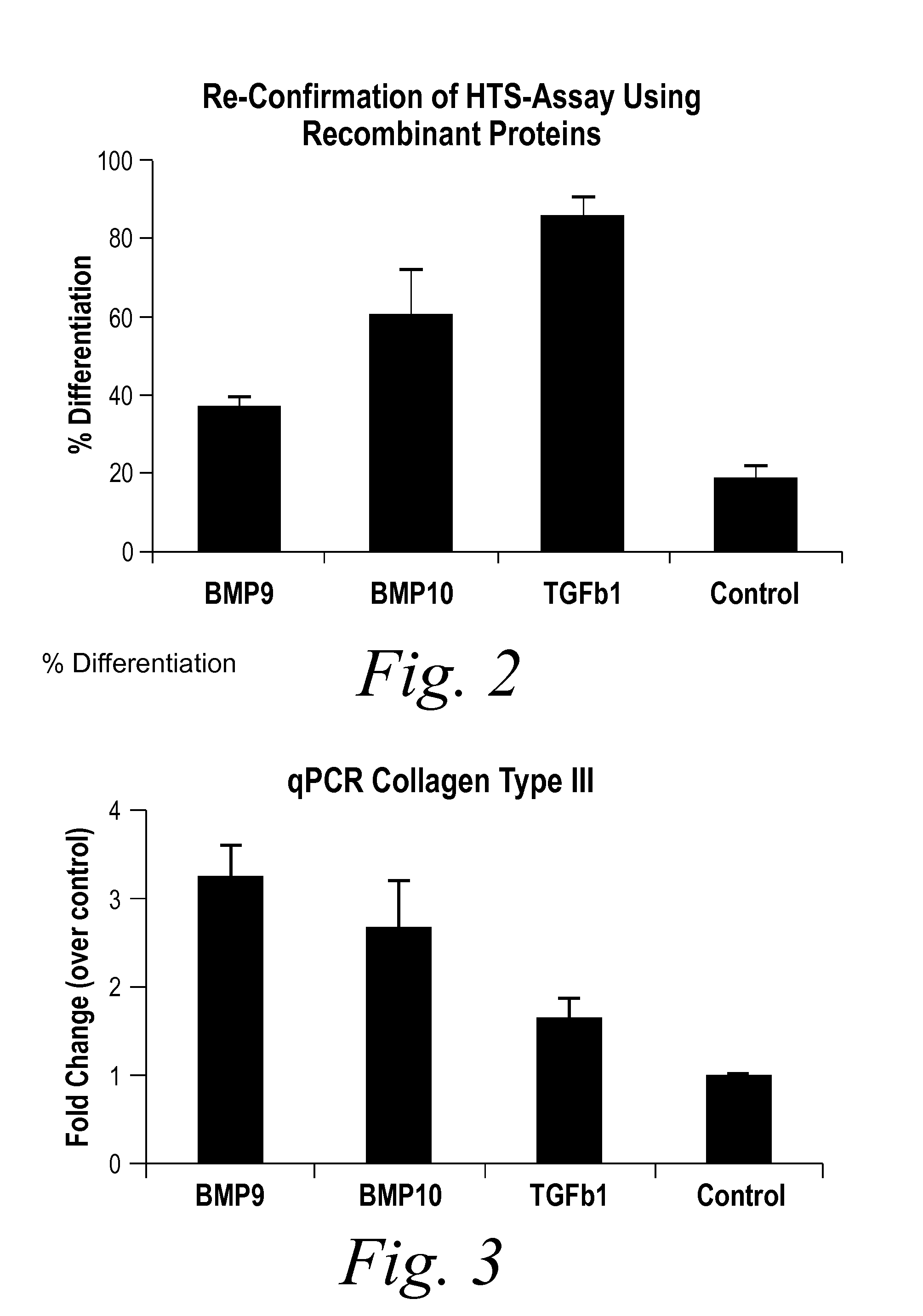
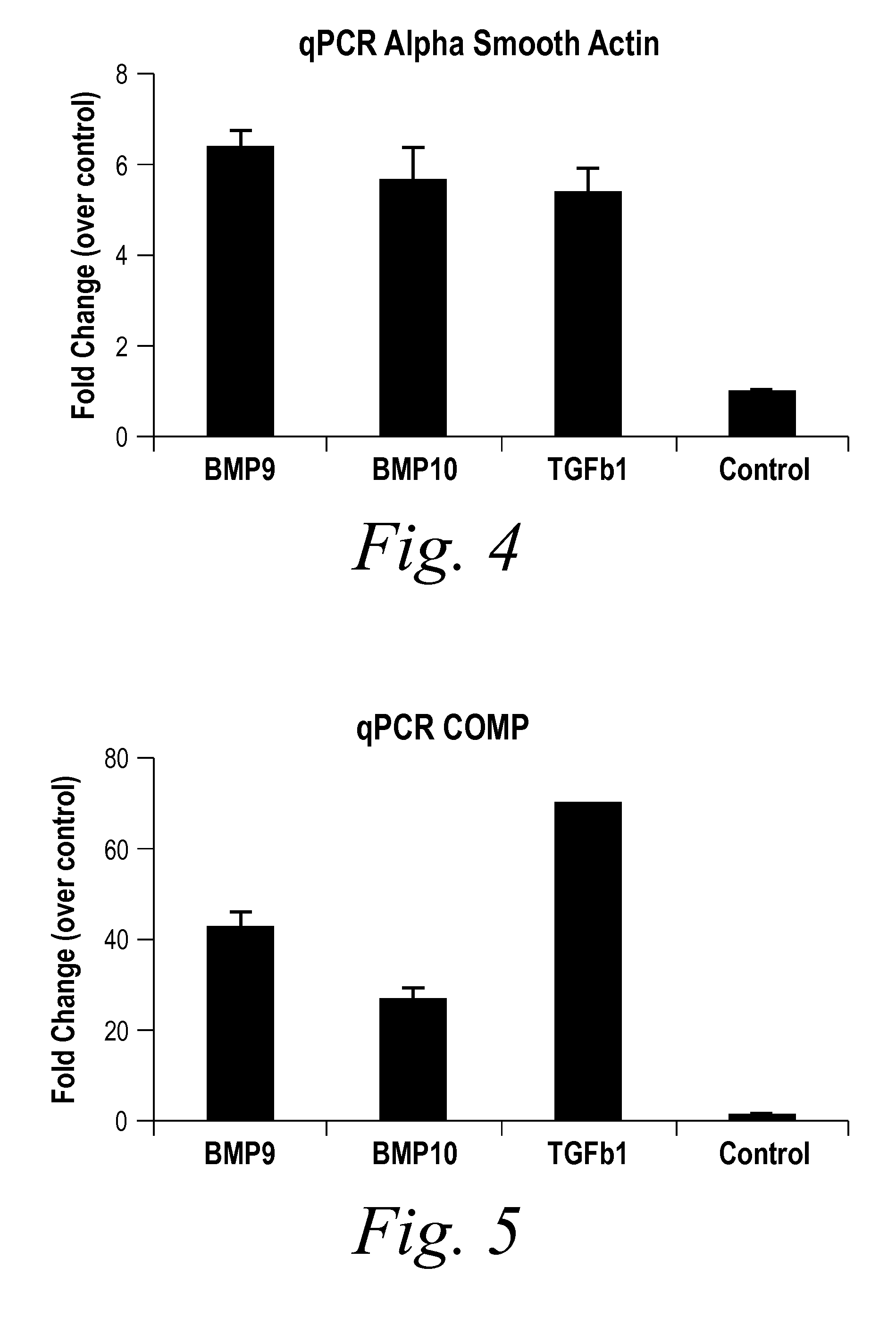
[0235]BMP9 and BMP10 were identified as indicators of fibrosis in a phenotypic screen and, therefore, as potential therapeutic targets capable of treating fibrotic disorders. The experiment was performed in 384 well plate format and in triplicate. Briefly, a reverse transfection in Hek293 cells of approximately 1500 human cDNA expression clones encoding predicted secreted proteins was performed using Fugene 6 (Roche) at a ratio of 4:1 (Fugene:DNA) in complete DMEM (Invitrogen) medium. Hek293 cells were washed 24 hours post transfection and incubated for an additional 48 hours to enrich for secreted proteins. Conditioned media from the transfected Hek293 cells were transferred onto human primary dermal fibroblast at passage 3 (Lonza) that were synchronized by starvation for 24 hours in absence of serum. The differentiation of fibroblast into myofibroblast was scored 96 hours later by immunostaining for the presence of alpha smooth muscle actin (αSMA [Sigma], a known marker of fibrobl...

example 2

[0237]To further assess the role of BMP9 in fibrosis, a reporter gene assay (RGA) was established. Specifically, a BMP9 reporter construct (ID-BRE) was generated using Id1 promoter BMP-response element (BRE) fused with firefly luciferase gene. The BMP9 RGA was conducted on HEK293 cells stably transfected with ID-BRE. HEK293 ID-BRE cells were seeded at 5×10⁵ cells / 35 mm-dish one day before transfection. 2.5 μg DNA constructs of constitutively active ALK1 and GFP (10:1) were co-transfected into HEK293 ID-BRE cells via Fugene 6 (Roche, 8 μl), followed by manufacturer's protocol on day 2. Transfection efficiency was measured by visualization of GFP. Transfected cells were then seeded into 96-well plate at 1×10⁴ cells / well on day 3. BMP9 was added to the 96-well plate at different concentration with or without a BMP9 neutralizing antibody or a soluble receptor, ALK1-Fc, on day 4. Cells were harvested on day 5 and luciferase activity was measured using Bright-Glo Luciferase Assay System (...

example 3

[0238]An Epithelial-Mesenchymal Transdiferentiation (EMT) assay was performed to assess whether BMP9 has any effect on EMT in hepatocytes. Specifically, Hep3B cells (ATCC) were cultured in DMEM-F12, supplemented with 10% FBS, 10 U / ml penicillin, and 10 U / ml streptomycin, at 37° C. in a humidified atmosphere containing 5% CO2. 2×10⁵ Hep3B cells were seeded in complete DMEM-F12 into each well of 24-well plates.

[0239]The medium was replaced with low serum (1%) medium with or without 10 ng / ml of BMP9 (R&D).

[0240]In one experiment, the cells were fixed 24 hours post-treatment with or without BMP9, fixed for image analysis and subjected to immunohistochemistry staining with E-cadherin and α-SMA. As evidenced by the cell morphological changes depicted in FIG. 6, BMP9 induced EMT, decreased E-cadherin and increased α-SMA, as compared to the control.

[0241]For quantitative PCR analysis, the cells were harvested 48 hours post-BMP9 treatment and the total RNA was extracted and subjected to RT-P...


PUM

PropertyMeasurementUnit
Therapeuticaaaaaaaaaa
Fluorescenceaaaaaaaaaa
Levelaaaaaaaaaa

Abstract

The present invention provides novel methods of inhibiting fibrosis, as well as methods of treating or inhibiting fibrotic disorders, using BMP9 and / or BMP10 antagonists. The present invention also provides methods of assessing whether a subject has or is at risk of developing a fibrotic disorder by detecting levels of BMP9 and / or BMP10. Further provided are methods of assessing the efficacy of a treatment regimen for treating a fibrotic disorder by detecting and comparing pre-treatment levels of BMP9 and BMP10 with post-treatment levels of BMP9 and BMP10.

Description

BACKGROUND OF THE INVENTION[0001]Fibrosis is a pathological process that refers to the aberrant formation or development of excess fibrous connective tissue by cells in an organ or tissue. Although processes related to fibrosis can occur as part of normal tissue formation or repair, dysregulation of these processes can lead to altered cellular composition and excess connective tissue deposition that progressively impairs tissue or organ function.[0002]For example, more than five million people worldwide are afflicted with pulmonary fibrosis, which occurs when the air sacs of the lungs gradually become replaced by fibrotic tissue, causing an irreversible loss of the tissue's ability to transfer oxygen into the bloodstream. Additionally, hepatic fibrosis represents a major worldwide healthcare burden and results from excessive connective tissue formation in the liver which can lead to portal hypertension or even cirrhosis (see Murphy et al., Expert Opin Investig Drugs. 2002 November; ...

Claims


Application Information

Patent Timeline
IPC IPC(8): A61K38/18A61K38/16A61K31/7088A61K45/06A61K39/395G01N33/50
CPCA61K38/1875A61K39/395G01N33/5091A61K31/7088A61K45/06A61K38/16G01N2800/7052G01N33/74G01N2333/495A61K2300/00
Inventor BUCKLER, ALANCHEN, CHAO-MINGUY, CHANTALE T.HEWETT, JEFFREYLU, CHRISWU, JING
Owner NOVARTIS AG
Learn More