Devices and methods for treatment of tissue

a tissue and device technology, applied in the field of medical systems and methods, can solve the problems of ineffectiveness of all medications, significant symptoms, pain in the pelvis, etc., and achieve the effect of enhancing the therapeutic

Active Publication Date: 2013-11-07
GYNESONICS
View PDF5 Cites 20 Cited by
  • Summary
  • Abstract
  • Description
  • Claims
  • Application Information

AI Technical Summary

Benefits of technology

[0029]In some embodiments, methods further include ablating a uterine fibroid within the uterus with the selected curved needle. In those cases, the needle may be a radiofrequency (RF) electrode, a microwave antenna, a cryogenic probe, or other energy delivery or mediating element intended for ablating or otherwise treating tissue. The distal tip of the needle will usually be adapted so that it will self-penetrate into the tissue as it is advanced from the needle guide. The direction of advancement will be coordinated with the imaging field of the ultrasound insert so that the penetration of the curved needle can be viewed by the physician, usually in real time. Further, an electrolyte (e.g., saline) or other agent may be infused within the uterus prior to or concurrently with fibroid ablation so as to enhance the therapeutic effect provided by the treatment needle. This is preferably accomplished by providing at least one or more (e.g., two, three, four, five, etc.) infusion holes or apertures on the needle body. In still other cases, the needle could be a hollow core needle intended for sampling, biopsy, otherwise performing a diagnostic procedure.

Problems solved by technology

Fibroid tumors affect up to 30% of women of childbearing age and can cause significant symptoms such as discomfort, pelvic pain, mennorhagia, pressure, anemia, compression, infertility, and miscarriage.
All medications are relatively ineffective and are palliative rather than curative.
Often these surgical procedures are associated with the typical surgical risks and complications along with significant blood loss and can only remove a portion of the culprit tissue.
However, laparoscopic myomectomy remains technically challenging, requiring laparoscopic suturing, limiting its performance to only the most skilled of laparoscopic gynecologists.
While effective, hysterectomy has many undesirable side effects such as loss of fertility, open surgery, sexual dysfunction, and long recovery time.
There is also significant morbidity (sepsis, hemorrhage, peritonitis, bowel and bladder injury), mortality and cost associated with hysterectomy.
The procedure requires the filling of the uterus with fluid for better viewing, and thus has potential side effects of fluid overload.
Hysteroscopic ablation is limited by its visualization technique and thus, only appropriate for fibroids which are submucosal and / or protrude into the uterine cavity.
In addition, long term MRI data suggests that incomplete fibroid infarction may result in regrowth of infarcted fibroid tissue and symptomatic recurrence.
Endometrial ablation destroys the endometrial tissue lining the uterus, and although an excellent choice for treatment of dysfunctional uterine bleeding, it does not specifically treat fibroids.
This technique is also not suitable treatment of women desiring future childbearing.
As with all laparoscopic techniques, myolysis treatment is limited by the fact that it can only allow for visualization of subserosal fibroids.
The aim of needle myolysis is to coagulate a significant volume of the tumor, thereby cause substantial shrinkage.
However, the desirability of multiple passes is diminished by the risk of adhesion formation which is thought to escalate with increasing amounts of injured uterine serosa, and by the operative time and skill required.
Myolysis is generally limited by its usage with direct visualization techniques, thus being limited to the treatment of subserosal fibroids.

Method used


Image

  • Devices and methods for treatment of tissue
  • Devices and methods for treatment of tissue
  • Devices and methods for treatment of tissue

Examples


Embodiment Construction

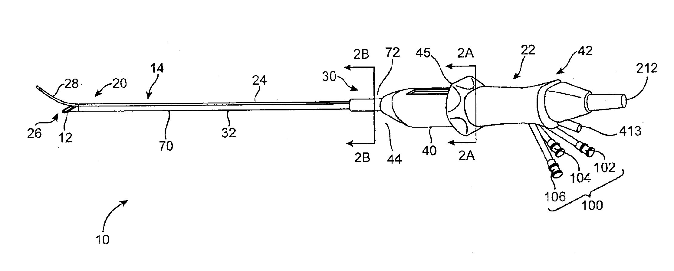
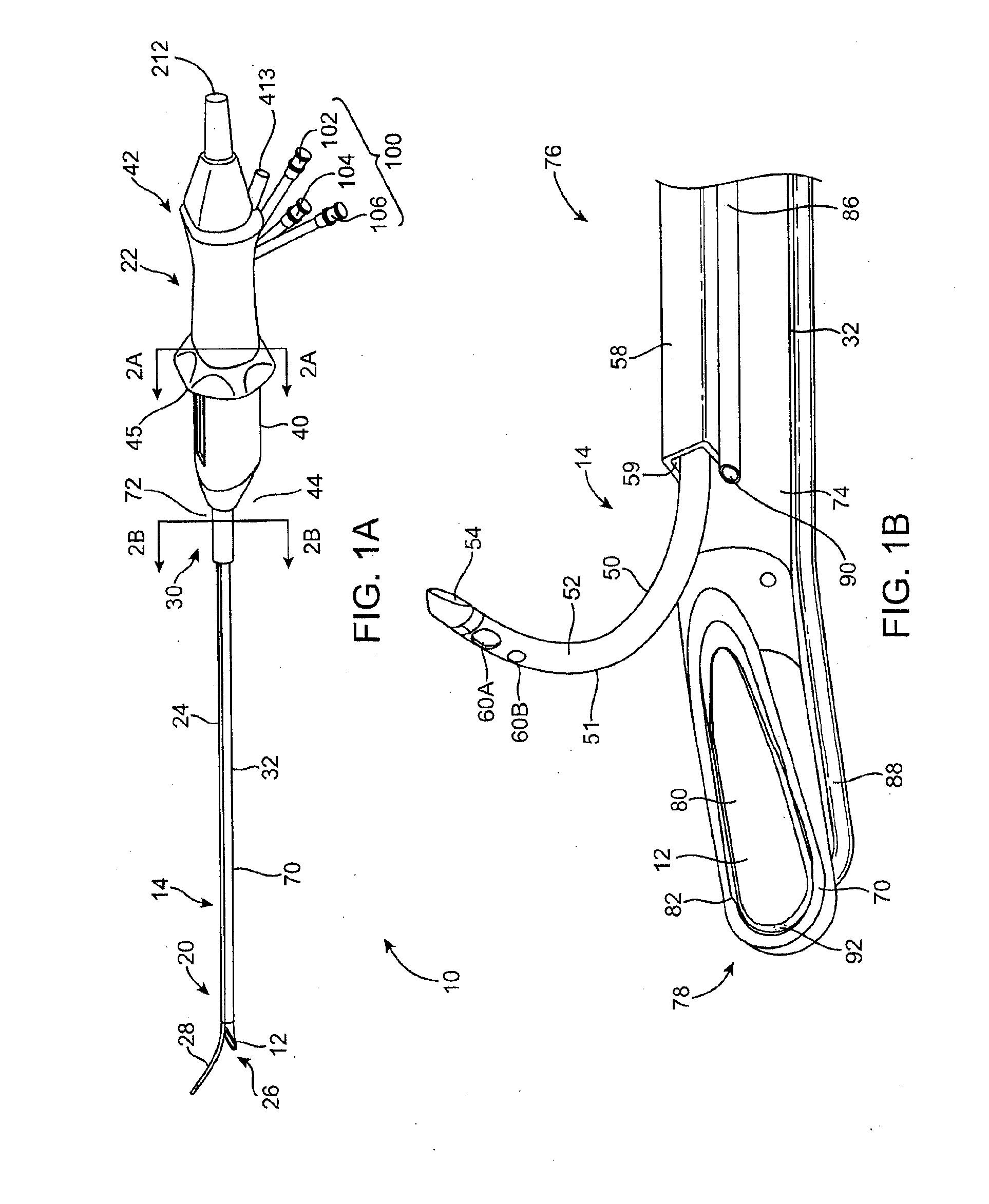
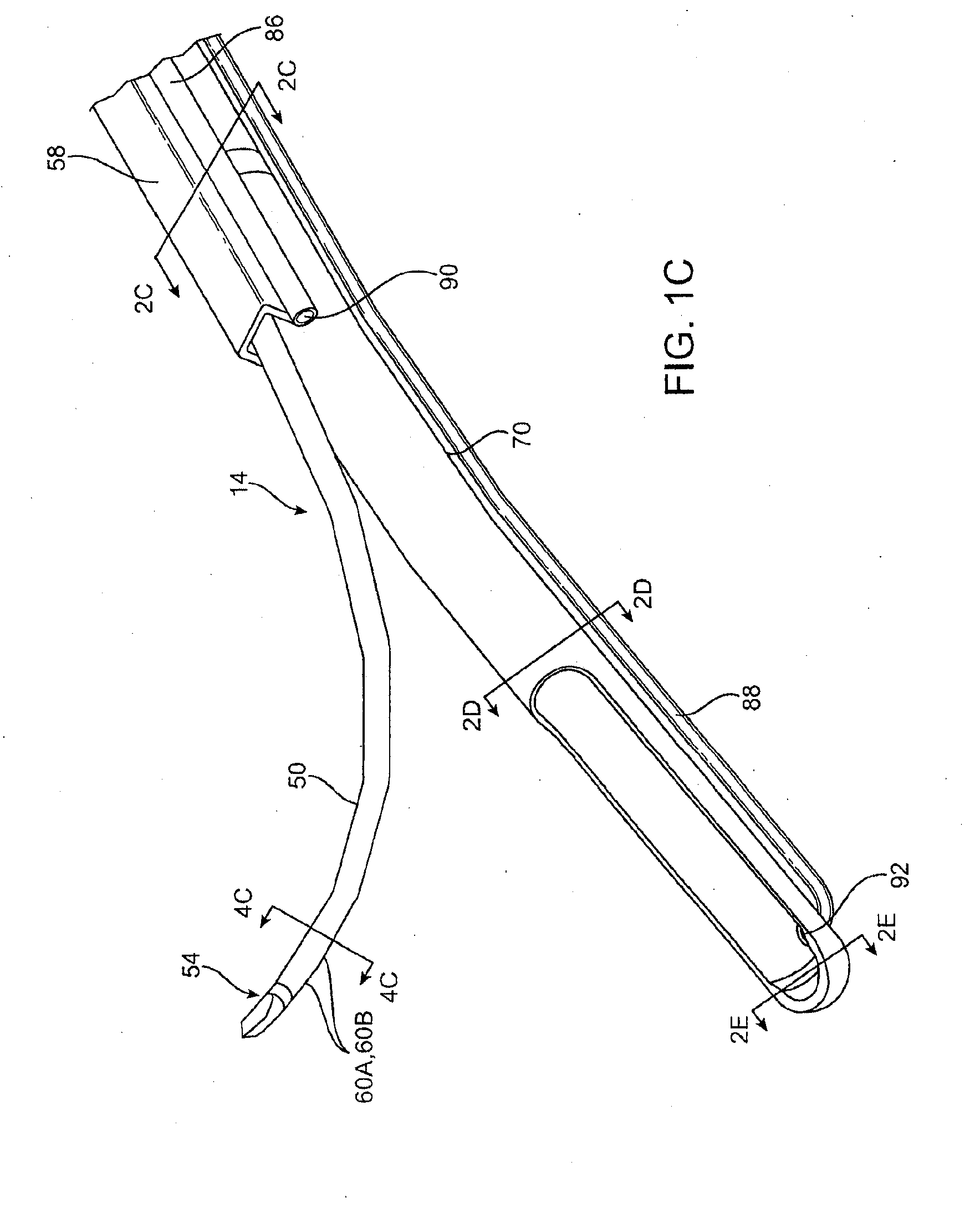
[0052]Referring to FIGS. 1A through 1C, an exemplary delivery system 10 embodying features of the present invention is shown having a shaft inclined viewing window 12 for improved imaging and a curved needle 14 for ablation treatment of a target site 16 such as fibroid tissues 18 (FIG. 5E) within a female's reproductive system. The delivery system 10 includes a system distal end 20, a system proximal end 22, and a rigid delivery shaft 24. Delivery shaft 24 includes a shaft distal end 26 with a bent or deflectable shaft distal tip 28, a shaft proximal end 30, and an axial passage 32 extending longitudinally through at least a portion of the delivery shaft 24. A handle 40 with handle proximal and distal ends 42 and 44, is attachable to the shaft proximal end 30. The handle 40 further includes a longitudinally movable slider 45 for enabling the advancement and retraction of the needle 14 to and from within a needle guide 58.

[0053]The curved needle 14 has a needle body 50 with a shaped ...


PUM


Abstract

Delivery systems, and methods using the same, having an ultrasound viewing window for improved imaging and a needle for ablation treatment of target tissues. In an embodiment, the target tissue is a fibroid within a female's uterus. In an embodiment, the delivery system includes a rigid shaft having a proximal end, a distal end, and an axial passage extending through the rigid shaft. In an embodiment, the axial passage is configured for removably receiving the ultrasound imaging insert having an ultrasound array disposed a distal portion.

Description

CROSS-REFERENCE TO RELATED APPLICATION[0001]This application is a continuation-in-part of U.S. application Ser. No. 12 / 973,587 (Attorney Docket No. 31992-706.301), filed Dec. 20, 2010, which is a continuation of U.S. application Ser. No. 11 / 564,164 (Attorney Docket No. 31992-706.501), filed on Nov. 28, 2006, which is a continuation-in-part of U.S. application Ser. No. 11 / 409,496 (Attorney Docket No. 31992-706.201), filed on Apr. 20, 2006, the full disclosures of which are incorporated herein by reference; this application is also a continuation-in-part of U.S. application Ser. No. 11 / 620,594 (Attorney Docket No. 31992-704.201), filed on Jan. 5, 2007, which claimed the benefit of Provisional Application 60 / 758,881 (Attorney Docket No. 31992-704.101), filed on Jan. 12, 2006, the full disclosures of which are incorporated herein by reference.BACKGROUND OF THE INVENTION[0002]1. Field of the Invention[0003]The present invention relates generally to medical systems and methods. More parti...

Claims


Application Information

Patent Timeline
Patent Type & Authority Applications(United States)
IPC IPC(8): A61B17/225
CPCA61B17/2256A61B8/0833A61B8/0841A61B8/12A61B2017/4216A61B2018/00559A61B8/445A61B2090/3784A61B18/1477A61B8/085A61B8/44A61B18/18A61B2018/00577A61B8/4411A61B2017/00106
Inventor DECKMAN, ROBERT K.GERBI, CRAIGMUNROW, MICHAELGROSSMAN, JESSICA
Owner GYNESONICS
Why Eureka
Try Eureka