

Inflammatory eye disorders
a technology for eye disorders and inflammation, applied in the field of inflammation eye disorders, can solve problems such as irritation, change of vision, and symptoms of irritation, and achieve the effect of increasing reflectivity and siz
- Summary
- Abstract
- Description
- Claims
- Application Information
AI Technical Summary
Benefits of technology
Problems solved by technology
Method used
Image

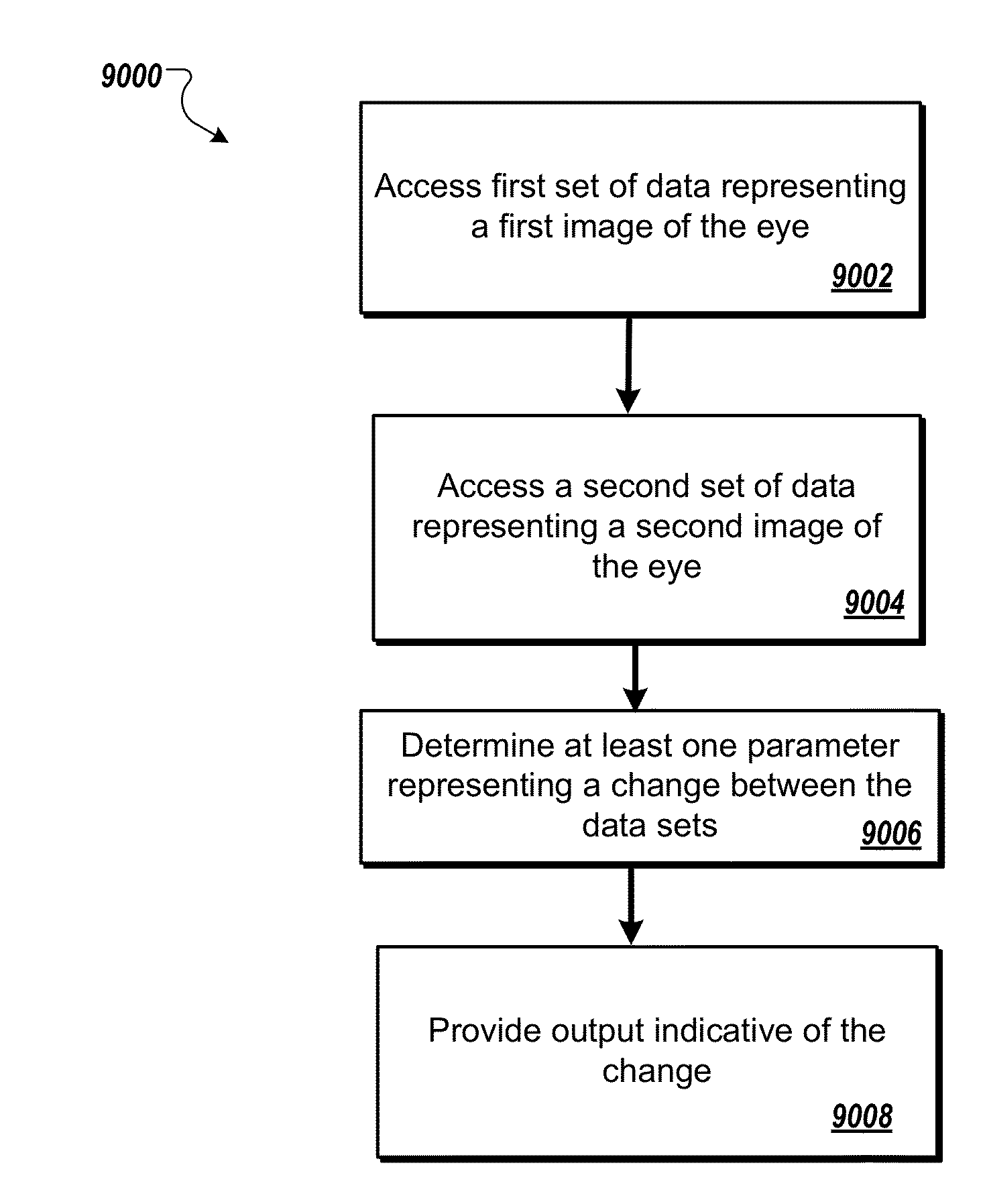
Examples
example 1
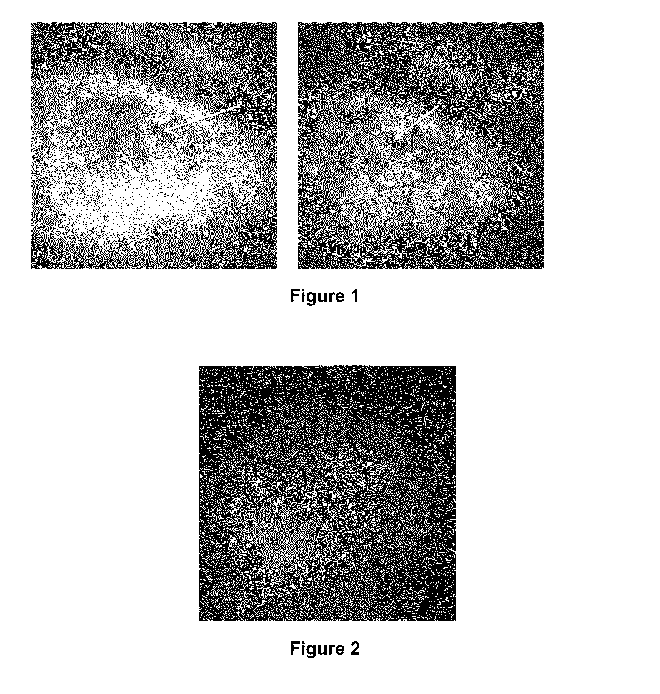
Detection of Changes in the Superficial Epithelial Cells Present in the Central Cornea of Patients Having Dry Eye Syndrome Upon Treatment
[0230]In vivo confocal microscopy was performed to image the superficial epithelial cells present in the central corneas of three subjects having dry eye syndrome (i) before or at an early time point in therapy, and (ii) at a second later time point. Each of these patients demonstrated a good response to treatment for his or her dry eye syndrome.
[0231]Images were gathered from subject #1 prior to treatment and at 6-weeks after the initiation of treatment (topical loteprednol four times a day for four weeks, followed by twice daily administration of topical loteprednol, with daily administration of artificial tears and Refresh PM ointment at bedtime throughout the treatment period).
[0232]Images were gathered from subject #2 early in treatment (four weeks after the initiation of treatment) and at a later time point during treatment (twelve weeks afte...
example 2
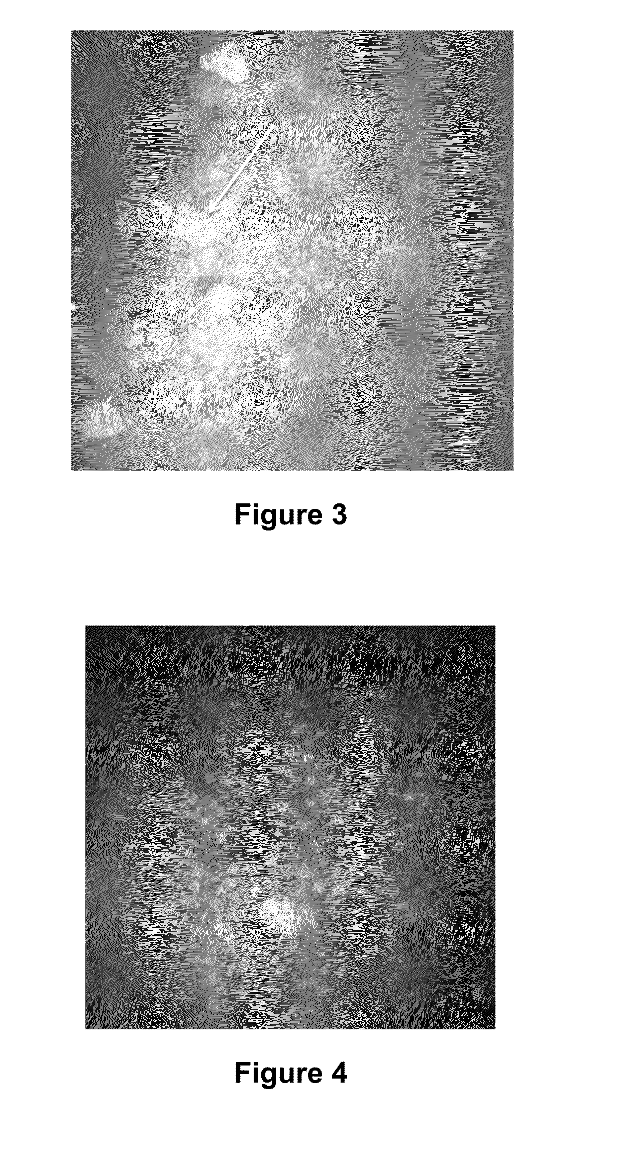
Detection of Changes in the Dendritic Cells Present in the Central Cornea of Patients Having Dry Eye Syndrome Upon Treatment
[0239]In vivo confocal microscopy was performed to image the dendritic cells present in the center of the corneas of two subjects (human subjects #1 and #2; described above) having dry eye syndrome (i) before or at an early time point in therapy, and (ii) at a later time point in therapy. Each of these patients demonstrated a good response to treatment for his or her dry eye syndrome.
[0240]Laser scanning in vivo confocal microscopy in these subjects was performed as described in Example 1. The resulting confocal images from two subjects were evaluated for density of immune cells present in the center of the cornea, the average size of dendritic immune cells present in the center of the cornea, and the average area covered by dendritic immune cells present in the center of the cornea. In vivo confocal microscopy images at a depth of 45 to 72 μm at the level of b...
example 3
Detection of Changes in the Corneal Nerves in Patients Having Dry Eye Syndrome Upon Treatment
[0243]In vivo confocal microscopy was performed to image the nerve cells present in the corneas of two subjects (human subjects #1 and #2; described above) having dry eye syndrome (i) before or at an early time point in therapy, and (ii) at a later time point in therapy. Each of these patients demonstrated a good response to treatment for his or her dry eye syndrome.
[0244]Laser scanning in vivo confocal microscopy in these subjects was performed as described in Example 1. The nerve analysis can be done using the semi-automated tracing program NeuronJ (Meijering et al., Cytometry A 58:167-176, 2004), a plug-in for ImageJ (available at the imagescience.org website). Nerve density can be assessed by measuring the total length of the nerve fibers in micrometers per frame (160,000 μm2). Main nerve trunks are defined as the total number of main nerve trunks in one image after analyzing the images ...
PUM
| Property | Measurement | Unit |
|---|---|---|
| Time | aaaaa | aaaaa |
| Size | aaaaa | aaaaa |
| Density | aaaaa | aaaaa |
Abstract
Description
Claims
Application Information