Regenerative peripheral nerve interface

a peripheral nerve and interface technology, applied in the field of regenerative peripheral nerve interface, can solve the problems of axonal damage threatening the fidelity of the device, signal degradation and failure gradually occurring, and difficult to interface these machines with living tissues

Active Publication Date: 2014-01-02
RGT UNIV OF MICHIGAN +1
View PDF2 Cites 22 Cited by
  • Summary
  • Abstract
  • Description
  • Claims
  • Application Information

AI Technical Summary

Problems solved by technology

While modern robotic technology has produced prosthetics capable of replicating complex movement and sensing environmental stimuli on par with the human arm, interfacing these machines with living tissues has proved challenging.
Current-generation neural interfaces that control prosthetic devices show promise early on, but signal degradation and failure gradually occur from scarring, inflammation, and axonal pruning.
In the acute phase, edema, hemorrhage, and axonal damage threaten the fidelity of the device.
In the chronic phase, macrophage and fibroblast responses result in robust scarring which causes signal loss and interface failure.
This detrimental response at the proximal end of a divided peripheral nerve can be a source of pain for the patient as well as a source of signal interference when decoding action potentials.
Metallic components are highly conductive and easy to fabricate, but have an unfavorable biocompatibility profile.
The mechanical stiffness of these components, mated with the soft surfaces of biologic tissues at the biotic-abiotic interface, likely exacerbates scar formation, which can cause signal interference and nerve damage.
These figures may not precisely reflect the characteristics of any given embodiment, and are not necessarily intended to fully define or limit specific embodiments within the scope of this technology.

Method used


Image

  • Regenerative peripheral nerve interface
  • Regenerative peripheral nerve interface
  • Regenerative peripheral nerve interface

Examples


example 1

Nerve Regeneration in the Presence of Conductive Polymers

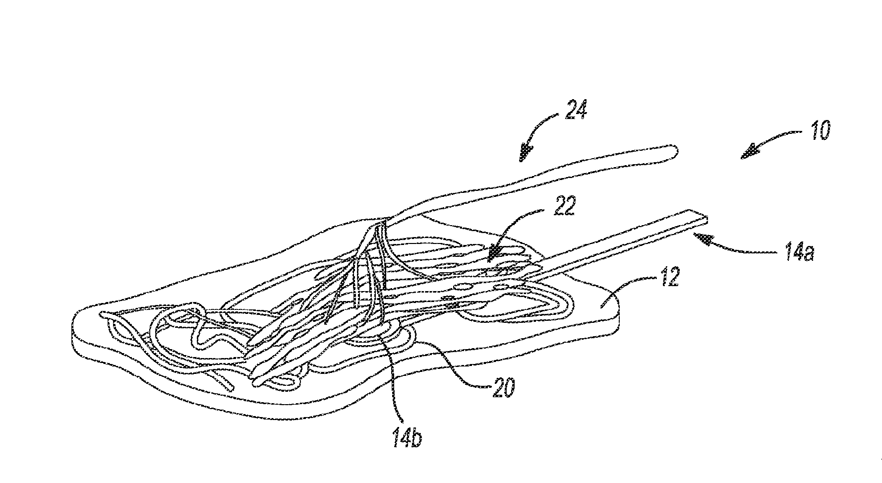
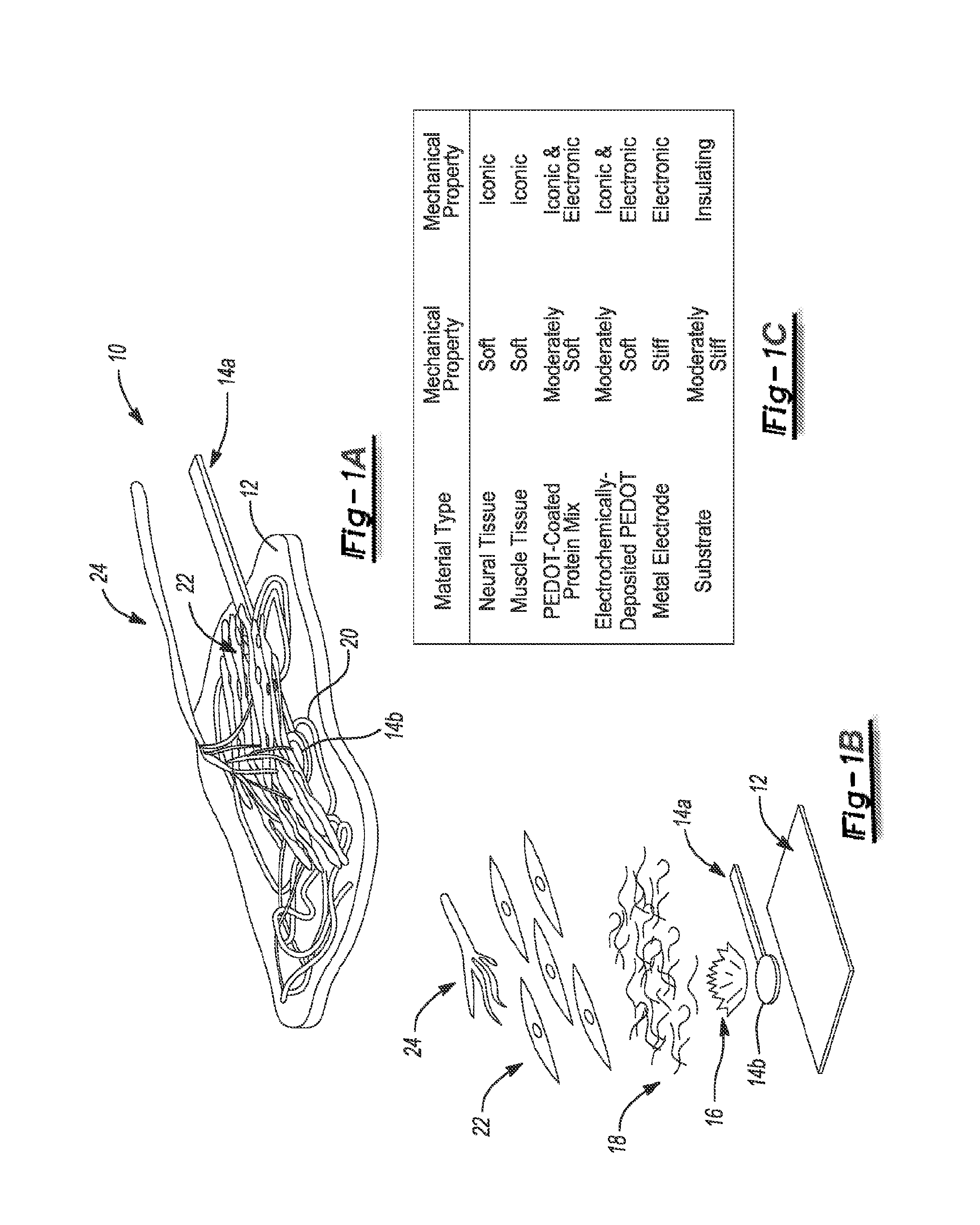
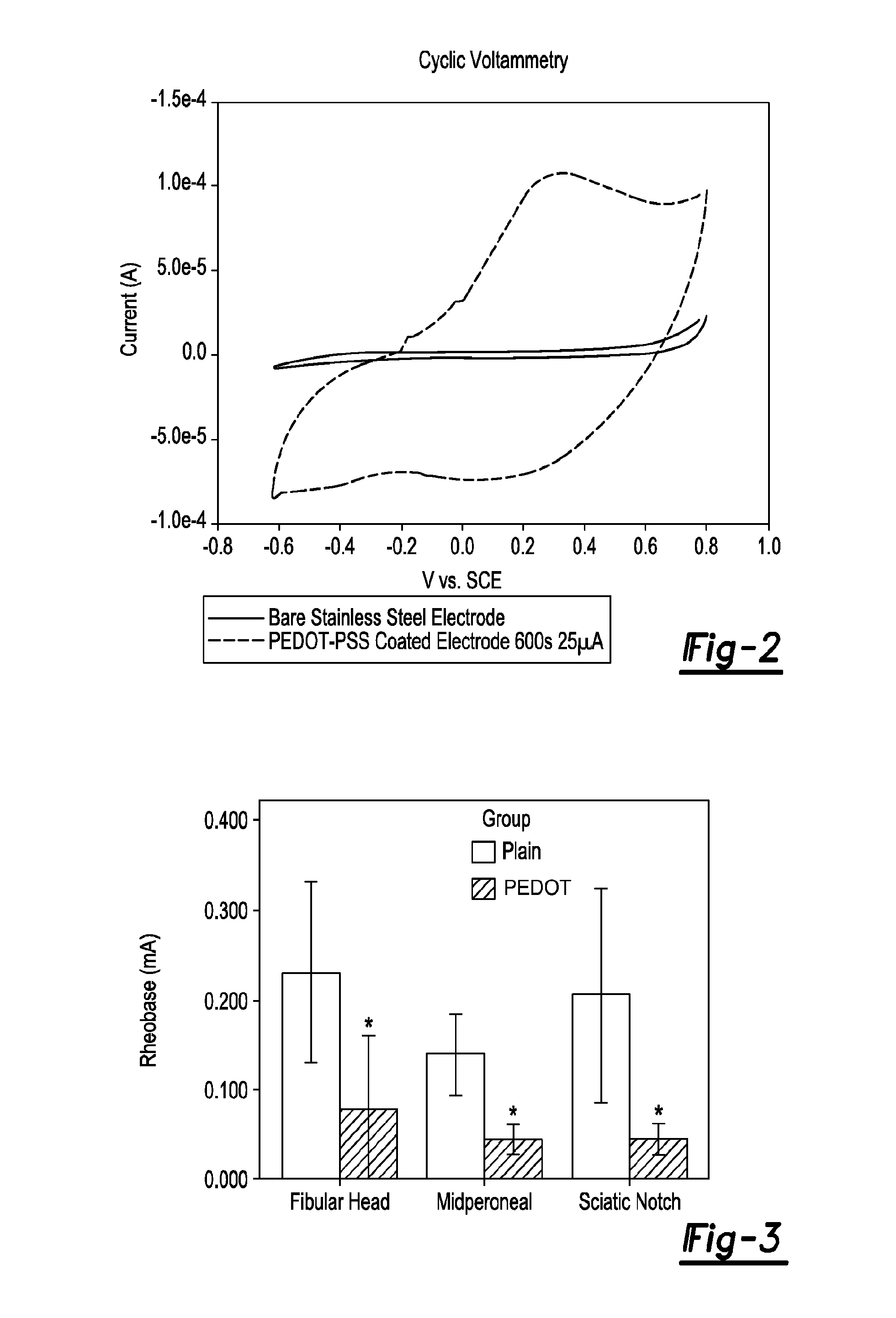
[0075]In the following example, an amputee's residual peripheral nerves are interfaced with processors that receive and transmit signals for prostheses control. PEDOT (3,4-polyethylenedioxythiophene) is an anisotropic, conductive polymer that beneficially reduces tissue impedance. PEDOT can be polymerized into nonconductive, processed decellular nerve (DN) leaving it highly conductive but stiff (DryPEDOT), or, less conductive but pliable (WetPEDOT). The nerve regeneration was tested in a rat peripheral nerve interface (PNI) environment and PEDOT biocompatibility was also evaluated.

[0076]A 15 mm, peroneal nerve gap was reconstructed with one of five nerve derived materials: Sham, Autograft, DN, DryPEDOT, and WetPEDOT, or not reconstructed: Gap, (n=8 / group). A single layer of acellular submucosa surrounded each graft as in peripheral nerve interfaces. After 90 days, evaluations were completed for muscle force, nerve conduction, ...

example 2

Long Term Stability of Regenerative Peripheral Nerve Interfaces (RPNI)

[0079]Stability of Regenerative Peripheral Nerve Interfaces

[0080]An amputee's peripheral nerves with neuro-prosthetic arms possessing the latest robotic technology are interfaced. The regenerative peripheral nerve interface (RPNI) is constructed by implanting syngeneic muscle cells from one of two stages of muscle development, immature (myoblast) or mature (transferred muscle). A construct is formed from a decellular muscle with or without the conductive polymer poly(3,4-ethylenedioxythiophene), or PEDOT, which supports the muscle cells and allows implantation of the transected residual nerve within the RPNI. For all RPNI, spontaneous EMG, nerve conduction, and neuromuscular junction formation following in-situ maturation occurred. RPNI were sustained long term for both the myoblast and the muscle transfer groups.

[0081]Animal care and operative procedures were performed in accordance with the United States Public ...

example 3

Peripheral Nerve Interface to Thin-Film Polyimide Electrode Arrays

[0084]In the following example, a peripheral nerve interface to provide motor signals to and record sensor feedback from prostheses is developed. In this study, a thin film polyimide electrode array as a peripheral nerve interface-signal recording probe was tested. The feasibility of array in terms of biocompatibility, material integrity (electrical and mechanical) and surgical availability was also tested.

[0085]The thin-film electrodes are microfabricated polyimide thin-film arrays developed and provided by NeuroNexus Technologies, Inc. The design space of this technology provides a large variability in terms of electrode size, placement and packaging for chronic preparations. The arrays were flexible, planar, 15 microns thick with 32 regularly spaced electrode channels. In the surgical procedure, the rat peroneal nerve was exposed and divided. The thin-film polymer arrays were implanted around the proximal stump as ...


PUM

PropertyMeasurementUnit
lengthaaaaaaaaaa
lengthaaaaaaaaaa
lengthaaaaaaaaaa

Abstract

The present disclosure provides a regenerative peripheral nerve interface (RPNI) for a subject comprising an insulating substrate, at least one metallic electrode deposited onto the insulating substrate forming a thin-film array; a portion of the at least one metallic electrode surface having a layer of a first conductive polymer and a layer of decellularized small intestinal submucosa (SIS) coating a portion of the electrode, wherein a second conductive polymer is electrochemically polymerized through the SIS to form the regenerative peripheral nerve interface. The present disclosure also provides that a layer of muscle tissue contacts the regenerative peripheral nerve interface.

Description

CROSS-REFERENCE TO RELATED APPLICATIONS[0001]This application is a National Stage of International Application No. PCT / US2012 / 021311 filed on Jan. 13, 2012, which claims the benefit and priority of U.S. Patent Application No. 61 / 432,787 filed on Jan. 14, 2011. The entire disclosures of each the above applications are incorporated herein by reference.FIELD[0002]The present technology relates to a regenerative peripheral nerve interface, including methods and devices relating to regenerative peripheral nerve interfaces.INTRODUCTION[0003]This section provides background information related to the present disclosure which is not necessarily prior art.[0004]Statistics estimate there are about 1.7 million people in the US alone living with limb loss. This corresponds to about 1 in every 200 people. There is a need to create reliable neural interfaces that can seamlessly and naturally connect patients with artificial arms and legs. The peripheral nerve system is viewed as the ideal locatio...

Claims


Application Information

Patent Timeline
Patent Type & Authority Applications(United States)
IPC IPC(8): A61F2/72
CPCA61F2/72A61L27/34A61L2430/32C08L89/00A61F2/0811A61N1/0551
Inventor CEDERNA, PAUL S.URBANCHEK, MELANIE G.MARTIN, DAVID C.
Owner RGT UNIV OF MICHIGAN
Why Eureka
Try Eureka