Muscle tissue regeneration using muscle fiber fragments

Active Publication Date: 2014-08-28
WAKE FOREST UNIV HEALTH SCI INC
View PDF2 Cites 11 Cited by
  • Summary
  • Abstract
  • Description
  • Claims
  • Application Information

AI Technical Summary

Benefits of technology

[0012]This invention describes uniformly structured small fragments of muscle fiber for efficient functional muscle regeneration as well as methods of making and using these muscle fiber fragments. The use of small

Problems solved by technology

Reconstruction of skeletal muscle tissue lost by traumatic injury, tumor excision or ablation, or functional damage is hindered by the lack of available functional muscle tissue or muscle tissue substitutes.
Autografts have been used clinically but require the presence of sufficient donor muscle tissue nearby and can leave muscle defects at the

Method used


Image

  • Muscle tissue regeneration using muscle fiber fragments
  • Muscle tissue regeneration using muscle fiber fragments
  • Muscle tissue regeneration using muscle fiber fragments

Examples


example 1

Preparation of Muscle Fiber Fragments

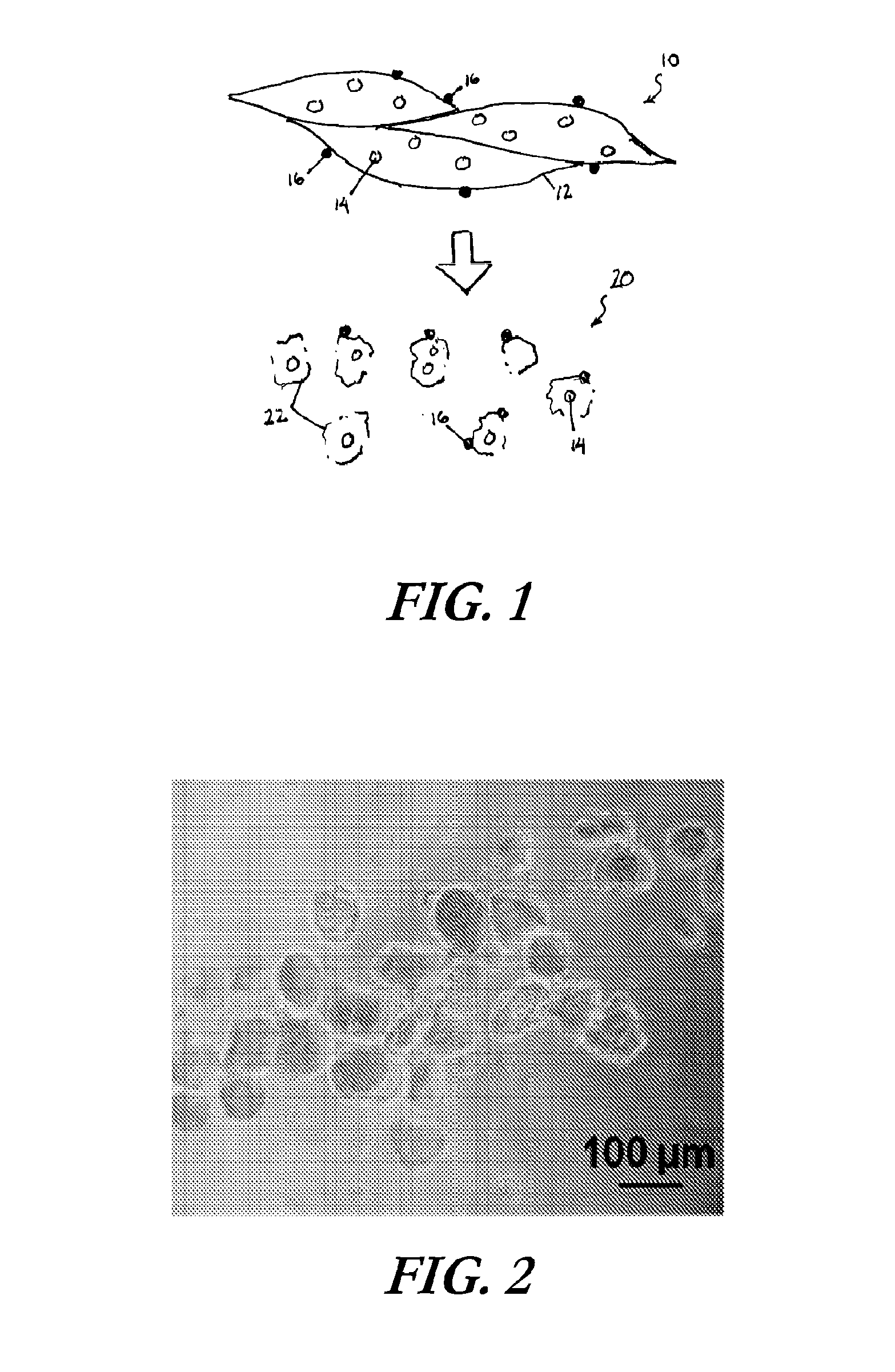
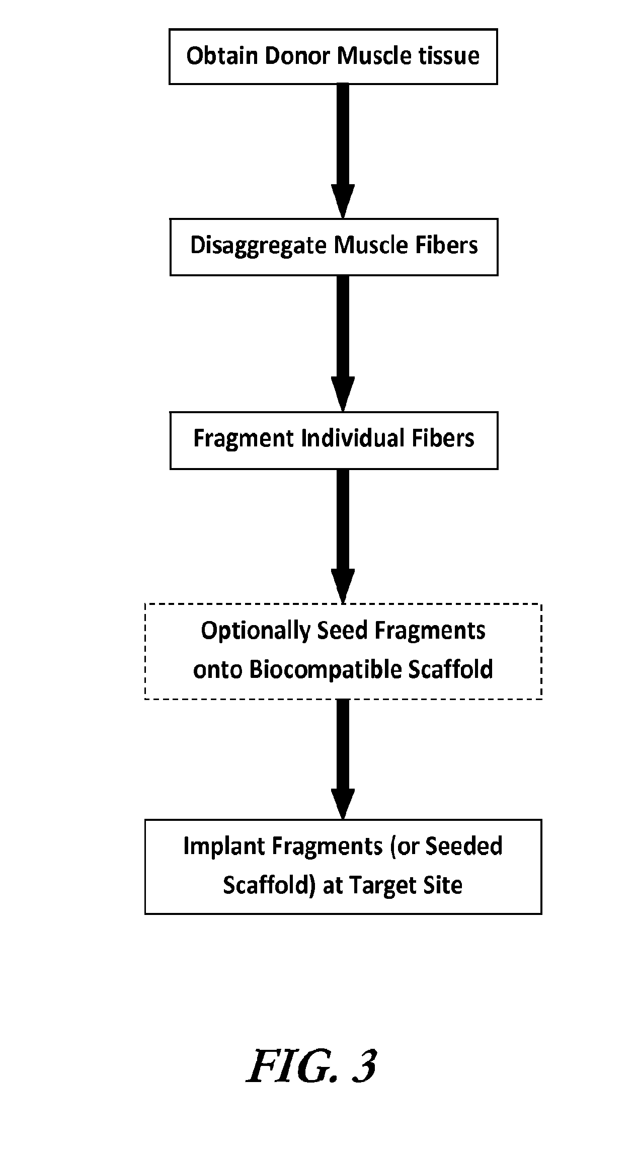
[0098]Uniformly structured muscle fiber fragments were isolated from tibialis anterior (TA) and gastrocnemius (GN) muscles of C57BL6 mice (male, 10-12 weeks). Isolated TA or GN muscles were minced with two disposable scalpels. The muscle tissue was minced into quite small sizes. After mincing, the fragments were digested with 0.2% collagenase type I in serum free DMEM with shaking at 37° C. for 1 hr. After enzymatic digestion, myofibers were pipetted to dissociate into individual fragments. Subsequently, the myofibers solution was immediately filtered through a cell strainer (100 μm size, BD falcon) to obtain uniform sized myofiber fragments.

Small myofiber fragments were labeled with Vybrant fluorescent dye (DiD, Invitrogen) for fiber tracking after implantation in animals. Confocal microscopy confirmed that isolated fiber fragments including cells on their surface were fluorescently labeled with Vybrant and the size of fiber fragments showed app...

example 2

Transplant of Muscle Fiber Fragments

[0099]To determine whether the processed muscle fragments can be developed as an injection therapy, isolated muscle fiber fragments were injected to the gluteal region of C57BL6 mice (10-12 weeks). 10 μl of fiber fragments solution in PBS (500 in fiber fragment numbers / injection) were injected twice toward the incision site (0.5-1 cm) on the gluteal region with a hamilton syringe (26 G, 10 μl). After transplantation, the incision was closed by suture. At 1, 2, 3, and 4 weeks after transplantation, the muscle tissue with incision site was harvested and further frozen in OCT compounds.

[0100]To examine whether injected muscle fiber fragments would reassemble into host muscle fibers and integrate with vascular and neural system of host animal, histological and immunohistological studies were performed using the harvest tissues. The frozen tissues were sectioned and fixed with 10% formalin solution for H&E staining. For immunohistochemical study of the...

example 3

Comparison of Transplantation Efficiencies of Muscle Fiber Fragments, Long Fibers and Acellularized Tissue Components

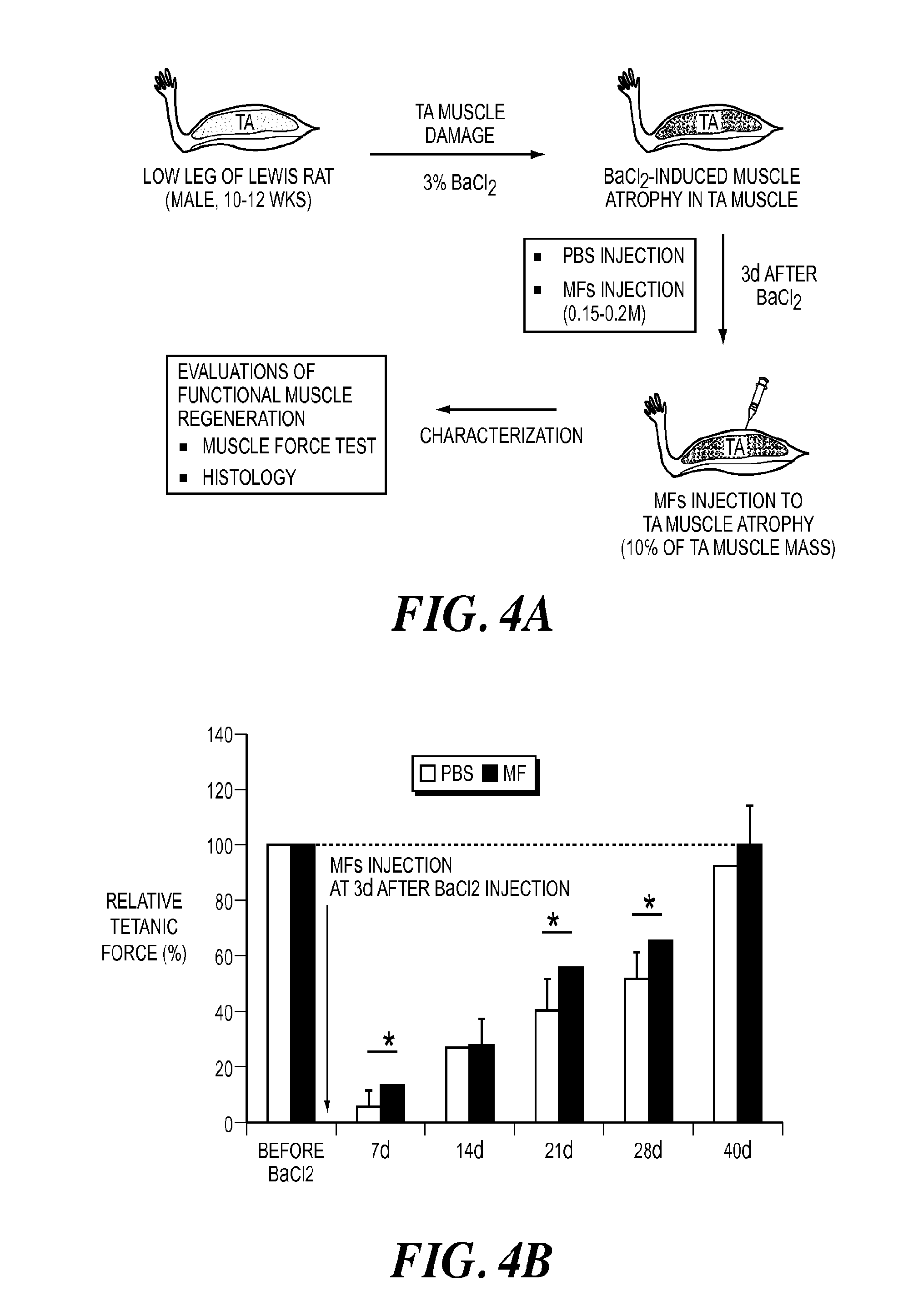
[0102]A study to compare the in vivo effect of decellularized fragments and fragments containing satellite cells was performed. Encouraged by the promising results from in vivo mouse study, an extended animal study using rats was performed in order to determine efficient engraftment of small fiber fragments when compared to that of long fibers using various injection volume and numbers of fiber fragments. Additionally, acellular fragments without cellular components were transplanted to examine possibility of their use as “off-the shelf” components for allogeneic transplantation. Muscle fiber fragments from rats (Lewis rats, 10-12 weeks) were isolated using the same protocols as described above for mice. Various volumes (100, 200, and 300 μL) of fiber fragments (7×10⁴ fibers / ml) were injected to gastrocnemius (GN) muscles without any muscle defects using a syringe wit...


PUM

PropertyMeasurementUnit
Fractionaaaaaaaaaa
Timeaaaaaaaaaa
Sizeaaaaaaaaaa

Abstract

The invention is directed to methods and compositions for obtaining uniform sized muscle fiber fragments for transplantation. These muscle fiber fragments are able to reconstitute into long fibers that are oriented along native muscle. The implanted muscle cells integrate with native vascular and neural network, as confirmed by histology and immunohistochemistry. This invention is particularly advantageous because autologous muscle can be harvested from a donor site, processed and injected into target sites in the operating room. The fragmented muscle fibers can be readily integrated within the host.

Description

GOVERNMENT RIGHTS[0001]This invention was made with Government support under Grant No. W81XWH-08-1-0333, awarded by the Department of Defense—Orthopedics Trauma Research Program (OTRP). The Government has certain rights in this invention.BACKGROUND OF THE INVENTION[0002]The technical field of this invention relates to methods and compositions for the regeneration of muscle tissue. The invention also relates to constructs containing muscle tissue.[0003]Reconstruction of skeletal muscle tissue lost by traumatic injury, tumor excision or ablation, or functional damage is hindered by the lack of available functional muscle tissue or muscle tissue substitutes. Autografts have been used clinically but require the presence of sufficient donor muscle tissue nearby and can leave muscle defects at the donor site. Herter et al., (J. Plast Reconstr. Aesthetic Surgery 60, 760-768 (2007)) provides an analysis of the various locations providing donor tissue and the benefits and disadvantages of th...

Claims


Application Information

Patent Timeline
IPC IPC(8): A61K35/34A61K47/34A61K45/06A61K47/42
CPCA61K35/34A61K47/42A61K47/34A61K45/06A61K38/18A61K9/0034A61L27/18A61L27/24A61L27/3683A61L27/3834A61L2300/414A61L2400/06A61L2430/30A61L27/3604A61P13/02A61P21/00A61P21/06A61K2300/00C08L67/04A61L27/3687A61L27/54C12N5/0062C12N5/0659C12N2513/00C12N2533/30C12N2533/54
Inventor ATALA, ANTHONYYOO, JAMESKO, IN KAP
Owner WAKE FOREST UNIV HEALTH SCI INC
Why Eureka
Try Eureka