Artificial placenta
a placenta and placenta technology, which is applied in the field of infant respiratory failure treatment, can solve the problems of high risk of lung damage using such methods, life-long dependency on mechanical ventilation, and mortality of very low birth weight infants, and achieves the effects of preventing coagulation, and reducing the risk of lung damag
- Summary
- Abstract
- Description
- Claims
- Application Information
AI Technical Summary
Benefits of technology
Problems solved by technology
Method used
Image
Examples
example 1
Preparation of an Artificial Placenta Device
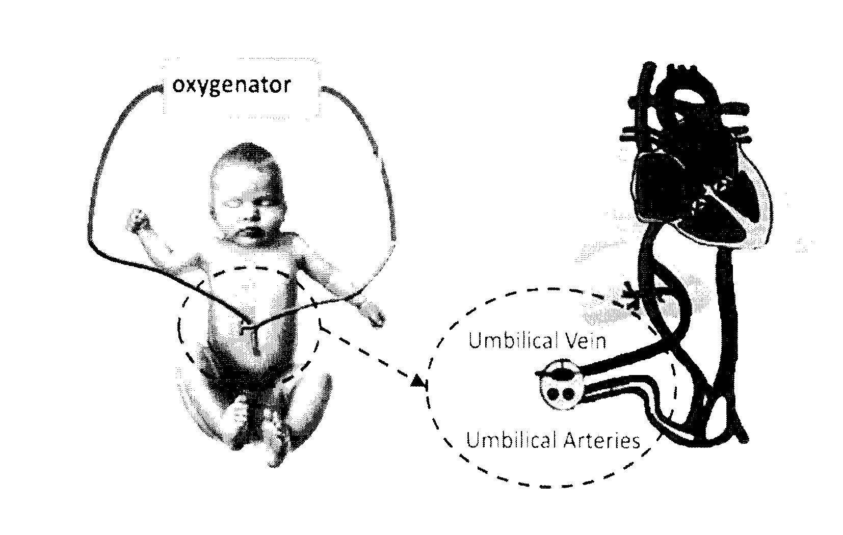
[0051]A miniature artificial lung / placental device was developed for use in preterm babies. The device improves the efficiency of oxygenation in preterm babies since it is based on passive flow, is coated with heparin to prevent clotting and provides appropriate resistance with sufficient flow and small volume.
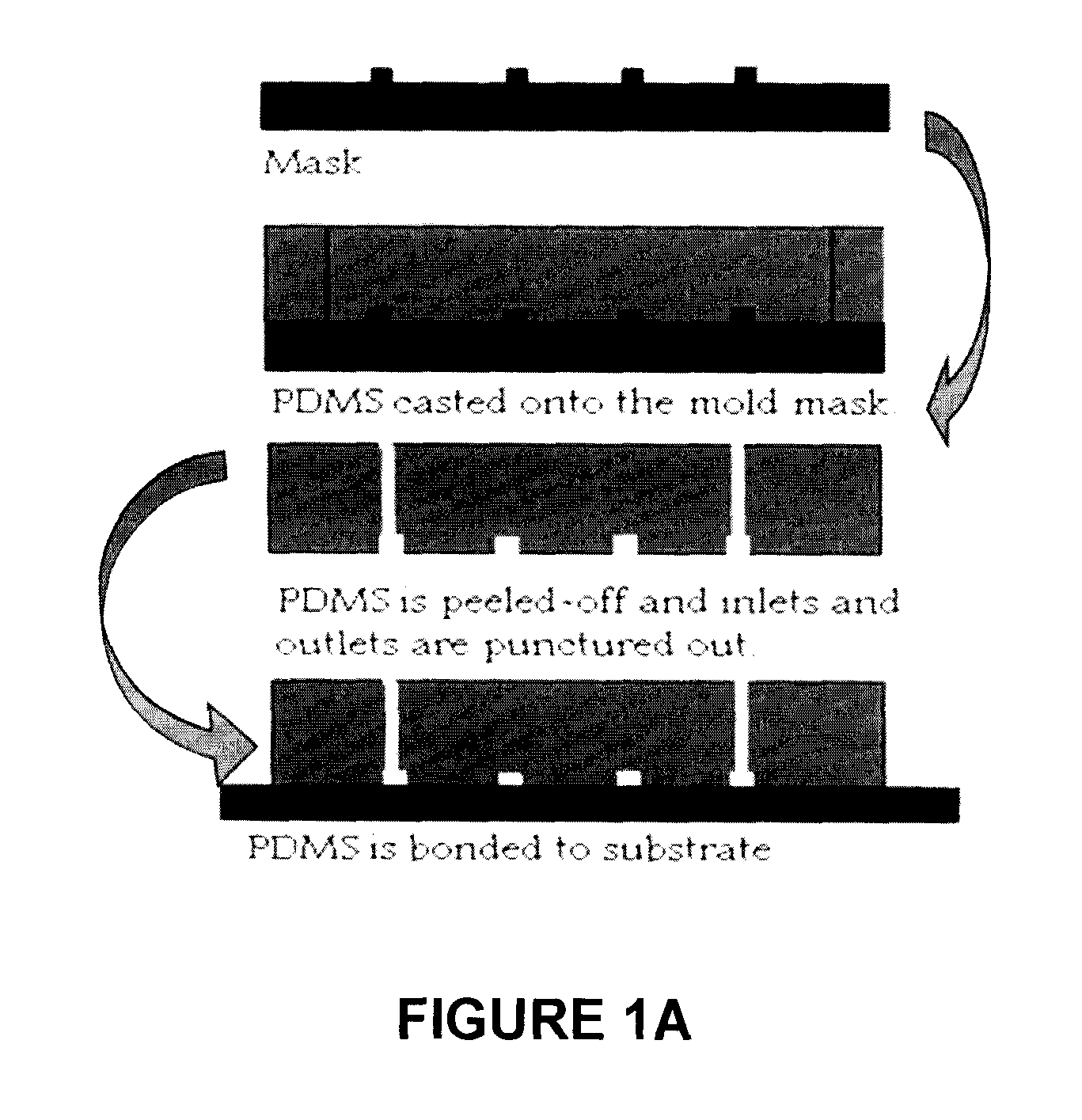
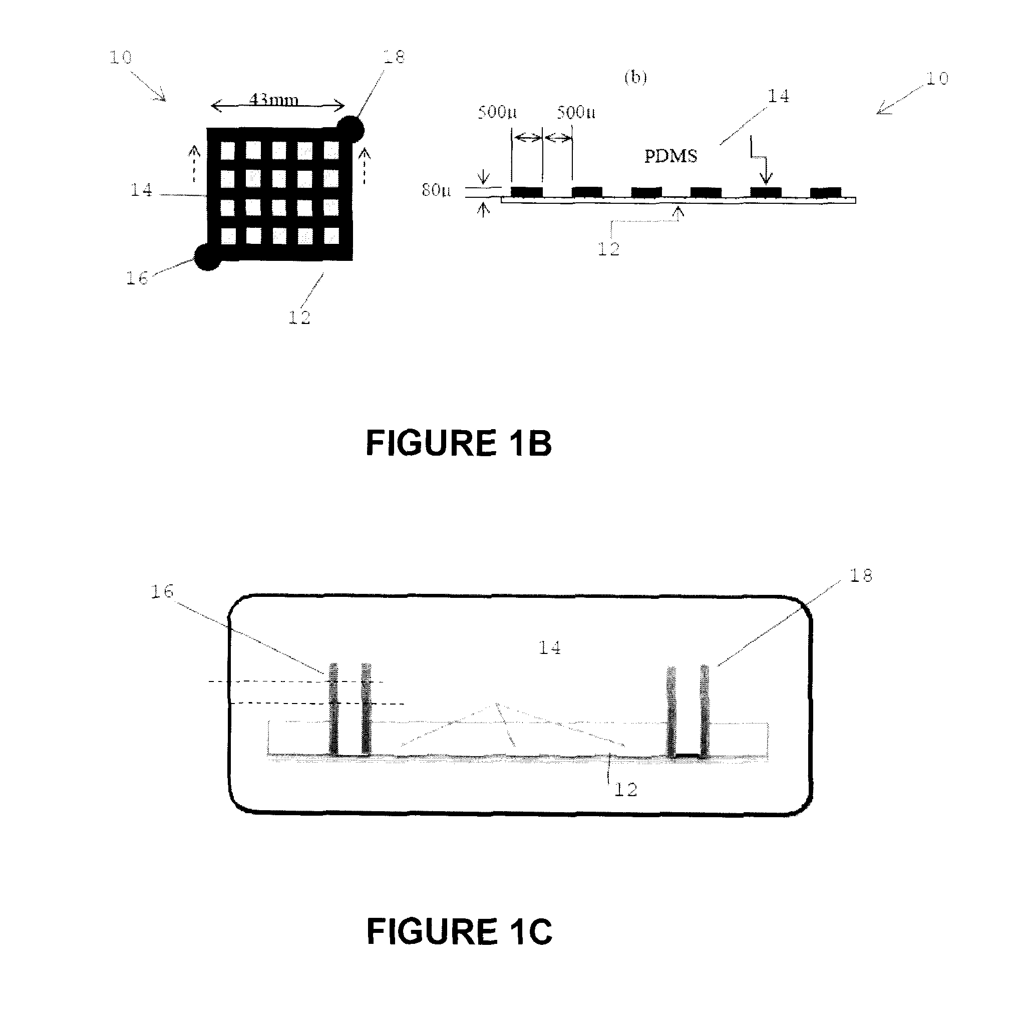
[0052]An approximately 3-inch microchannel mould was created to cast a PDMS (a silicone-based organic polymer) microchannel and then a membrane is attached to seal the channel. The specific process used to create a mold for the vascular microchannel network, prepare the vascular network and attach the membrane was as follows. The silicone substrate was cleaned as follows: rinsed with acetone for 15 sec., rinsed with methanol for 15 seconds and rinsed with de-ionized (DI) water for 5 min. The sample was dried using compressed nitrogen and dehydrated at 110° C. for 2 min.
[0053]The microchannel mould was prepared as follows. The silicon...
example 2
In Vitro Testing of Artificial Placenta Device
[0058]For in-vitro testing of a placenta device as described in Example 1, human erythrocyte concentrates were adjusted with plasma for a hematocrit of 50%. pH was adjusted by adding NaHCO3 and aerating with nitrogen. Heparin (3 units / mL) was added for anticoagulation. The blood was pumped through the gas exchange device at flow rates of 1-4 mL / min while the pressure was measured (FIG. 2a). Blood samples were collected before and after flowing through the membrane device according to the flow diagram of FIG. 3 to determine the gas exchange. The results show that the gas flux decreased with decrease in residence time (or increased flow rate) (FIG. 4). Both membranes were comparable in O2 exchange. Both microfluidic devices with PDMS and PC membranes were compared to a commercial hollow-fiber based oxygenator OXR® and found to exhibit about a five-fold increase in O2 flux.
[0059]Fluids of various viscosities were flowed through the device t...
example 3
[0066]To test gas-exchange, two channels were attached together such that the membranes involved in diffusion faced each other as shown in FIG. 17. The top channel was attached to a pump that pumped blood through the device. The bottom channel was attached to a gas supply of varying oxygen levels, e.g. 30% O2, 40% O2, 50% O2, no gas, room air and atmospheric conditions. The gas exchanger served to simulate blood in umbilical arteries before entering the placenta. As blood passed through the device, it passed along a concentrated supply of gases that diffused through and interacted with the blood. The resistance of the device was measured. If not enough blood passed through the device, adequate oxygenation did not occur. If a large volume of blood flowed through the device, sufficient blood did not circulate through the body, the carbon dioxide concentration decreased and oxygen concentration in the blood increased. A basic visualization is displayed in FIG. 16 modeling d...
PUM
 Abstract
Description
Claims
Application Information
 - R&D
- Intellectual Property
- Life Sciences
- Materials
- Tech Scout
- Unparalleled Data Quality
- Higher Quality Content
- 60% Fewer Hallucinations
Browse by: Latest US Patents, China's latest patents, Technical Efficacy Thesaurus, Application Domain, Technology Topic, Popular Technical Reports.
© 2025 PatSnap. All rights reserved.Legal|Privacy policy|Modern Slavery Act Transparency Statement|Sitemap|About US| Contact US: help@patsnap.com


