Nanoparticles with enhanced mucosal penetration or decreased inflammation

a technology of nanoparticles and mucosal surface, applied in the field of nanoparticles, can solve the problems of limited scope of biodegradable mpps prepared by nanoprecipitation, limited control of drug delivery at mucosal surface, and limited efficacy, and achieve the effect of effective drug delivery and high drug loading

Inactive Publication Date: 2014-11-06
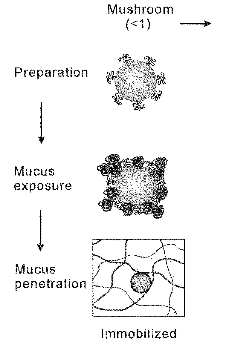
THE JOHN HOPKINS UNIV SCHOOL OF MEDICINE
View PDF4 Cites 71 Cited by
  • Summary
  • Abstract
  • Description
  • Claims
  • Application Information

AI Technical Summary

Problems solved by technology

However, controlled drug delivery at mucosal surfaces has been limited by the presence of the protective mucus layer.
As a result of mucus turnover, most therapeutics delivered locally to mucosal surfaces suffer from poor retention and distribution, which limits their efficacy.
However, the scope of biodegradable MPPs prepared by nanoprecipitation is limited because it requires dissolution of the drug and polymer in water-miscible solvents.
Numerous hydrophobic drugs may benefit from local delivery by MPPs, due to serious systemic side effects, however their poor solubility in water-miscible organic solvents limits efficient encapsulation into MPPs by nanoprecipitation.
However, these hydrophilic drugs cannot be easily formulated into MPPs by nanoprecipitation.

Method used


Image

  • Nanoparticles with enhanced mucosal penetration or decreased inflammation
  • Nanoparticles with enhanced mucosal penetration or decreased inflammation
  • Nanoparticles with enhanced mucosal penetration or decreased inflammation

Examples


example 1

Preparation of Nanoparticles

[0296]Materials and Methods

[0297]Biodegradable nanoparticles were prepared by either o / w single emulsion or w / o / w double emulsion method as described in R. C. Mundargi et al, J. Control. Release 125, 193 (2008), M. Li et al., Int. J. Pharm. 363, 26 (2008), C. E. Astete and C. M. Sabliov, J. Biomater. Sci. Polymer Ed. 17, 247 (2006), and R. A. Jain, Biomaterials, 21, 2475 (2000).

[0298]Nanoparticles were characterized for size, surface property and drug loading (for drug encapsulated nanoparticles). The displacements of nanoparticles were tracked in fresh, undiluted human CVM using multiple particle tracking.

[0299]Nanoparticles Prepared with Different Amounts of PEG

[0300]PLGA-PEG nanoparticles were prepared with varying target PEG contents (0, 2, 3, 5, 8, 10 and 25 wt %, referred to as PLGA, PLGA-PEG2%, PLGA-PEG3%, PLGA-PEG5%, PLGA-PEG8%, PLGA-PEG10% and PLGA-PEG25%) using emulsification. The PEG molecular weight 5 kDa was selected since at the same PEG con...

example 2

Nanoparticles Prepared with Different Emulsifiers

[0305]Materials and Methods

[0306]Alexa Fluor 555 cadaverine (AF555) was chemically conjugated to polymers. Nanoparticles were prepared using emulsification. Typically, a mixture (total 50 mg) of PLGA-PEG5k and AF555-labeled PLGA-PEG5k was dissolved in 1 mL dichloromethane (DCM). The oil phase was poured into 5 mL aqueous solution containing 1% emulsifier under sonication (VibraCell, Sonics & Materials Inc., Newtown, Conn.) at 30% amplitude for 2 mins in an ice-water bath to form the oil-in-water emulsion.

[0307]The emulsion was poured into another 40 mL aqueous phase of emulsifier solution under magnetic stirring at 700 rpm for at least 3 hours to allow the solvent to evaporate. The solvent was further evaporated by placing the solution in a vacuum chamber for 30 mins. The final nanoparticle suspensions were filtered through 1 μm syringe filter, centrifuged at 20,000 g for 25 mins and thoroughly washed with water.

[0308]Emulsifiers incl...

example 3

Preparation of Drug Encapsulated Nanoparticles

[0314]Materials and Methods

[0315]Curcumin was selected as a model hydrophobic drug which was dissolved with polymer in DCM. The procedure was similar to that for preparation of unloaded nanoparticles. The prepared curcumin-nanoparticles can be visualized in mucus because of curcumin's intrinsic fluorescence.

[0316]BSA was used as a model hydrophilic drug because it is representative of large molecule biologics. BSA-FITC and BSA (10% ratio of BSA-FITC) were dissolved in 0.2 mL 16% w / v aqueous solution at 37° C. This solution was added to 1 mL of 100 mg / ml PLGA-PEG5k in DCM solution during probe sonication (30% amplitude, 1 min with is pulse) in the ice-water bath. The resultant W / O primary emulsion was immediately added to a second water phase (5 mL 1% saponin solution) under sonication (20% amplitude for 2 min) The double emulsion was transferred to another 40 mL 1% saponin solution with magnetic stirring for 3 hours. Nanoparticles were f...


PUM

PropertyMeasurementUnit
diameteraaaaaaaaaa
diameteraaaaaaaaaa
diameteraaaaaaaaaa

Abstract

Nanoparticles formed by emulsion of one or more core polymers, one or more surface altering materials, and one or more low molecular weight emulsifiers have been developed. The particles are made by dissolving the one or more core polymers in an organic solvent, adding the solution of the one or more core polymers to an aqueous solution or suspension of the emulsifier to form an emulsion, and then adding the emulsion to a second solution or suspension of the emulsifier to effect formation of the nanoparticles. In the preferred embodiment, the molecular weight of the emulsifiers is less than 1500, 1300, 1200, 1000, 800, 600, or 500 amu. Preferred emulsifiers include cholic acid sodium salt, dioctyl sulfosuccinate sodium, hexadecyltrimethyl ammonium bromide, saponin, TWEEN® 20, TWEEN® 80, and sugar esters. The surface altering materials are present in an amount effective to make the surface charge of the particles neutral or essentially neutral when the one or more emulsifiers are charged. The emulsifiers have an emulsification capacity of at least about 50%, preferably at least 55, 60, 65, 70, 75, 80, 85, 90, or 95%.

Description

FIELD OF THE INVENTION[0001]This invention is in the field of nanoparticles, particularly nanoparticles that rapidly penetrate, mucus, such as human mucus, and methods of making and using thereof.BACKGROUND OF THE INVENTION[0002]Localized delivery of therapeutics via biodegradable nanoparticles often provides advantages over systemic drug administration, including reduced systemic side effects and controlled drug levels at target sites. However, controlled drug delivery at mucosal surfaces has been limited by the presence of the protective mucus layer.[0003]Mucus is a viscoelastic gel that coats all exposed epithelial surfaces not covered by skin, such as respiratory, gastrointestinal, nasopharyngeal, and female reproductive tracts, and the surface of eye. Mucus efficiently traps conventional particulate drug delivery systems via steric and / or adhesive interactions. As a result of mucus turnover, most therapeutics delivered locally to mucosal surfaces suffer from poor retention and ...

Claims


Application Information

Patent Timeline
Patent Type & Authority Applications(United States)
IPC IPC(8): A61K47/34A61K9/00A61K9/14
CPCA61K47/34A61K9/146A61K9/0019A61K9/0014A61K9/0048A61K9/0036A61K9/5138A61K9/5146A61K9/5153A61K31/12A61K31/7088A61P11/00A61P29/00A61K47/50A61K9/16A61K9/51A61K47/30A61K49/00B82B3/00B82Y5/00
Inventor HANES, JUSTINXU, QINGGUOBOYLAN, NICHOLAS
Owner THE JOHN HOPKINS UNIV SCHOOL OF MEDICINE
Why Eureka
Try Eureka