Radiation exposure diagnostic marker igfbp-5, composition for radiation exposure diagnosis by measuring the expression level of the marker, radiation exposure diagnostic kit comprising the composition, and method for diagnosing radiation exposure using the marker
a radiation exposure and diagnostic marker technology, applied in the direction of peptides, biological material analysis, dna/rna fragmentation, etc., can solve the problems of tissue damage or visible body change, cells lose normal function, and no nuclear acids function normally, so as to increase the expression level, enhance radiation sensitivity, and reduce the expression level
- Summary
- Abstract
- Description
- Claims
- Application Information
AI Technical Summary
Benefits of technology
Problems solved by technology
Method used
Image
Examples
example 1
Cell Line Incubation and Irradiation Thereto
[0116]Human umbilical vein endothelial cells (HUVECs) were added with 2 wt % FBS and several growth factors in endothelial cell basal medium-2(EBM-2), and then incubated in an incubator under 37° C. with 5 wt % Co2. Irradiation was carried out using gamma rays, i.e., caesium-137 gamma ray sources (137Cs γ-ray, Atomic Energy of Canada, Ltd, Canada). The growing cells were irradiated by 4 Gy γ-rays (for 1 minutes and 25 seconds) at a rate of 2.82 Gy / min, and then used in experiments.
example 2
Discovery of Radiation Exposure Diagnostic Gene by Microarray Analysis
[0117]1. RNA Extraction
[0118]According to the manufacturer's instructions, 1 ml TRIzol (Invitrogen, Carlsbad, Calif.) was added to a plate on which the cultured and irradiated cell lines of Example 1 were dispensed, and then, 0.2 ml of chloroform was added thereto so as to extract mRNA. The sample was shaken for 15 to 20 seconds, incubated at room temperature for 2 to 3 minutes, treated with ice-cold isopropanol to separate an aqueous layer therefrom, and washed in ice-cold 70% ethanol. RNA pellets were then air-dried to be resuspended in diethylprocarbonate (DEPC)-treated water, and a small amount of the sample was extracted for the purpose of agarose gel analysis and spectrometric quantitative analysis.
[0119]2. Microarray Analysis
[0120]Each total RNA (10 μg) sample that was previously prepared was labelled with Cy5-conjugated dCTP (Amersharm, Piscataway, N.J.) using SuperScrip II reverse transcriptase (Invitroge...
example 3
Confirmation of Expression of Genes Discovered
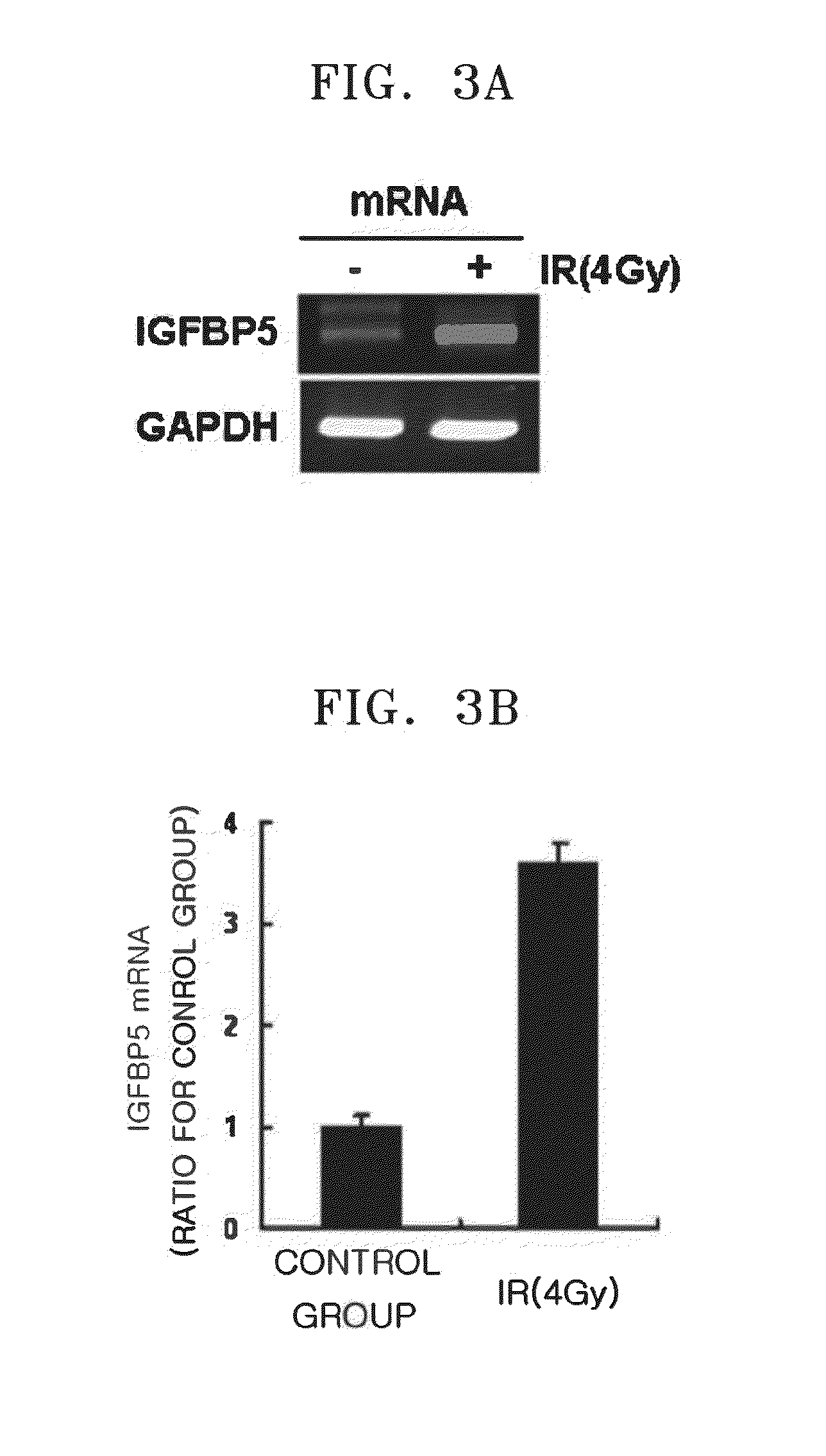
[0126]For the purpose of re-confirmation of microarray data of Example 2, RT-PCR and real-time PCR were performed.
[0127]1. RT-PCR Analysis
[0128]2 ug of the extracted and quantified RNA sample had oligo dT and dNTP added thereto, and was then heated at a temperature of 65° C. for 5 minutes, followed by being cooled on ice. Here, 5× First strand buffer (invitrogen superscript II kit), 0.1M DTT, and RNase inhibitor were added to the sample. Following heating at a temperature of 42° C. for 2 minutes, 1 ul of superscript II was added to the sample, and then the sample was allowed to stand at a temperature of 42° C. for 1 hour. Then, the sample was heated at a temperature of 70° C. for 15 minutes, thereby completing the reaction to synthesize cDNAs. These synthesized cDNAs were used to perform PCR at an appropriate annealing temperature for each primer.
[0129]2. Real-Time PCR Analysis
[0130]RNA was extracted and quantified so as to perform real-...
PUM
| Property | Measurement | Unit |
|---|---|---|
| temperature | aaaaa | aaaaa |
| temperature | aaaaa | aaaaa |
| temperature | aaaaa | aaaaa |
Abstract
Description
Claims
Application Information
 - R&D
- Intellectual Property
- Life Sciences
- Materials
- Tech Scout
- Unparalleled Data Quality
- Higher Quality Content
- 60% Fewer Hallucinations
Browse by: Latest US Patents, China's latest patents, Technical Efficacy Thesaurus, Application Domain, Technology Topic, Popular Technical Reports.
© 2025 PatSnap. All rights reserved.Legal|Privacy policy|Modern Slavery Act Transparency Statement|Sitemap|About US| Contact US: help@patsnap.com


