

Fluorescent protein-based indicators
a technology of fluorescent protein and calcium indicator, which is applied in the direction of transferases, peptide sources, instruments, etc., can solve the problems of limiting the usefulness of synthetic dyes, bleaching, and invading the environment, and achieving poor temporal resolution
- Summary
- Abstract
- Description
- Claims
- Application Information
AI Technical Summary
Benefits of technology
Problems solved by technology
Method used
Image

Examples
example 1
[0073]This Example describes procedures used to design and synthesize novel fluorescent proteins. This Example further describes procedures used to optimize and characterize the novel fluorescent proteins.
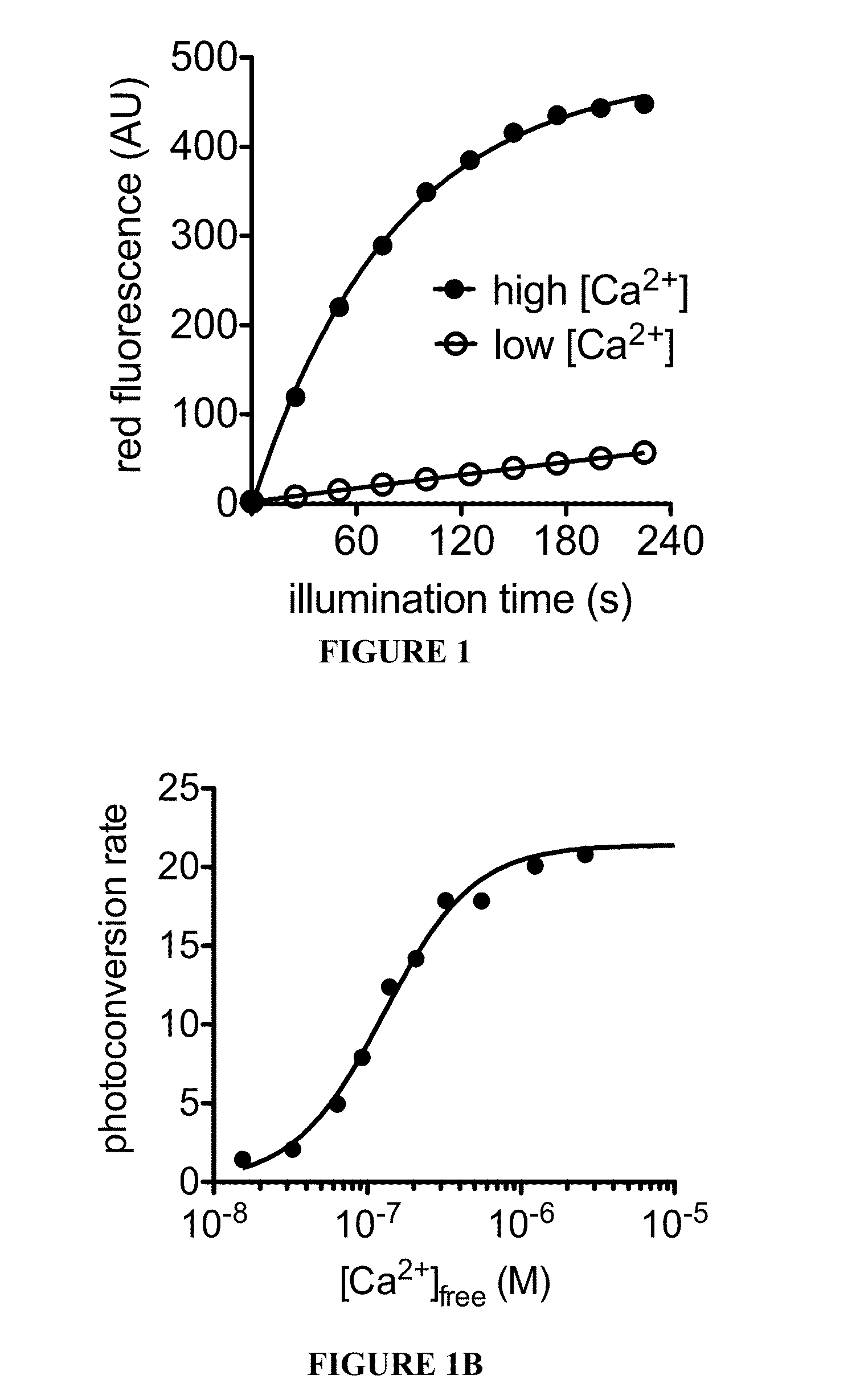
[0074]Circular permutation of fluorescent protein domains and attachment to ligand binding domains can allow modulation of fluorescence intensity through conformational rearrangement of a FP chromophore chemical environment driven by ligand binding. Thus, the procedures described herein utilized a circular permutation of a photoconvertible fluorescent protein, EosFP, to attempt to permit modulation of photoconversion efficiency of the protein in a ligand-dependent manner.
[0075]To construct a fluorescent protein that would undergo more efficient green-to-red photoconversion in the presence of calcium, libraries were created and screened for circularly permuted EosFP variants fused at the termini to calmodulin (CaM) and the calmodulin-interacting peptide M13. Circularly permuted vari...
example 2
[0082]This Example describes procedures used to determine the crystal structure of the novel fluorescent proteins. The crystal structures of this Example were used to, among other things, identify mutations that may enhance the properties of the fluorescent proteins.
[0083]To develop a crystal structure, purified CaMPARI v0.2 (CaMPARI v0.1 with E380M mutation) protein in 10 mM Tris, 100 mM NaCl, 10 mM EGTA was mixed with an equal volume of a precipitant solution of 200 mM ammonium sulfate, 100 mM HEPES pH 7.5, 25% PEG 3350 at room temperature in a sitting-drop vapor diffusion setup. A single yellow-green dagger-shaped crystal was cryoprotected in the precipitant solution supplemented with 20% glycerol, and x-ray diffraction data were collected at 100 K. Data were processed using MOSFLM and SCALA within the CCP4 software package. The structure was solved by molecular replacement searching first for the EosFP fragment using a single EosFP molecule from PDB ID 1ZUX, followed by portions...
example 3
[0085]This Example describes procedures performed to characterize the in vitro properties of the fluorescent proteins. First, CaMPARI protein was expressed from the pRSET plasmid (Life Technologies, Carlsbad, Calif.) in T7 Express E. coli cells cultured for 36 h in 100 mL of autoinduction medium supplemented with 100 mg / L ampicillin. Cell pellets were lysed using B-PER (Pierce, Rockford, Ill.) supplemented with 1 mg / mL lysozyme and 1 minute of sonication. After removing insoluble material by centrifugation, CaMPARI protein was purified by immobilized metal affinity chromatograpy on nickel-charged Profinity resin (Bio-Rad, Hercules, Calif.), washing with 10 mM imidazole, and eluting with 100 mM imidazole.
[0086]Protein concentration was quantitated by denaturing in 0.1 M NaOH and using the extinction coefficient 44,000 M−1 cm−1 at 447 nm for denatured GFP-like chromophores. Quantum yields were measured directly with an integrating-sphere spectrometer (Quantaurus-QY, Hamamatsu, Japan)....
PUM
| Property | Measurement | Unit |
|---|---|---|
| Time | aaaaa | aaaaa |
| Wavelength | aaaaa | aaaaa |
| Wavelength | aaaaa | aaaaa |
Abstract
Description
Claims
Application Information