
Composition and method for sulfated non-anticoagulant low molecular weight heparins in cancer and tumor metastasis

a low molecular weight, heparin technology, applied in the direction of synthetic polymeric active ingredients, capsule delivery, microcapsules, etc., can solve the problems of poor oral absorption, limited use of heparin as an anti-metastatic agent, and poor anti-coagulation

Active Publication Date: 2015-05-14
MOUSA SHAKER A
View PDF5 Cites 6 Cited by
  • Summary
  • Abstract
  • Description
  • Claims
  • Application Information

AI Technical Summary

Benefits of technology

The present invention provides a nanoformulation that includes nanoparticles made of a shell made of materials like PLGA or chitosan, which encapsulates a glycosaminoglycan (GAG) like heparin. The GAG is bonded to the shell using either ionic or covalent bonds. The nanoformulation can be used to treat cancer in humans by administering it to the patient. The technical effect of this invention is the creation of a new and effective way to treat cancer using nanoparticles and GAGs.

Problems solved by technology

Although the therapeutic effects of these compounds are well documented, their use is limited because of their poor oral absorption and short half-life.
The use of heparin as an anti-metastatic agent is limited due to the bleeding side effect.
Sialylated fucosylated glycans are the ligands for P-selectin, and tumors with high expression of these ligands typically have poor prognosis due to high rates of metastasis.
Low molecular weight heparin (LMWH) has been shown to decrease tumor metastasis in animal experiments and clinical trials, but the use of heparin and its derivatives as anti-metastatic agents is limited because of its risk in inducing adverse bleeding complications.
Tumor excision of a primary cancer with no metastasis has been the cornerstone treatment for most cancers, but scientific evidence has revealed that cancer excisional surgery itself can increase the risk of metastasis.

Method used


Image

  • Composition and method for sulfated non-anticoagulant low molecular weight heparins in cancer and tumor metastasis
  • Composition and method for sulfated non-anticoagulant low molecular weight heparins in cancer and tumor metastasis
  • Composition and method for sulfated non-anticoagulant low molecular weight heparins in cancer and tumor metastasis

Examples


example 1

Preparation of Sulfated Non-Anticoagulant Heparin (S-NACH)

[0075]Unfractionated porcine Heparin or bioengineered heparin was fragmented by periodate oxidation based on a procedure from Islam et al. See Islam T, Butler M, Sikkander S A, Toida T, Linhardt R J, Further evidence that periodate cleavage of heparin occurs primarily through the anti-thrombin binding site, Carbohydr Res. 2002, 337(21-23):2239-43.

[0076]Heparin, sodium salt (20 g, 1.43 mmol) was dissolved in 175 mL of distilled water. The pH was adjusted to 5.0 using 1 N HCl, NaIO4 (15 g, 0.07 mol), dissolved in 500 mL water, which was added in a single portion with stirring. The pH was readjusted to 5.0 using 1 N HCl and left for 24 hours at 4° C. in the dark. The solution was dialyzed against 4 volumes of water (with one change of water) for 15 hours at 4° C. To the approximately 1.5 L of solution obtained after dialysis, 32 mL of 10 N NaOH was added. The solution was stirred at room temperature for 3 hours. To prevent the d...

example 2

Cancer Cells

[0077]Pancreatic cancer cell line MPanc96 and its luciferase transfected Mpanc96-luc cells were grown in DMEM supplemented with 5% fetal bovine serum, 1% penicillin, and 1% streptomycin. Cells were cultured at 37° C. to sub-confluence and treated with 0.25% (w / v) trypsin / EDTA to affect cell release from culture flask. After washing cells with culture medium, cells were suspended in DMEM (free of phenol red and fetal bovine serum) and counted. These cells were also genetically modified to contain luciferase, which emits signals that can be detected by in vivo imaging system (IVIS).

[0078]Other human pancreatic cancer cell lines MIA-PaCa-2 and BxPC-3 were obtained from the American Type Culture Collection (Rockville, Md.). Two different pancreatic cancer cell lines were chosen for study because each cell line exhibits different characteristics in vivo with respect to rate of growth and metastasis, with the former showing more rapid and extensive dissemination. Cells were ma...

example 3

Platelet-Cancer Cell Adhesion Assay

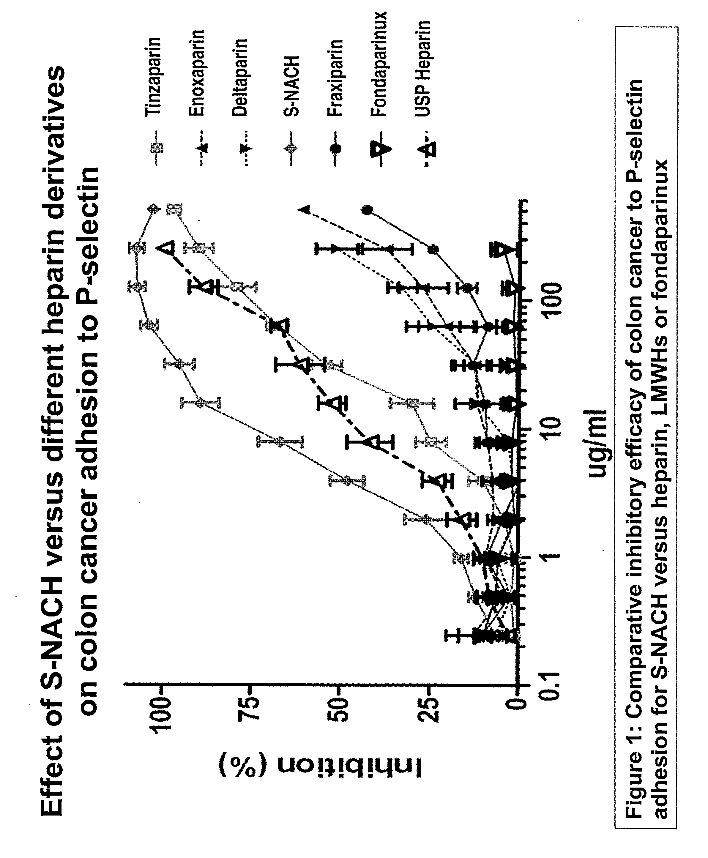
[0079]A cancer cells-platelets adhesion assay was used to test the efficacy of the test compounds in inhibiting the adhesion between P-selectin and tumor cells. This was done by growing the Mpanc96 cancer cells in 96-well plate to 100% confluence. Ten ml of human blood was drawn by vacutainer tube containing citrate acid and dextrose and stored at 4° C. overnight. After 24 hours, the supernatant of the blood was collected (platelet-rich plasma). The platelet rich plasma was centrifuged for 10 minutes at 1000×g and resuspended in 10 ml PBS. The platelets were labeled with Calcien AM fluorescence dye and incubated for 30 minutes at 37° C. The platelets were then washed five times with PBS by centrifuging at 2,000×g for 10 minutes. The test compounds were mixed with the platelets 30 minutes prior to adding them to the cancer cells to allow the compounds to bind to the P-selectin of the platelets. The concentrations ranged from 5 ug / ml to 20 ug / ml for ...


PUM

PropertyMeasurementUnit
Chemotherapeutic propertiesaaaaaaaaaa

Abstract

A nanoformulation that includes nanoparticles. Each nanoparticle includes a shell in which a glycosaminoglycan (GAG is encapsulated. The GAG is ionically or covalently bonded to the shell. The GAG is selected from the group consisting of sulfated non-anticoagulant heparin (SNACH), super-sulfated non-anticoagulant heparin (S-SNACH), and a combination thereof. The shell includes Poly (lactic-co-glycolic acid) (PLGA), Polyethylene Glycol (PEG)-PLGA, maleimide-PEG-PLGA, chitosan, chitosan-PLGA, methoxy-polyethyleneglycol-poly (lactide-co-glycolide) (MPEG-PLGA)-(maleimide-PEG-PLGA), PLGA-Polycaprolate, or calcium alginate. A method of using the nanoformulation to treat a cancer in a subject includes administering to the patient a therapeutically effective amount of the nanoformulation for treating the cancer.

Description

RELATED APPLICATIONS[0001]The present invention claims: priority to U.S. Provisional No. 61 / 896,738, filed on Oct. 29, 2013, which is incorporated herein by reference in its entirety: and priority to U.S. Provisional No. 61 / 896,770, filed on Oct. 29, 2013, which is incorporated herein by reference in its entirety.TECHNICAL FIELD[0002]The present invention relates to compositions and associated methods for suppression of primary tumor growth and metastasis by use of sulfated, super-sulfated non-anticoagulant low and ultra-low molecular weight heparins, and their nanoformulations.BACKGROUND[0003]The Glycosaminoglycans (GAG's) are copolymers of uronic acid and amino hexose with a sequence that has unique biological activity. When prepared from natural sources, this family of heterogeneous macromolecules has sequences that bind specific proteins providing them with various biological activities. Some of the interactions that are of therapeutic utility are between the serine proteases ge...

Claims


Application Information

Patent Timeline
IPC IPC(8): A61K31/727A61K31/704A61K47/48A61K33/243
CPCA61K31/727A61K47/48215A61K47/482A61K47/48869A61K31/704A61K31/7068A61K47/6935A61K47/6937A61K33/243
Inventor MOUSA, SHAKER A.
Owner MOUSA SHAKER A
Learn More