Novel labeling composition for cancer lesion

a technology for cancer lesion and composition, applied in the direction of biological testing, peptide analysis, biological material analysis, etc., can solve the problems of rare and difficult to clearly distinguish the lesion, the diagnostic method may not be used during surgery, and the operating surgeon has little method to accurately identify the lesion in real time during surgery, etc., to achieve high in vivo compatibility, easy to see, and easy to degrade

Active Publication Date: 2015-11-19
NAT CANCER CENT
View PDF6 Cites 1 Cited by
  • Summary
  • Abstract
  • Description
  • Claims
  • Application Information

AI Technical Summary

Benefits of technology

[0011]The wording “macroaggregated albumin (MAA)” used herein means proteinous particles which have a diameter of 10 to 50 μm and are prepared by heating and coagulating human serum albumin. A structure and physical property of MAA differs from human serum albumin having a diameter less than 10 nm. When the MAA is intravenously injected, the MAA may remain in pulmonary capillary, which is 8 μm, thereby causing microembolus. By using such a property, the MAA labeled with a radioactive isotope has been used for lung scintigram (for diagnosis of disorders in pulmonary blood flow, right-to-left shunt or lung increased venous pressure such as pulmonary embolism, pulmonary blood clot, pulseless disease, pneumonia, and lung cancer), venous scan (for in situ diagnosis of venous blood of central nerves) or venous scan (for diagnosis of peripheral artery blood flow disorders such as faza disease). The MAA of the present invention is injected into a cancer lesion tissue, and used as a mediator for binding a labeling material to the cancer lesion tissue. The MAA of the present invention may be synthesized by using recombinant HSA or non-autologous HSA. Also, commercially available MAA may be purchased and used. The MAA of the present invention is injected into a cancer lesion tissue and used as a mediator for binding a labeling material to the cancer lesion tissue, wherein the mediator adsorbs the labeling material to prevent the labeling material from diffusion to the cancer lesion tissue.
[0031]In addition, the complex included in the composition of the present invention may remain in a cancer lesion in a body for a long period of time relative to a complex in which a pigment for staining a living tissue binds to other materials, and thus accuracy of cancer lesion excision can be easily verified during a surgical procedure, as well as surgical excision of the cancer lesion. For example, through ultrasound, a microlesion site is identified before surgery. Then the complex of the present invention is injected into the lesion region to make the lesion region to be stably and accurately identified during surgery which is performed few hours after.

Problems solved by technology

However, in actual clinical surgery, there is little method to allow an operating surgeon to precisely identify the lesion in real time during surgery.
Although a very precise diagnostic method has been developed, the diagnostic method may not be used during surgery.
However, in such a case, it is rare to clearly distinguish the lesion.
In particular, in the case where a lesion is small, it is more difficult to distinguish.
Although the method of drawing a picture on skin by using a pen to mark a site of a lesion can be easily used, the method has a drawback of low accuracy, because, due to a very flexible characteristic of breast tissue, a shape of breast is greatly changed during surgery from a shape at the point of diagnosis, and, in the case of a lesion at deep breast, a mark on a skin surface is insufficient.
Also, a drawback of the method of inserting a wire into a breast lesion is lower accuracy than expected because essentially a wire should be vertically inserted into a skin surface, but the vertical insertion of the wire may affect an ultrasonic probe, so that the wire is inevitably inserted obliquely; and because a location of the wire may be moved according to movement of breast.
In addition, another drawback of the method is that the inserted wire interferes with surgery; and a procedure to excise a site for inserting the wire should be additionally performed.
However, in the case of a lesion in deep breast, there are drawbacks in that a black pigment may not be identified at the exterior, and a surgical region may be contaminated by the pigment.
These drawbacks also become a limitation in a surgical operation to remove cancer tissue beside breast cancer.
Thus, through technique developed so far, it is difficult to precisely determine an extent of surgery for surgically removing a cancer lesion.

Method used


Image

  • Novel labeling composition for cancer lesion
  • Novel labeling composition for cancer lesion
  • Novel labeling composition for cancer lesion

Examples


example 1

Construction of Macroaggregated Albumin (MAA)

[0048]10 ml and of 2% human serum albumin diluted in 0.1 M acetate buffer (pH 5.4) was mixed with 50 mg of tin chloride, and vigorously stirred for 10 minutes at room temperature followed by additional stir for 20 minutes at 70° C. for reaction. After the reaction was stopped, the reactant was cooled. Then, 0.35 ml and of 20% human serum albumin was added, and the resultant was stirred again for 10 minutes. The reactant was aliquot to a glass vial (2 mg for each, based on MAA) and lyophilized to prepare thiol MAA.

example 2

Radioactive Isotope-Bound MAA Complex and Investigation of Availability Thereof

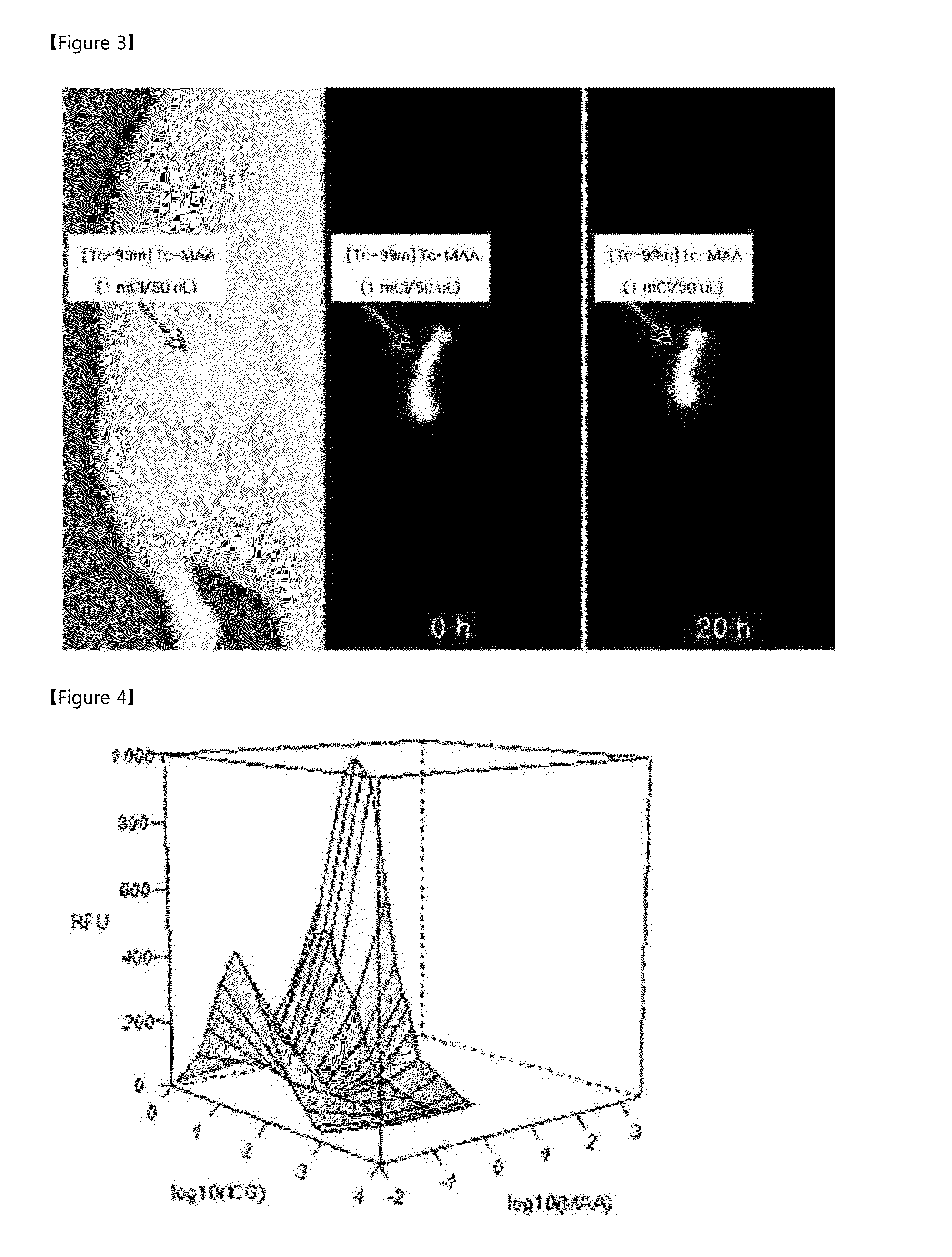
[0049]2 ml and of [Tc-99m]TcO4− (10 mCi / ml), which is a radioactive isotope, was added to the thiol MAA constructed in Example 1. The resultant mixture was reacted for 10 minutes at room temperature to construct a radioactive isotope-bound MAA complex ([Tc-99m]Tc-MAA). To investigate whether the radioactive isotope normally binds to MAA, the complex is applied on instant thin layer chromatography (ITLC), and developed by using acetone as a solvent, and, as a result, it has been found that at least 99% of thiol MAA binds to the radioactive isotope, thereby forming a complex.

[0050]In addition, to investigate whether the constructed complex may be used as an in vivo labeling agent, an experiment was performed as follows: the constructed [Tc-99m]Tc-MAA 1 mCi / 50 μl was injected into a left buttock of a nude mouse. A gamma image of the nude mouse was taken by using an animal SPECT device (NanoSPECT, Bioscan) at...

example 3

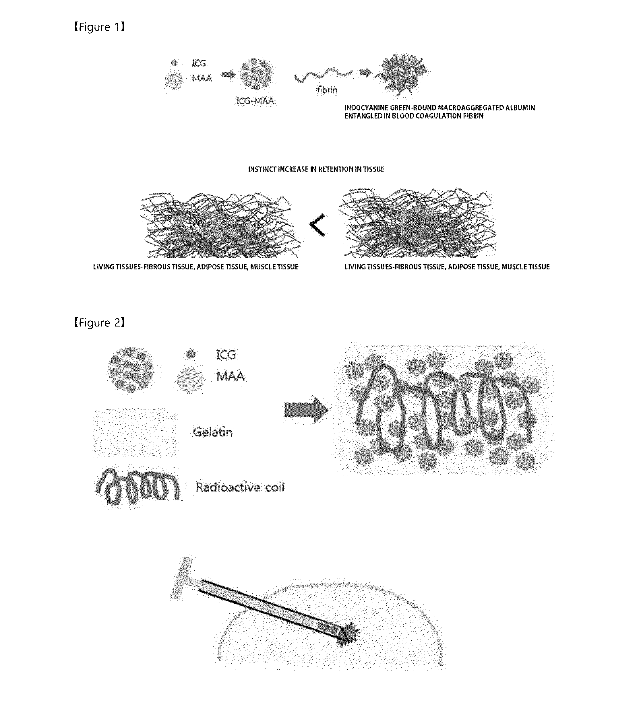
Preparation of Indocyanine Green (ICG)-Bound MAA-Based Labeling Agent and Investigation of Availability Thereof

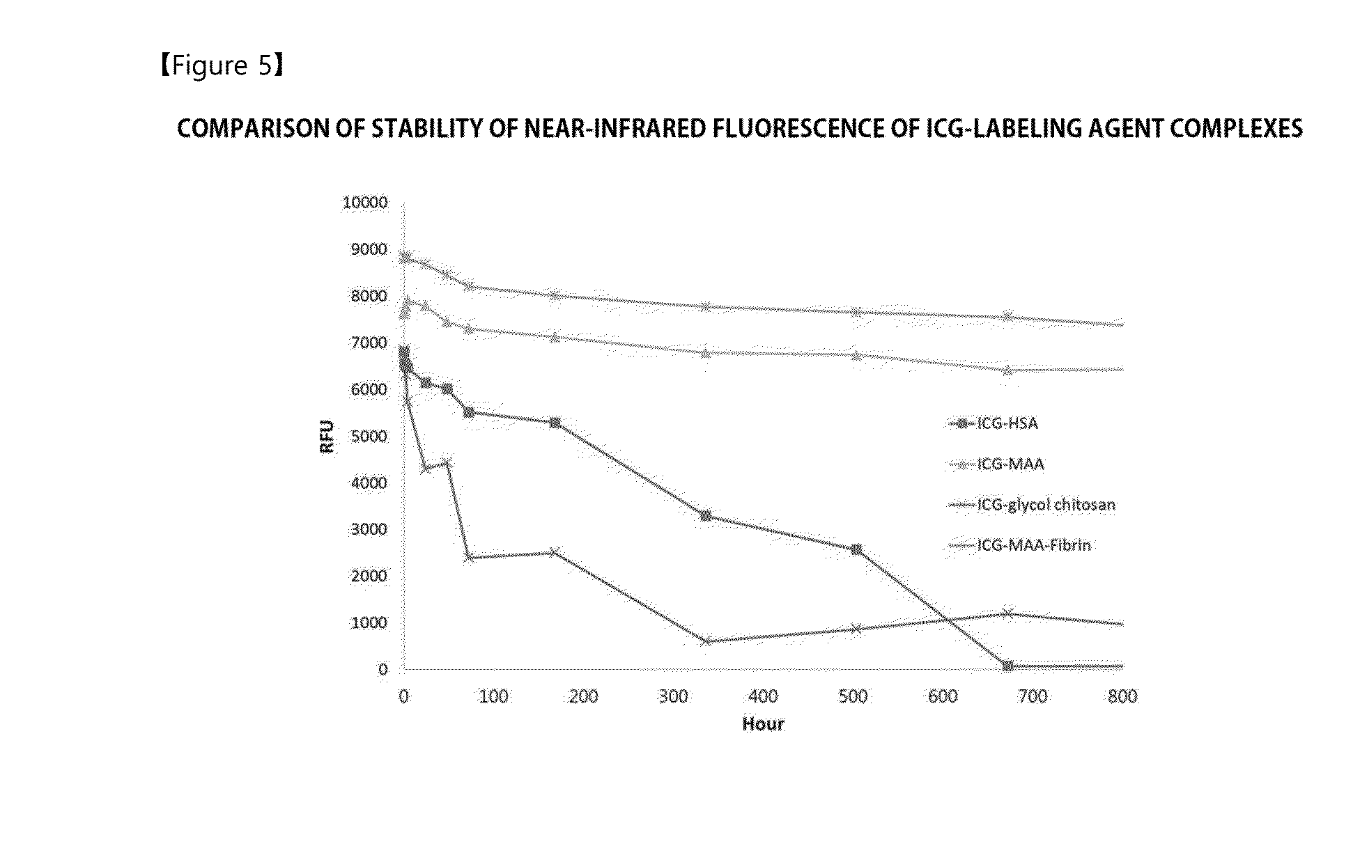
[0051]Since it has been expected that a complex, in which MAA binds to indocyanine green (ICG) capable of generating a near-infrared fluorescent signal, may be used as a labeling agent stably acting in vivo, the complex was constructed and availability thereof as an in vivo labeling agent was investigated.


PUM

PropertyMeasurementUnit
diameteraaaaaaaaaa
diameteraaaaaaaaaa
near-infrared wavelengthaaaaaaaaaa

Abstract

The present invention relates to a labeling composition for a cancer lesion, having a complex in which a pigment for straining living tissues, a radioactive isotope, or a combination thereof binds to macro aggregated albumin (MAA). A method for providing information regarding a cancer lesion site using the labeling composition for a cancer lesion. A labeling kit for a cancer lesion having the labeling composition for a cancer lesion; and a complex in which a pigment for straining living tissues binds to MAA included in the labeling composition for a cancer lesion. The labeling composition for a cancer lesion according to the present invention binds to a cancer lesion to detect a site, size, and the like of the cancer lesion in real time, thereby improving the success rate of a surgical operation for the cancer lesion and also preventing excessive loss of normal tissues.

Description

TECHNICAL FIELD[0001]The present invention relates to a novel labeling composition for a cancer lesion. More particularly, the present invention relates to a labeling composition, for a cancer lesion, including a complex in which macroaggregated albumin (MAA) is bound to a pigment for staining living tissues, radioactive isotope, or combination thereof; a method for providing information about a cancer lesion site by using the labeling composition for a cancer lesion; a kit including the labeling composition for a cancer lesion for labeling a cancer lesion; and a complex in which a pigment for staining living tissues binds to MAA included in the labeling composition for a cancer lesion.BACKGROUND ART[0002]For anticancer therapy, a method using various anticancer agents has been developed, however a surgical operation method of removing cancer cells is still most frequently used method. When the surgical operation method is used, technique to minimize surgical extent during surgery i...

Claims


Application Information

Patent Timeline
Patent Type & Authority Applications(United States)
IPC IPC(8): A61K51/08A61K49/00
CPCA61K51/081A61K49/0056A61K49/006G01N33/574G01N33/58G01N2333/765G01N33/60
Inventor KIM, SEOK KIKANG, SE HUNKIM, SEOK WONJUNG, SO YOUN
Owner NAT CANCER CENT
Why Eureka
Try Eureka