Method and apparatus for biomolecule analysis

a biomolecule and apparatus technology, applied in the field of biomolecule analysis, can solve the problems of recollecting and measuring the signals of microspheres, requiring extra equipment operation, and long methods, and achieves the effects of quick and efficient collection, simplified washing step, and improved efficiency

Inactive Publication Date: 2015-11-19
MAI JUNYU
View PDF13 Cites 8 Cited by
  • Summary
  • Abstract
  • Description
  • Claims
  • Application Information

AI Technical Summary

Benefits of technology

[0006]The present disclosure describes an apparatus or a device and its usage for quick and efficient collection of beads and wash of the beads and for signal detection in a bead-based biomolecule analysis. The same apparatus or device can also be applied for cell-based analysis and membrane-based dot blot assays. Advantageously, the apparatus and methods of the present disclosure simplify the washing step without using centrifuge, vacuum, or magnetic force; provide more efficient washing with less volume of buffer and less time; are capable of condensing the beads and biomolecules in a small detection area to generate stronger signal; use significantly less amount of assay reagent than traditional assays; and enable quantitative signal detection and analysis using a plate reader or an imaging device.

Problems solved by technology

All of these methods are lengthy, repetitive, and need extra equipment operation.
After the assay, recollecting and measuring the signals of the microspheres is also a big challenge, especially for assays using small amount of beads and multiple washing steps, because loss of beads is inevitable in most cases.
The entire process, including multiple lengthy shaking and washing steps, takes more than two hours or even overnight to finish.
A commonly used apparatus for dot blot assay relies on a vacuum source to suck solution through the membrane, but the apparatus set up is complicated, expensive, and need to be assembled, disassembled and cleaned for every use.
Only a limited number of membranes can be processed using such device.
Cross-contamination is hard to avoid in these methods because one continuous piece of membrane is used for multiple assay dots.

Method used


Image

  • Method and apparatus for biomolecule analysis
  • Method and apparatus for biomolecule analysis
  • Method and apparatus for biomolecule analysis

Examples


example 1

Human Alpha-Fetoprotein (AFP) Fluorescence Immunoassay

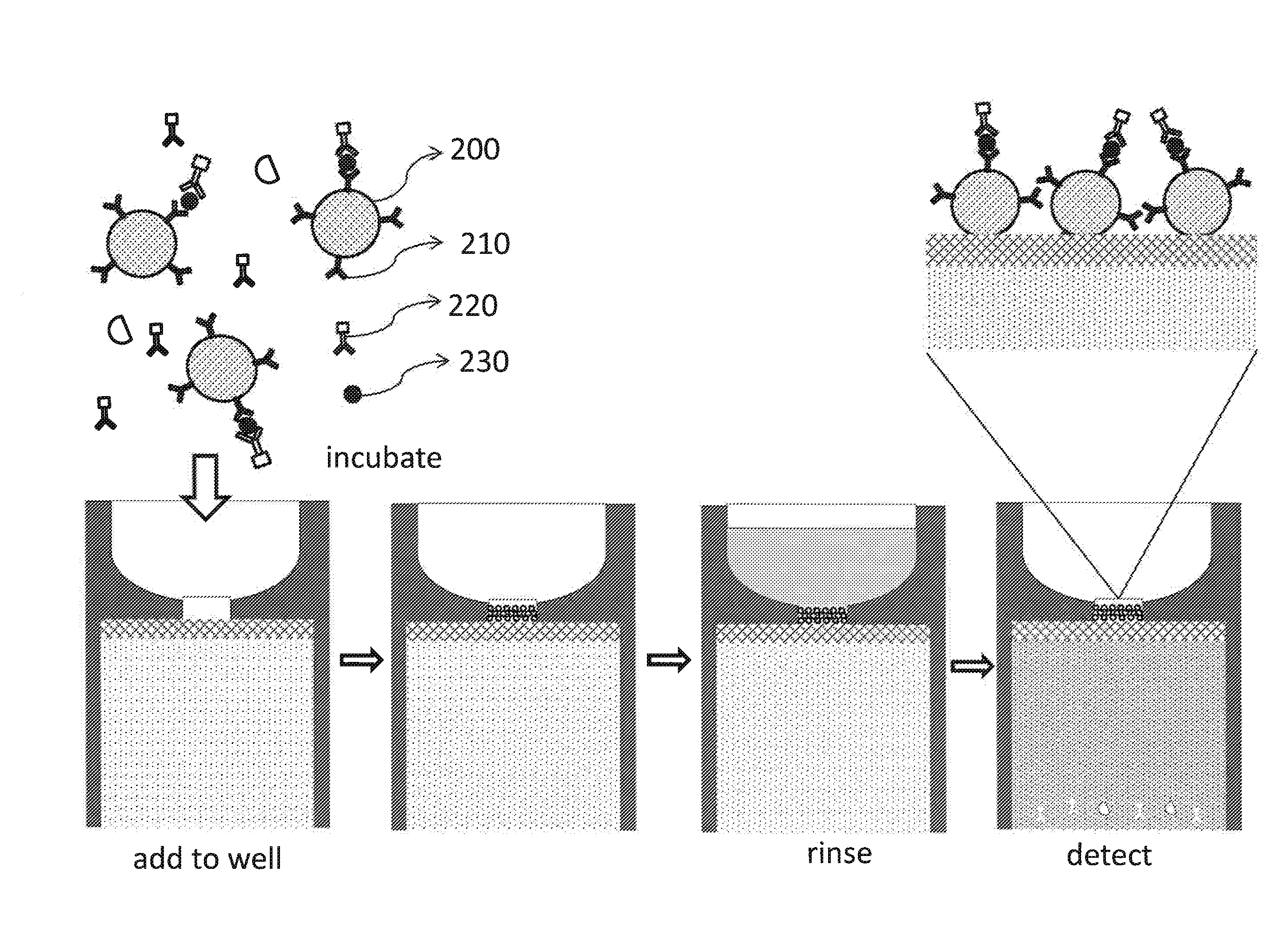
[0054]This example shows how a device described in this invention can be used for a rapid silica bead-based fluorescent immunoassay to detect AFP from human serum sample. The process can be better understood with reference to FIG. 6.

[0055]The assay will need the following reagents: (1) silica beads 200 functionalized with AFP capture antibody 210; (2) fluorescently-labeled AFP detection antibody 220; (3) serum sample containing the target AFP molecule 230; and (4) rinsing buffer.

[0056]The capture antibody, mouse monoclonal anti-human AFP antibody, is available from R&D Systems. It can be covalently attached to 5 μm carboxylated silica beads through carboxyl-amine crosslinking reaction. Functionalized silica beads can be blocked with 10% bovine serum albumin (BSA) and stored at 1% solid in phosphate buffered saline (PBS) solution containing 1% BSA with 0.5% Tween-20 (1% BSA-PBST). The detection antibody, polyclonal chicken anti-AF...

example 2

Human Alpha-Fetoprotein (AFP) Chemiluminescence Immunoassay

[0059]This example shows how a device described in this invention can be used for a rapid agarose bead-based chemiluminescent immunoassay. The process can be better understood with reference to FIG. 7.

[0060]The assay will need the following reagents: (1) Agarose beads 300 functionalized with AFP capture antibody 310; (2) AFP detection antibody labeled with horse radish peroxidase (HRP-antiAFP antibody) 320; (3) serum sample that may contain the target AFP molecule 330; (4) rinsing buffer 1% BSA-PBST; and (5) HRP chemiluminescence substrate 340.

[0061]The capture antibody, such as the mouse monoclonal anti-human AFP antibody from R&D Systems can be crosslinked to 20 μm glyoxal agarose beads through glyoxal-amine chemistry. The anti-AFP agarose beads can be stored at 20% slurry 1% BSA-PBST. The detection antibody, polyclonal chicken anti-AFP antibody can be labeled with HRP using commercially available labeling kits. In some ca...

example 3

Botulinum Neurotoxin Type a (BoNT / A) Fluorescence Resonance Energy Transfer (FRET) Assay

[0064]This example shows how the device in this invention can be used to rapidly detect active BoNT / A in a large volume of serum sample using polyacrylamide beads and a FRET substrate (see FIG. 8).

[0065]The assay will need to use the following reagents: (1) Polyacrylamide beads 400 functionalized with anti-BoNT / A antibody 410; (2) serum sample that may contain the active BoNT / A molecule 420; (3) assay buffer 20 mM HEPES at pH 8.0; and (4) SNAPtide fluorogenic BoNT / A substrate 430 from EMD Millipore.

[0066]BoNT / A antibody (Santa Cruz Biotechnology) can be crosslinked to pre-activated polyacrylamide (PA) beads such as Ultralink Biosupport from Thermo Scientific. The anti-BoNT / A PA beads can be stored at 20% slurry in PBS solution containing 1% BSA with 0.5% Tween-20. 10 μl anti-BoNT / A PA beads will be added into 500 μl serum sample and incubated at 37° C. for 1 hour with slow rotation. The mixture w...


PUM

PropertyMeasurementUnit
heightaaaaaaaaaa
heightaaaaaaaaaa
volumeaaaaaaaaaa

Abstract

Disclosed are devices or apparatus and methods for membrane-, bead- or cell-based biomolecule analysis, where a detectable signal can be measured after affinity binding and/or enzymatic reaction occurs on the membranes, beads or cells. The apparatus or device can rapidly separate the beads or cells from the liquid solution without using centrifugation, vacuum, or magnetic force. The apparatus or device includes a sample well for receiving a sample containing cells or beads, as well as other aqueous solutions, a porous filter membrane at the bottom of the well which is capable of retaining the cells or beads on its upper surface by size exclusion, and an absorbing plug in touch with the filter membrane for removing the liquid solution. Multiple devices can be arranged in a multi-well array in a plate so that signals from multiple assays can be directly analyzed in a plate reader or an imaging instrument.

Description

CROSS REFERENCE TO RELATED APPLICATIONS[0001]This application claims the benefit under 35 U.S.C. §119(e) of U.S. Provisional Application No. 61 / 994,645, filed on May 16, 2014, which is incorporated by reference in its entirety.FIELD[0002]Disclosed are novel apparatus and uses thereof in biomolecule analysis. Particular embodiments contemplate membrane, cell or bead-based biomolecule analysis, where after a biochemical reaction such as an enzymatic reaction or an affinity binding, the signals carried by the membranes, cells or beads are to be analyzed to determine the result of the assay.BACKGROUND[0003]Microspheres, microparticles, or beads, have become an important form of solid state substrate for detecting, analyzing and identifying a wide range of biomolecules. Advantages of using microspheres compared to traditional plate-based assay methods include their large surface area, easiness of mixing with a solution and re-collecting, availability of different chemistry to functionali...

Claims


Application Information

Patent Timeline
Patent Type & Authority Applications(United States)
IPC IPC(8): G01N1/40G01N33/542G01N33/543
CPCG01N1/4077G01N33/54386G01N2001/4088G01N33/54393G01N33/542G01N33/525
Inventor MAI, JUNYU
Owner MAI JUNYU
Why Eureka
Try Eureka