Materials and Methods for Rescue of Ischemic Tissue and Regeneration of Tissue Integrity During Resection, Engraftment and Transplantation
- Summary
- Abstract
- Description
- Claims
- Application Information
AI Technical Summary
Benefits of technology
Problems solved by technology
Method used
Image
Examples
example 1
Materials and Methods
Cell Culture and Seeding of MEECs
[0083]MEECs or the implantable material is prepared as described in the following sections:
[0084]Human umbilical vein endothelial cells (HUVECs) pooled from 3 donors or HUVECs constitutively expressing GFP were grown in endothelial growth medium supplemented with EGM-2 growth supplements (Lonza). HUVECs (passage 3-5) were first cultured on gelatin-coated tissue culture plates (0.1% gelatin type A, Sigma, St. Louis, Mo.) and then cells were seeded in 3D matrix. For cell-matrix engraftment, compressed denatured collagen matrices (Gelfoam, Pfizer, New York, N.Y.) were cut into 1×1×0.3 cm blocks and hydrated in culture medium at 37° C. for 2 h. Then 4.5×10⁴ ECs (suspended in 50 μL media) were seeded onto one surface of the hydrated matrix and allowed to attach for 1.5 h. Subsequently, the matrix was turned over and additional 4.5×10⁴ ECs were added to infiltrate from the second side. After an additional 1.5 h incubation period to ena...
example 2
Animal Model of 70% Hepatectomy and Liver Engraftment
[0141]Male C57BL / 6 mice (9-12 weeks old) were purchased from Charles River Laboratories (Wilmington, Mass.). The animals were maintained in a temperature-controlled room (22° C.) on a 12 h light-dark cycle. After arrival, mice were continuously fed ad libitum until euthanasia. Partial hepatectomy was performed as previously described.24
[0142]Remaining ischemic median liver lobe was used to attach the implants and the right lobe to assess paracrine effects. For liver engraftment, excised mouse left lobes from a group of ten mice were maintained in warm EGM-2 medium (37° C.) until engraftment to the remaining median lobe of a same or different group of ten mice in the presence or the absence of MEECs or acellular matrices at the interface between recipient and donor liver. Animals were sacrificed after one week. Mouse blood samples were collected by intracardiac puncture. Serum was separated by centrifugation at 3,000×g for 10 min ...
example 3
Whole-Mount Multiphoton Imaging of Macrophage Presence and Angiography in Liver, Gene Expression Analysis by Real-Time PCR, TUNEL Assay and Western Blotting
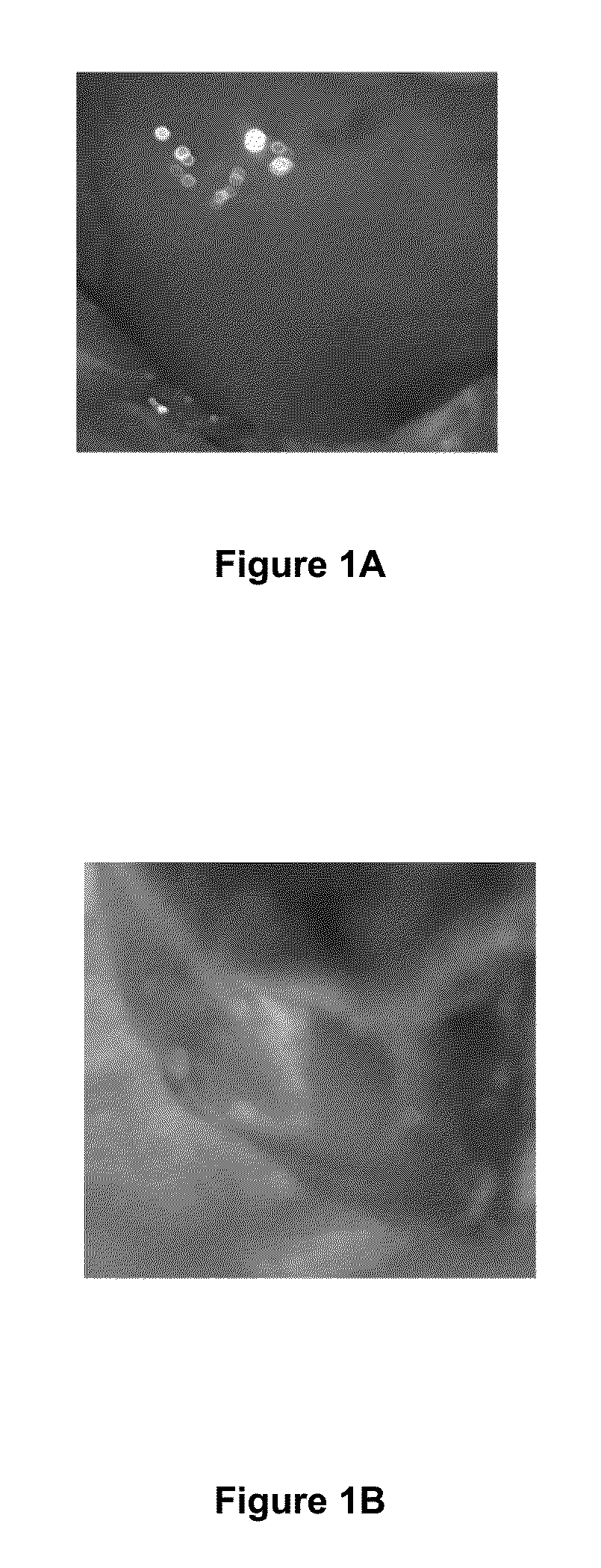
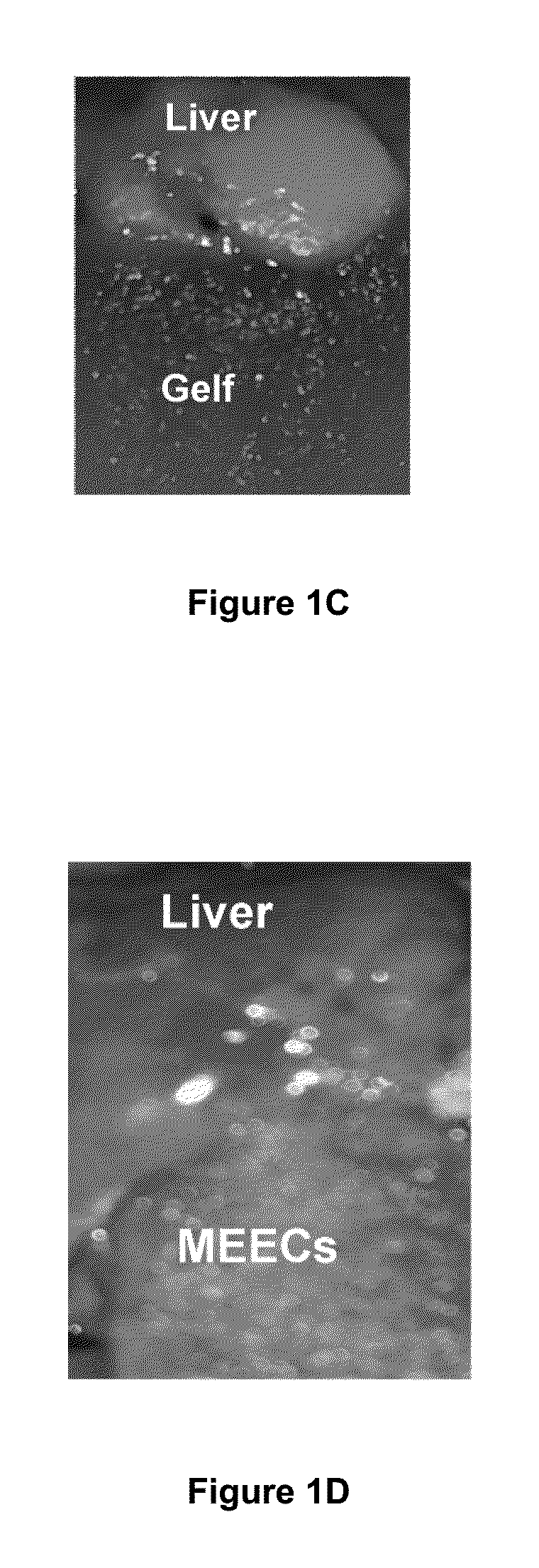
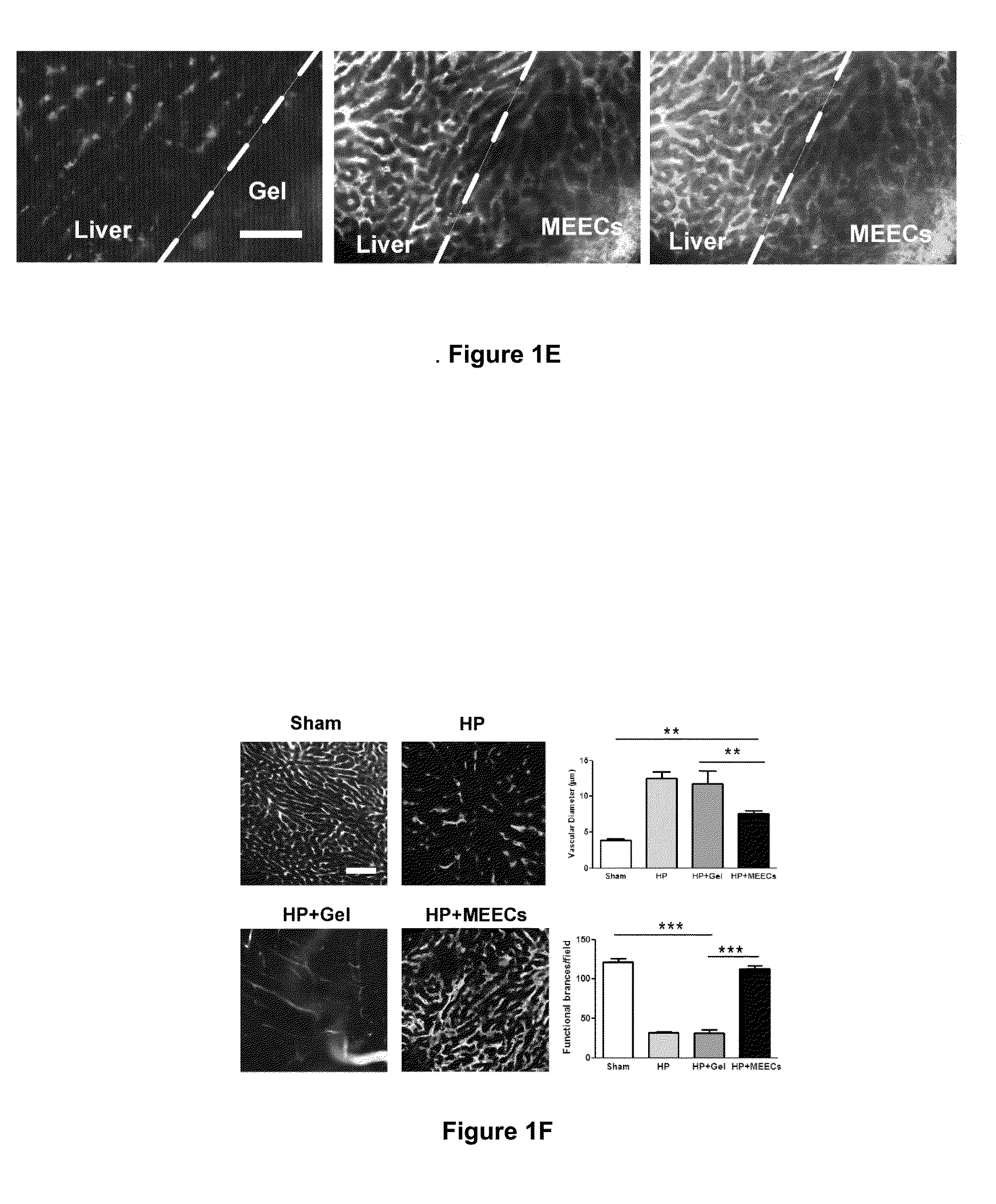
[0144]In short, FIG. 7 exemplifies the source of angiogenesis into implants of MEECs. HUVECs constitutively expressing GFP were seeded in gelfoams and grown in endothelial growth medium supplemented with EGM-2 growth supplements for 2 weeks under standard culture conditions (37° C. humidified environment with 5% CO2). Vascularity was analyzed in the interface between implants of HUVECs expressing GFP and implanted median liver lobes of hepatectomized mice by angiography (intracardiac perfusion of Texas red-dextran 70 kda using intravital multiphoton microscopy. Representative image of new vascular anastomoses in the implant interface coming from the extension of hepatic vessels (in red) and from MEEC-generated vessels (in yellow) are shown in the left panel. Control of expression of GFP in HUVEC-GFP cells is shown in the middle p...
PUM
| Property | Measurement | Unit |
|---|---|---|
| Fraction | aaaaa | aaaaa |
| Fraction | aaaaa | aaaaa |
| Composition | aaaaa | aaaaa |
Abstract
Description
Claims
Application Information
 - R&D
- Intellectual Property
- Life Sciences
- Materials
- Tech Scout
- Unparalleled Data Quality
- Higher Quality Content
- 60% Fewer Hallucinations
Browse by: Latest US Patents, China's latest patents, Technical Efficacy Thesaurus, Application Domain, Technology Topic, Popular Technical Reports.
© 2025 PatSnap. All rights reserved.Legal|Privacy policy|Modern Slavery Act Transparency Statement|Sitemap|About US| Contact US: help@patsnap.com


