
Anti-trop2 antibody-drug conjugate

Active Publication Date: 2016-10-13
DAIICHI SANKYO CO LTD +1
View PDF3 Cites 46 Cited by
  • Summary
  • Abstract
  • Description
  • Claims
  • Application Information

AI Technical Summary

Benefits of technology

The patent describes an anti-TROP2 antibody-drug conjugate that has a specific structure and can effectively treat tumors. By using this compound, researchers have been able to create a safe and effective treatment for cancer.

Problems solved by technology

As a result, DE-310 was not also commercialized, and it remains unclear whether or not exatecan effectively acts as a drug directed to such targeting.
However, the antitumor effect of said complex is not known at all.
However, the strength or coverage of their activity is still insufficient, and there are unsatisfied medical needs for hTROP2 as a therapeutic target.

Method used


Image

  • Anti-trop2 antibody-drug conjugate
  • Anti-trop2 antibody-drug conjugate
  • Anti-trop2 antibody-drug conjugate

Examples


example 1

Immunization of Mouse and Obtainment of Hybridoma

1-1) Preparation of Cell to be Used in Mouse Immunization

[0391]5×10⁶ NCI-H322 cells (human non-small cell lung cancer cell line, ATCC CRL-5806; ATCC: American Type Culture Collection) were cultured in an RPMI-1640 (Roswell Park Memorial Institute-1640) medium (10 ml) for 5 days, then recovered, washed with PBS (phosphate-buffered saline) twice, and resuspended in PBS (500 μl).

1-2) Immunization of Mouse

[0392]For the first immunization, each BALB / c mouse (6 weeks old) was intraperitoneally immunized with NCI-H322 cells (1×10⁷ cells). For the second to fifth immunizations, the mouse was intraperitoneally immunized with 1×10⁶ NCI-H322 cells at 1-week intervals. For the sixth (final) immunization, the mouse was immunized through the tail vein and intraperitoneally with the NCI-H322 cells at 1×10⁶ cells / 200 μl PBS for each route. Spleen cells were excised 3 days after the final immunization.

1-3) Preparation of Spleen Cell of Immunized Mouse...

example 2

Purification of Antibody from Hybridoma

[0397]Pristane (2,6,10,14-tetramethylpentadecane; 0.5 ml) was intraperitoneally administered in advance to each 8- to 10-week old mouse or nude mouse, which was then raised for 2 weeks. Each monoclonal antibody-producing hybridoma obtained in Example 1 was intraperitoneally injected to the mouse. After 10 to 21 days, the hybridoma was allowed to cause ascitic canceration, and the ascites was then collected. The obtained ascites was centrifuged to remove solid matter. Then, antibodies were purified by salting out with 40 to 50% ammonium sulfate, a caprylic acid precipitation method, a DEAE-Sepharose column, and a protein G column, and IgG or IgM fractions were collected and used as purified monoclonal antibodies.

example 3

Identification of Antigen to which Antibody Produced by Hybridoma Binds

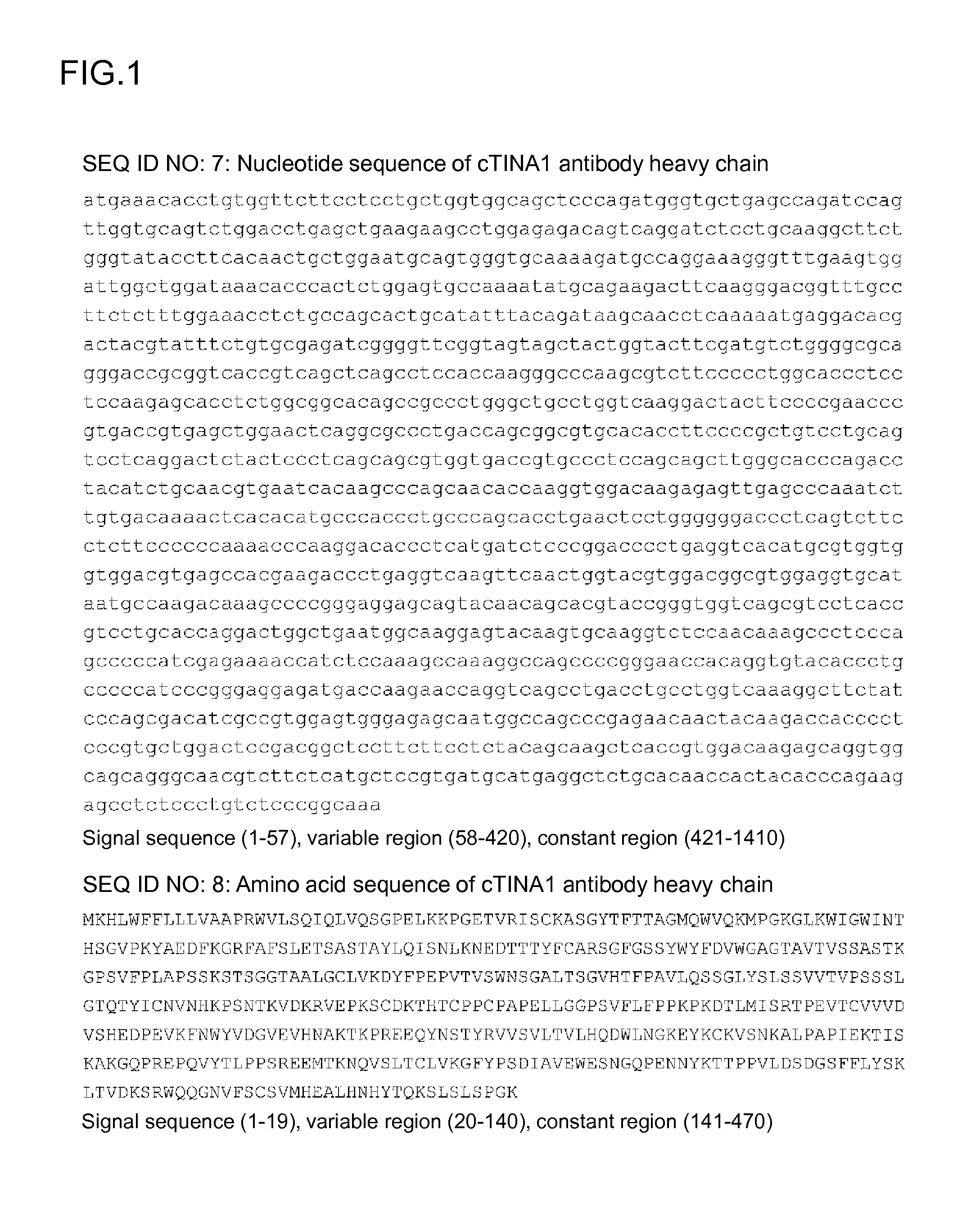
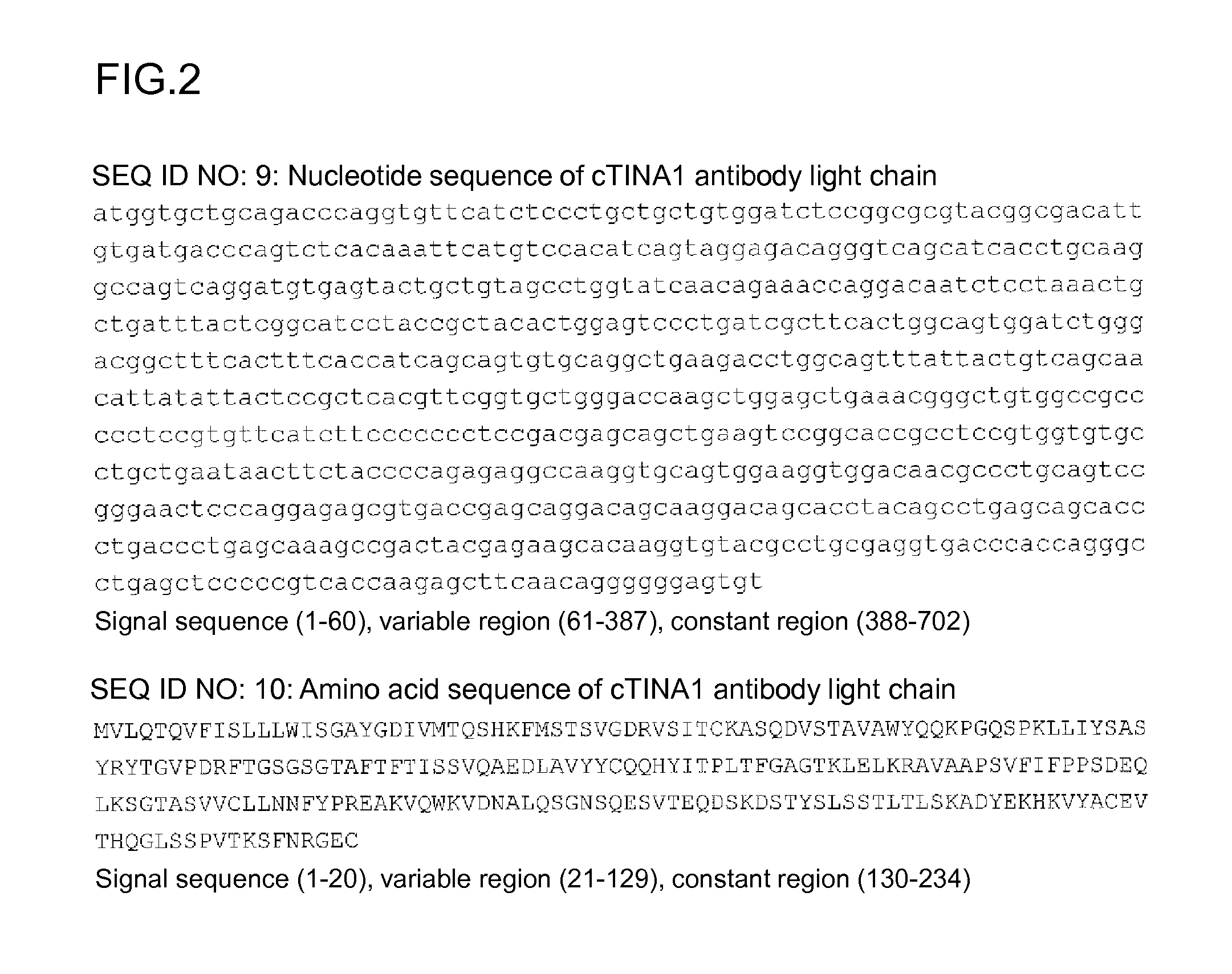
[0398]An antigen was identified for TINA1, an antibody produced by the hybridoma prepared in Example 2.

3-1) Immunoprecipitation of Biotin-Labeled Cell Surface Protein Using TINA1 Antibody

[0399]5×10⁶ NCI-H322 cells were recovered and washed with PBS three times. EZ-Link Sulfo-NHS-Biotin (Pierce / Thermo Fisher Scientific, Inc.) was suspended in PBS at a concentration of 0.1 mg / ml. The NCI-H322 cells were rotated at room temperature for 30 minutes in biotin / PBS solution, then washed with 100 mM glycine / PBS solution (25 ml) twice, and then washed with PBS (25 ml) three times. The cells thus washed were resuspended in a lysis buffer (150 mM NaCl, 50 mM Tris-HCl pH 7.6, 1% NP-40+Protease inhibitor, 1 tablet / 50 ml of Complete EDTA free (Hoffmann-La Roche Ltd.); 2 ml) and treated at 4° C. for 30 minutes. Protein G Sepharose / lysis buffer (50% slurry; 30 μl) obtained by replacing a buffer of Protein G Sepharose (Protein G S...


PUM

PropertyMeasurementUnit
Therapeuticaaaaaaaaaa

Abstract

It is intended to provide an antitumor drug having an excellent therapeutic effect, which is excellent in terms of antitumor effect and safety. There is provided an antibody-drug conjugate in which an antitumor compound represented by the following formula is conjugated to an anti-TROP2 antibody via a linker having a structure represented by the following formula: -L1-L2-LP-NH—(CH2)n1-La-(CH2)n2-C(═O)— wherein the anti-TROP2 antibody is connected to the terminal of L1, and the antitumor compound is connected to the carbonyl group of the —(CH2)n2-C(═O)— moiety with the nitrogen atom of the amino group at position 1 as a connecting position.

Description

CROSS-REFERENCE TO RELATED APPLICATIONS[0001]This application is a continuation application filed under 35 U.S.C. §111(a) claiming the benefit under 35 U.S.C. §§120 and 365(c) of International Application No. PCT / JP2014 / 006421, filed Dec. 24, 2014, which claims priority under 35 U.S.C. §119(e) to Japanese Patent Application No. 2013-267548, filed Dec. 25, 2013, the entire contents which are hereby incorporated by reference.TECHNICAL FIELD[0002]The present invention relates to an antibody-drug conjugate having an antitumor drug conjugated to an anti-TROP2 antibody via a linker structure moiety, the conjugate being useful as an antitumor drug.BACKGROUND ART[0003]An antibody-drug conjugate (ADC) having a drug with cytotoxicity conjugated to an antibody, whose antigen is expressed on a surface of cancer cells and which also binds to an antigen capable of cellular internalization, and therefore can deliver the drug selectively to cancer cells and is thus expected to cause accumulation of...

Claims


Application Information

Patent Timeline
IPC IPC(8): C07K16/30A61K31/4745A61K47/48
CPCC07K16/30C07K2317/92C07K16/3023C07K16/3069C07K16/3046C07K16/3053A61K47/48569A61K47/486A61K47/48592A61K47/48638A61K47/48615A61K47/48623A61K47/48715A61K31/4745A61K47/48384C07K2317/77C07K2317/24C07K16/303A61K47/6851A61K47/6803A61P35/00A61K47/6859A61K47/6865A61K47/6889A61K47/6869A61K47/6857A61K47/6863A61K39/395C07D491/22
Inventor AGATSUMA, TOSHINORITAKAHASHI, SHUHASEGAWA, JUNOKAJIMA, DAISUKEHAMADA, HIROFUMIYAMAGUCHI, MIKI
Owner DAIICHI SANKYO CO LTD
Learn More