
Reversibly switchable photoacoustic imaging systems and methods

a photoacoustic imaging and switchable technology, applied in medical science, health care informatics, surgery, etc., can solve the problems of gfp-like fps lacking strong optical absorption, substantial trade-off, and less well-suited for deep tissue pats

Inactive Publication Date: 2017-03-09
ALBERT EINSTEIN COLLEGE OF MEDICINE OF YESHIVA UNIV +1
View PDF7 Cites 22 Cited by
  • Summary
  • Abstract
  • Description
  • Claims
  • Application Information

AI Technical Summary

Benefits of technology

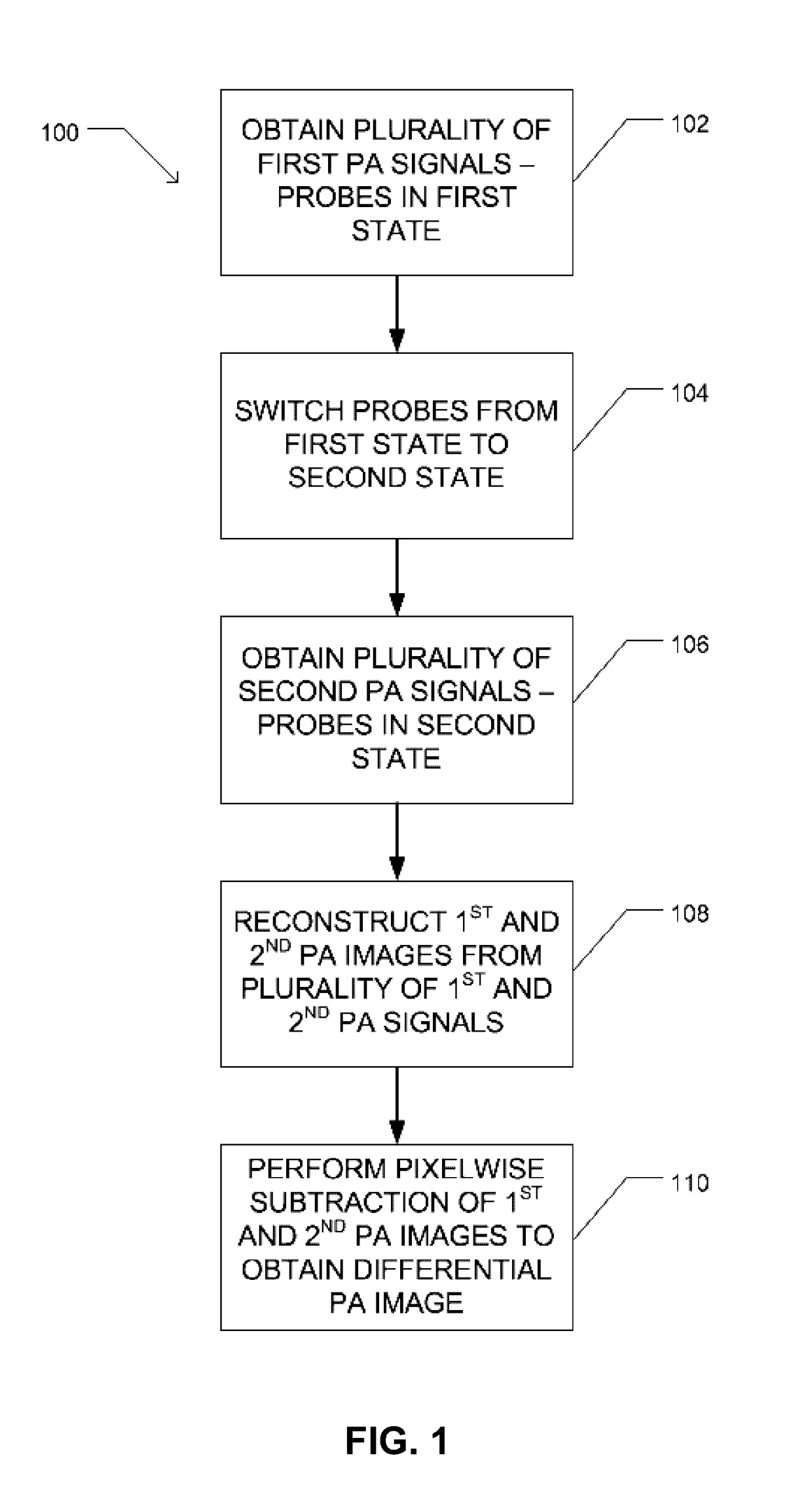
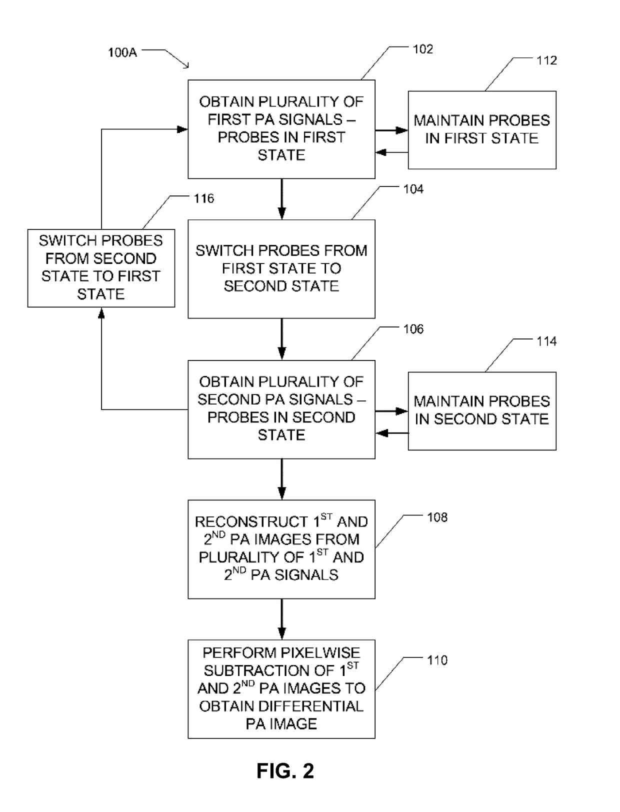
The patent describes a method and system for obtaining an image of a region of interest using reversibly-switchable probes that can be switched between a first state and a second state. The first state is a first laser pulse wavelength, and the second state is a second laser pulse wavelength. The method includes obtaining a first PA signal from the region of interest using the first laser pulse wavelength, switching the probes from the first state to the second state, obtaining a second PA signal from the region of interest using the first laser pulse wavelength, and reconstructing a first PA image of the region of interest from the first PA signals and a second PA image of the region of interest from the second PA signals to obtain the RS-PA image of the region of interest. The system includes a laser source for producing the first and second laser pulses, a controller for controlling the timing of the laser pulses, an optical combining module for directing the laser pulses into the region of interest, and an ultrasound transducer for receiving the PA signals. The technical effect is a method and system for obtaining higher-resolution images of a region of interest using RS-PA imaging.

Problems solved by technology

However, strong light scattering in tissue leads to a substantial tradeoff between the spatial resolution and penetration depth, even in advanced optical imaging techniques.
However, existing GFP-like FPs lack strong optical absorption at wavelengths beyond 610 nm and are prone to photobleaching and therefore are less well-suited for deep-tissue PAT.
Accurate spectral unmixing of these biomolecules is hampered by highly wavelength-dependent light attenuation in tissue at depths beyond the optical diffusion limit.

Method used


Image

  • Reversibly switchable photoacoustic imaging systems and methods
  • Reversibly switchable photoacoustic imaging systems and methods
  • Reversibly switchable photoacoustic imaging systems and methods

Examples


example 1

Comparison of BphP1 with Available Genetically Encoded Probes

[0173]To assess the characteristics of the bacterial phytochrome BphP1 compared to existing genetically encoded probes (rsTagRFP and iRFP720) for use as reversibly-switchable photoacoustic (RS-PA) probes, the following experiments were conducted.

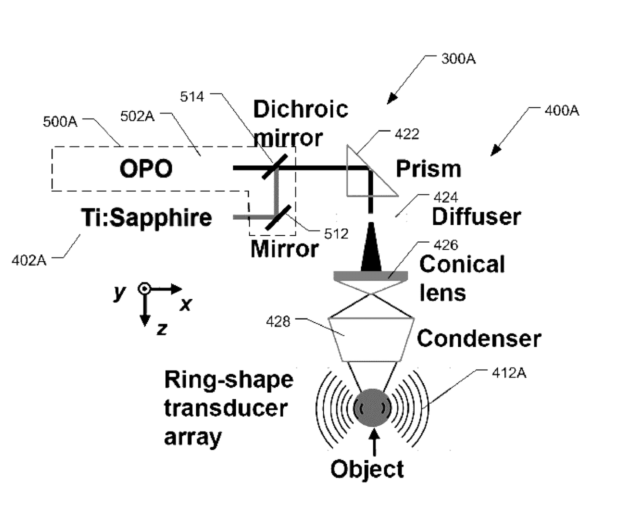
[0174]A whole-body reversibly-switchable PACT system (RS-PACT) was upgraded to utilize BphP1 as a RS-PA probe, as described herein previously and as illustrated in FIG. 5. In this experiment, the RS-PACT system included an optical parametric oscillator (OPO) laser and a Ti:Sapphire laser that were synchronized to provide an excitation optical wavelength range of 400-900 nm. 780 nm light from the Ti:Sapphire laser was used for both whole-body PA imaging and switching off BphP1 at the same time, while the 630 nm light from the OPO laser was used for switching on the protein. The flashlamps of the two pump lasers were synchronized, and the two lasers were individually triggered by an ...

example 2

Effect of Imaging Depth on Reversibly-Switchable Probe

[0179]To assess the ability of BphP1 to function as a RS-PA probe for deep PA imaging in scattering media representative of biological tissues, the following experiments were conducted. Samples of the purified proteins described in Ex. 1 were embedded at depths ranging from 0 mm to 10 mm in a scattering media. The scattering media included 1% intralipid, 10% gelatin, and 7% oxygenated bovine blood in distilled water. The scattering media had a reduced scattering coefficient of about 10 cm−1. The RS-PACT system described in Ex. 1 was used to obtain PA signals at a laser pulse wavelength match to each probe protein's maximum response: 567 nm (rsTagRFP), 715 nm (iRFP720) and 780 nm (BphP1).

[0180]FIG. 18 is a series of PA images of the protein samples obtained as described above at a depths of 0 mm and 10 mm. Each PA image is paired with an image of oxygenated whole bovine blood obtained under matched conditions for comparison. FIG. ...

example 3

Characterization of Reversible Photoswitching of BphP1

[0183]To characterize the reversible photoswitching of BphP1, the following experiments were conducted.

[0184]The optical absorbance of a sample of purified BphP1 at 780 nm was assessed during two transitions: i) a transition from the Pfr (ON) state to the Pr (OFF) state induced by 780 nm illumination; and ii) a transition from the Pr (OFF) state to the Pfr (ON) state induced by 630 nm illumination. FIG. 21 is a graph summarizing the normalized absorbance at 780 nm of the BphP1 over numerous switching cycles. As shown in FIG. 21, the optical absorbance of BphP1 showed an exponential decay from ON to OFF under 780 nm illumination, and an exponential recovery from OFF to ON under 630 nm illumination. For absorbance measurements, a Hitachi U-2000 spectrophotometer was used.

[0185]The BphP1 sample was further assessed during the transition from the Pr (OFF) state to the Pfr (ON) state induced by 630 nm illumination at illumination inte...


PUM


Abstract

Reversibly switchable photoacoustic tomography (RS-PAT), a photoacoustic technique with enhanced sensitivity and resolution, is disclosed. RS-PAT utilizes a subtractive process for the formation of a photoacoustic image of a region containing a plurality of switchable photoacoustic probes. In various aspects, the photoacoustic detection in RS-PAT imaging occurs minimally twice: a first image obtained when the photoacoustic probe is in active (absorbing or ON) state and a second image obtained when the photoacoustic probe is in an inactive (less-absorbing or OFF) state. Subtraction of the second image from the first image is used to obtain the RS-PAT image.

Description

STATEMENT REGARDING FEDERALLY SPONSORED RESEARCH & DEVELOPMENT[0001]This invention was made with government support under grants R01 CA186567 and DP1 EB016986 awarded by the National Institutes of Health. The government has certain rights to the invention.BACKGROUND[0002]Using reporter genes that can be selectively expressed in targeted biological processes, optical imaging has provided valuable information for biomedical studies at different length scales. However, strong light scattering in tissue leads to a substantial tradeoff between the spatial resolution and penetration depth, even in advanced optical imaging techniques. Photoacoustic (PA) tomography (PAT), on the other hand, breaks the depth and resolution limitations of pure optical imaging by acoustically detecting optical absorption contrast. In PAT, light-induced ultrasound waves are detected outside tissue to form an image that maps the original optical energy deposition inside the tissue. The weak ultrasonic scattering...

Claims


Application Information

Patent Timeline
Patent Type & Authority Applications(United States)
IPC IPC(8): A61B5/00
CPCA61B5/0095A61B5/7239A61B5/0037A61B5/201A61B2576/02G16H30/40
Inventor WANG, LIHONGVERKHUSHA, VLADISLAV V.YAO, JUNJIEKABERNIUK, ANDRII A.LI, LEI
Owner ALBERT EINSTEIN COLLEGE OF MEDICINE OF YESHIVA UNIV
Learn More