Non-radioactive method for determining the cytolitic activity of an agent with respect to target cells, use thereof and associated kit

Inactive Publication Date: 2018-02-22
CLEAN CELLS
View PDF0 Cites 5 Cited by
  • Summary
  • Abstract
  • Description
  • Claims
  • Application Information

AI Technical Summary

Problems solved by technology

Furthermore, by virtue of its radioactive nature, the signal generated is very strong, which results in a high signal/noise ratio (of about 5 to 12).
The method is thus very sensitive.
On the other hand, it has the drawback of requiring a 51Cr-labeling time (incubation and washes), that cannot be shortened, of approximately one to two hours depending on protocols, significantly extending the duration of the experiment.
Finally, the regulatory restrictions associated with the use of radioactivity are increasingly burdensome and expensive, in terms of administrative authorization, of source management, of waste elimination, of labor law, of medical monitoring or of exposure of handlers.
Moreover, several variants of this method have been described, using other radioisotopes, for example based on the use of tritium (3H) or indium (111In) but involving the same radioprotection restrictions and thus subjected to the same limitations.
On the other hand, the spontaneous release of calcein is very high (approximately 40% of the maximum release) reflecting a high calcein-permeability of the plasma membrane, which results in a high background noise of the method.
Since the fluorescent signal emitted by calcein is weak, the signal/noise ratio is not very favorable, at around 2.5.
All of these limitations generate quite a high variability of the Eu3+ assay, and also make it impossible to use it with a certain number of cell types, which makes its use and its validation difficult in an industrial context.
Although these methods are specific for the cell death mechanism, they do not enable a high-throughput analysis because of the technical restrictions associated with flow cytometry.
Nevertheless, these microscopic methods do not make it possible to discriminate between several distinct cell types that would have been mixed with the needs of the assay (in the case of an ADCC measurement for example), unless fluorescence microscopy methods are used.
This then amounts to methods similar to those described for flow cytometry, but which use laborious counting methods and the already mentioned drawbacks of which are further amplified.
These methods, which are s

Method used


Image

  • Non-radioactive method for determining the cytolitic activity of an agent with respect to target cells, use thereof and associated kit
  • Non-radioactive method for determining the cytolitic activity of an agent with respect to target cells, use thereof and associated kit
  • Non-radioactive method for determining the cytolitic activity of an agent with respect to target cells, use thereof and associated kit

Examples


example 1

Linearity of the Luminescent Signal and the Influence of the Amount of Cells on this Signal

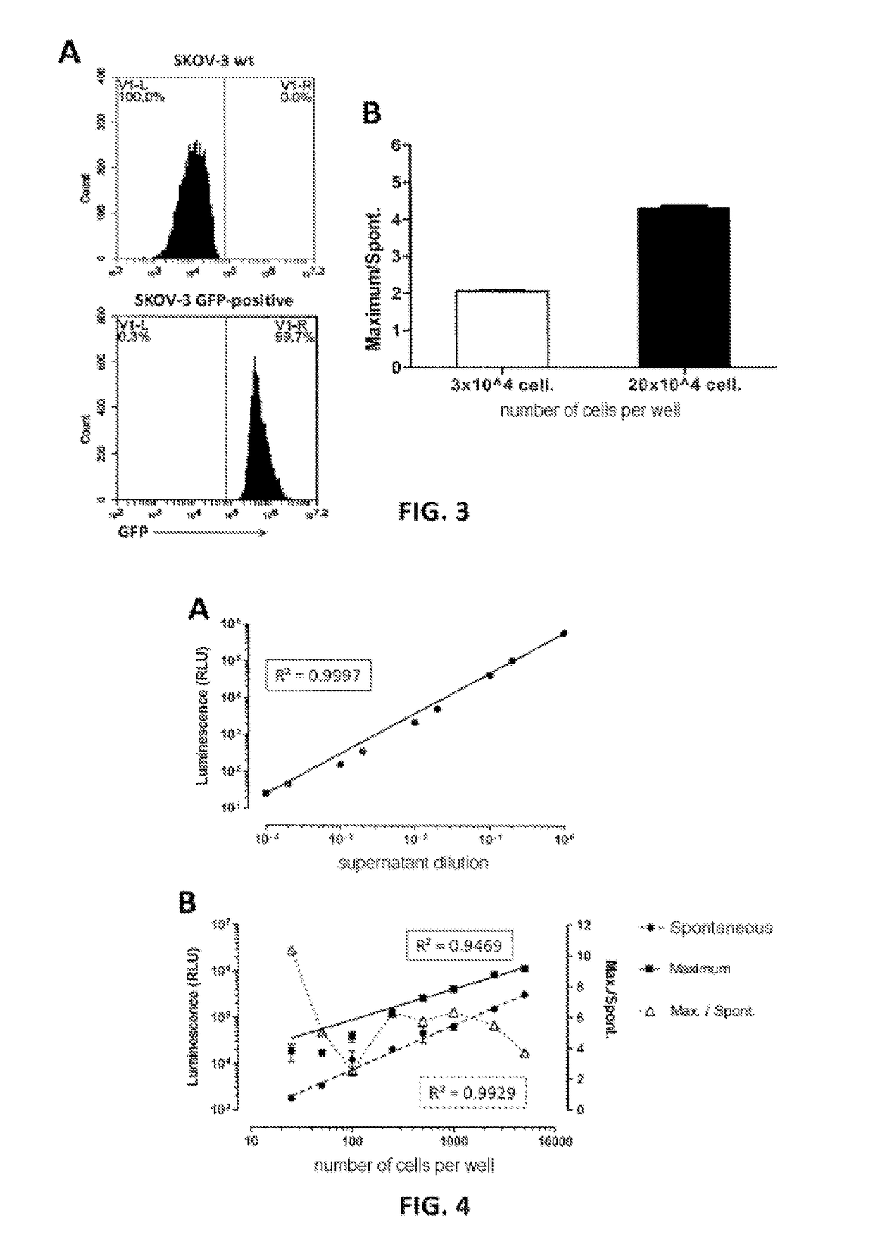
[0135]The luminescence of a supernatant containing nanoluciferase was measured, after lysis with triton X-100, serially diluted between 1 / 1 and 1 / 10 000. 25 μl of each dilution are mixed with 25 μl of Nano-Glo® substrate (Promega) in a “half-well” 96-well plate before reading on a luminometer (Mithras LB 940, Berthold Technologies, acquisition time 0.05 s). A linear regression of the values obtained is performed (see FIG. 4A: strength of the luminescent signal obtained, expressed in RLU (Relative Luminescence Unit) as a function of the dilution). It is noted, firstly, that this signal is linear (regression coefficient R2=0.9997) over the entire measurement range and extends approximately from 10 to 106 arbitrary luminescence units on the reader used.

[0136]Secondly, the linearity of the signals from spontaneous and maximum release of CHO-K1 cells expressing nanoluciferase was evaluated by incub...

example 2

Linear Relationship Between the Strength of the Luminescent Signal and the Number of Cells Actually Dead During a Cytotoxicity Assay

[0138]A complement lysis assay was carried out on Raji cells (expressing nanoluciferase) in the presence of a commercially available anti-CD20 therapeutic antibody, MabThera® (INN: rituximab).

[0139]These Raji cells expressing nanoluciferase are incubated for 4 h at 37° C. in flat-bottomed 96-well plates (3000 cells per well) in RPMI-1640 medium, in the presence of variable amounts of anti-CD20 antibodies (MabThera®, Roche, range of 9 concentrations from 1093.5 to 0.167 ng / ml per 1 / 3 dilution increment) and of 1 CH50 (hemolytic complement 50, usual unit of measurement of complement activity) of guinea pig complement (Sigma-Aldrich). Control conditions are also carried out, making it possible to measure the spontaneous release (3000 target cells in RPMI-1640, 1 CH50 of guinea pig complement and 1093.5 ng / ml of an antibody which does not bind to Raji cells...

example 3

[0141]FIG. 8, in which the specific lysis of adherent cells (SKOV3) or non-adherent cells (Raji) expressing nanoluciferase was measured in an ADCC assay according to the present invention, shows the dynamic nature of said method.

[0142]Non-adherent (Raji) or adherent (SKOV3) target cells transformed to stably express nanoluciferase were incubated (at 37° C. in a humidified atmosphere at 5% CO2) in flat-bottomed 96-well plates treated for cell culture (3000 target cells per well), in the presence of increasing concentrations (the logarithms of which are indicated along the x-axis) of the corresponding antibody (rituximab [MabThera®] or trastuzumab [Herceptin®], respectively) and of cytotoxic effector cells (T lymphocytes expressing CD16, 30 000 effector cells per well) in RPMI-1640 supplemented with 5% of FCS. Each range is deposited 4 times (for the 4 incubation times tested) with 3 wells per condition (triplicate). After 1 (black circles), 2 (black squares), 3 (white diamonds) or 4 ...


PUM


Abstract

A non-radioactive method for the direct in vitro determination (control and quantification) of the cytolytic activity of an active agent with respect to target cells and/or a medium surrounding target cells, comprising the steps of genetically transforming target cells to express an enzyme exogenous to said target cells, exposing said genetically transformed target cells to the active agent and/or to said surrounding medium to be tested, which can result in the lysis of at least a portion of the target cells by releasing said exogenous enzyme into the extracellular medium, and measuring the activity of the exogenous enzyme released during the lysis of said target cells, characterised in that said exogenous enzyme is an enzyme having a molar mass less than or equal to 45 kDa (e.g. an Oplophorus gracilirostris luciferase), and of which the activity can be detected by luminescence or fluorescence. Application to the measurement of antibody-dependent cellular cytotoxicity (ADCC), complement-dependent cytotoxicity (CDC) and/or the measurement of apoptosis.

Description

[0001]The present invention relates to the field of the measurement of cell death and more particularly to a method for the determination of the cytolytic action of active substances with respect to target cells and / or a medium surrounding said target cells.[0002]In the industrial field, the regulations make it obligatory to characterize as completely as possible any phenomenon or product that may have consequences on the health of human or animal populations. In the vast context of the setting up of tests which measure cell death, called cytotoxicity tests, these phenomena or products can group together environmental phenomena, such as exposure to a source of irradiation or of pollution, chemical or biological products which are not for health purposes, or else products for therapeutic purposes.[0003]It is in the field of biologically active substances, and more specifically in the context of substances for therapeutic purposes, that the characterization obligations are the most th...

Claims


Application Information

Patent Timeline
IPC IPC(8): C12Q1/66G01N33/50
CPCC12Q1/66G01N33/5014
Inventor BONNAUDET, VERONIQUEBRETAUDEAU, LAURENTROSSIGNOL, ALEXIS
Owner CLEAN CELLS
Why Eureka
Try Eureka