Peptide for inducing regeneration of tissue and use thereof

a technology of tissue regeneration and peptides, which is applied in the direction of peptides, drug compositions, cardiovascular disorders, etc., can solve the problems of limited research and development, gradual loss of proliferative ability and multipotency, and little understanding of the significance of the presence of mesenchymal stem cells in the living body

Active Publication Date: 2018-03-15
STEMRIM INC +1
View PDF1 Cites 3 Cited by
  • Summary
  • Abstract
  • Description
  • Claims
  • Application Information

AI Technical Summary

Benefits of technology

[0032]Further, the present inventors confirmed that the identified peptides have migration-promoting activity on skin fibroblasts, which are PDGFRαpositive, and have an effect of reducing the size of cerebral infarcts in a cerebral infarction model mouse.
[0035]Next, based on the peptide consisting of amino acids of positions 89 to 215 which was confirmed to have migration-promoting activity on MSC-1 and human bone marrow mesenchymal stem cells, the present inventors prepared peptides with the C-terminus increasingly shortened, i.e. a peptide consisting of amino acids of positions 89 to 205, a peptide consisting of amino acids of positions 89 to 195, and a peptide consisting of amino acids of positions 89 to 185, and examined each of them for migration-promoting activity. The result showed that among the prepared peptide fragments, those with a shorter C terminus demonstrated more enhanced migration-promoting activity on the PDGFRα-positive bone marrow mesenchymal stem cell line (MSC-1) and human bone marrow mesenchymal stem cells.

Problems solved by technology

However, little is understood about the significance of the presence of mesenchymal stem cells in the living body.
However, bone-marrow blood containing bone marrow mesenchymal stem cells is collected only by an invasive method where thick needles are repeatedly inserted into the iliac bone.
In addition, continuous passages of bone marrow mesenchymal stem cells outside the body lead to gradual loss of their proliferative ability and multipotency.
Moreover, since culturing bone marrow mesenchymal stem cells with high quality control for ensuring the safety of in vivo transplantation requires special cell culture facilities such as cell processing center (CPC), it can only be performed currently in very limited universities and companies.

Method used


Image

  • Peptide for inducing regeneration of tissue and use thereof
  • Peptide for inducing regeneration of tissue and use thereof
  • Peptide for inducing regeneration of tissue and use thereof

Examples


example 1

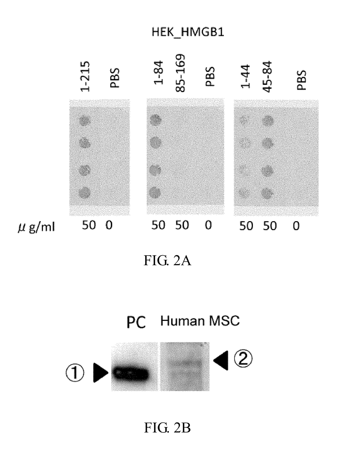
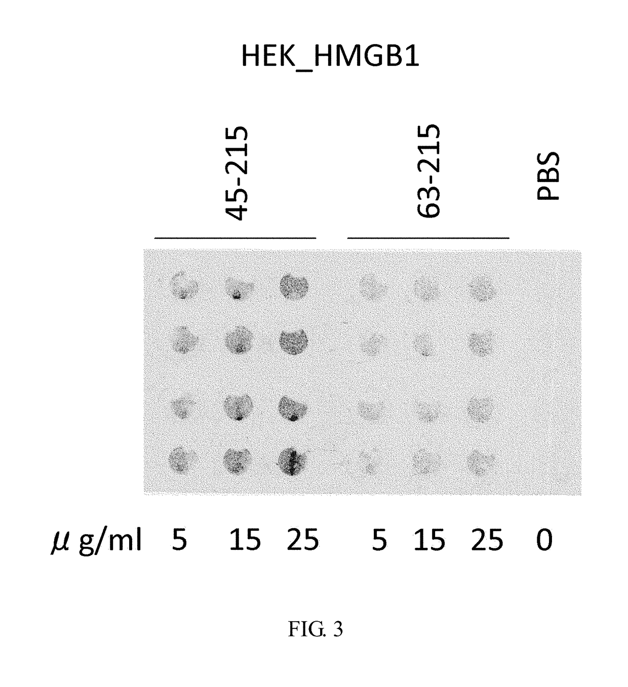
Purification of HMGB-1 and HMGB1-Derived Peptides Using HEK293

[0527]RNA was extracted from newborn mouse skin using Trizol (Invitrogen), and then cDNA was synthesized using SuperScript III cDNA synthesis kit (Invitrogen). Using this cDNA as a template,

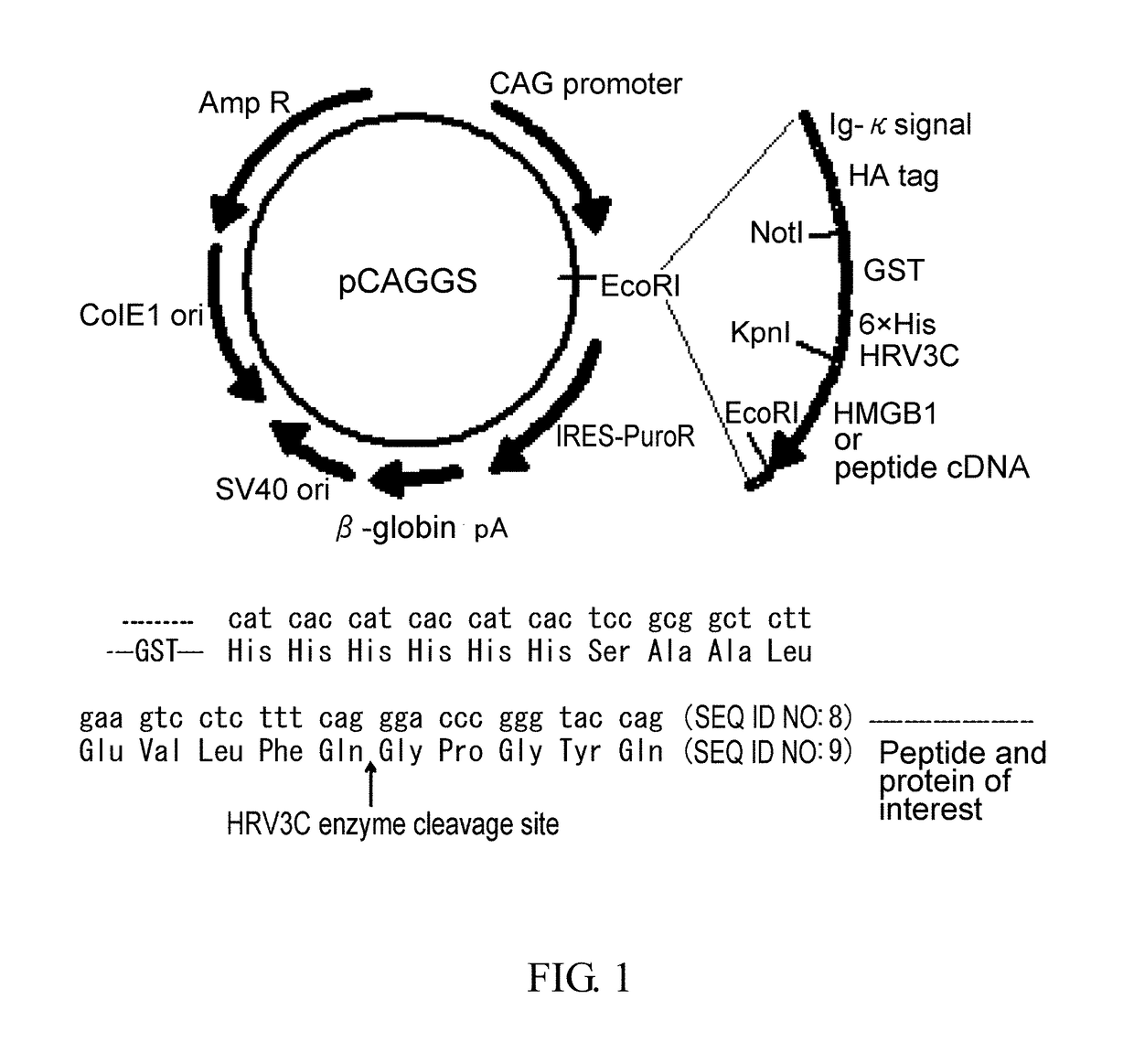
[0528]HMGB1 cDNA was amplified by polymerase chain reaction (PCR). The resulting cDNA was inserted into pCAGGS, a plasmid vector for protein expression in mammalian cells, such that the vector would express the protein attached with an IgG κ chain signal sequence as a secretion signal, and with an HA tag, GST tag, and 6× His tag sequences at the N terminus of its amino acid sequence for the convenience of purification (FIG. 1). In addition, a sequence cleaved by HRV3C was inserted between the His tag and the protein or peptide of interest. After digestion with HRV3C, a peptide fragment of Gly Pro Gly Thy Gin (SEQ ID NO: 7) will be attached to the N-terminal of the protein or peptide of interest. In the meantime, restriction sites were ...

example 2

Sorting of Primary Cultured Pdgfrα-Positive Bone Marrow Mesenchymal Stem Cells and Assessment of Migration-Promoting Activity

[0546]Thigh and tibial bones were excised from donor mice: B6.129S4-Pdgfratm11(EGFP)Sor / J (PDGFRα-GFP Mouse). After removing attached muscles and other tissues, the bones were crushed finely and incubated with 0.2% collagenase (Roche, REF: 10103586001) / DMEM / 2% FBS(filtrated) at 37° C. for 40 minutes. Then, cell aggregates and muscle tissues were removed by filtration through a 40-μm nylon mesh. After centrifugation at 1200 rpm for 10 minutes, the resulting cells were suspended in αMEM containing 10% FBS and 1% P / S and cultured in an incubator under 5% CO2 at 37° C. until they reached 100% confluence. The cells were harvested and the following experiment was carried out according to the protocol attached to CD11b MicroBeads (Miltenyi Biotec; order No: 130-049-601). The cells were adjusted to 107 cells / 90 μl with PBS(−), and CD11b MicroBeads were added at 10 μl / ...

example 3

Assessment of Primary Cultured Pdgfrα-Positive Bone Marrow Mesenchymal Stem Cells for Multipotency

[0553]FACS sorting of PDGFRα-positive, Lineage-negative, c-kit-negative cells

[0554]Under sufficiently deep anesthesia with isoflurane, C57B16 mice (male, 6 weeks old) were euthanized by carbon dioxide inhalation. Thigh and tibial bones were excised and fat and muscle tissues were removed from them. The bones were soaked in EtOH to thoroughly remove attached tissues from them. Bone marrow tissues were obtained using a syringe with 26G needle. The obtained bone marrow cells were combined with DMEM containing 0.2% Collagenase A and incubated at 37° C. for 40 minutes. After adding DMEM containing 10% FBS, the cells were centrifuged at 1500 rpm for 10 minutes. The supernatant was discarded and the precipitated bone marrow cells were collected.

[0555]The cells were plated in a culture dish with a diameter of 10 cm and cultured using D-MEM containing 10% FBS supplemented with lx streptomycin-pe...


PUM

PropertyMeasurementUnit
diameteraaaaaaaaaa
diameteraaaaaaaaaa
diameteraaaaaaaaaa

Abstract

(Objective) An objective of the present invention is to provide therapeutic agents that, in association with stimulation of PDGFRα-positive cells such as bone marrow mesenchymal stem cells, promote their mobilization into blood and accumulation in a damaged tissue, and induce tissue regeneration in a living body.(Means for solution) Multiple peptides were synthesized, and the migration-promoting activity of each peptide was evaluated. As a result, the present inventors successfully identified multiple peptides that have migration-promoting activity on a PDGFRα-positive bone marrow mesenchymal stem cell line (MSC-1). Further, the present inventors confirmed that the identified peptides also have migration-promoting activity on skin fibroblasts, which are PDGFRα-positive cells.

Description

CROSS-REFERENCE TO RELATED APPLICATIONS[0001]This application is a continuation Application of application Ser. No. 14 / 114,395, filed Feb. 6, 2014; which is a National Stage Application of International Application Number PCT / JP2012 / 059113, filed Apr. 3, 2012; which claims priority to Japanese Application No. 2011-098270, filed Apr. 26, 2011; and Japanese Application No. 2011-219454, filed Oct. 3, 2011; all of which are incorporated herein by reference in their entirety.TECHNICAL FIELD[0002]The present invention relates to peptides for inducing tissue regeneration and uses thereof.[0003]The Sequence Listing for this application is labeled “SeqList-18Feb14.txt”, which was created on February 18, 2014, and is 13 KB. The entire content is incorporated herein by reference in its entirety.BACKGROUND OF THE INVENTION[0004]It has been becoming clear that each organ or tissue in the living organism has tissue stem cells that maintain its structural and functional homeostasis. For example, c...

Claims


Application Information

Patent Timeline
Patent Type & Authority Applications(United States)
IPC IPC(8): C07K14/47
CPCC07K14/4705C07K14/4702A61K38/17A61P1/00A61P1/04A61P1/16A61P1/18A61P11/00A61P13/00A61P15/00A61P17/00A61P17/02A61P17/14A61P19/00A61P21/00A61P25/00A61P43/00A61P9/00A61P9/10A61K35/15A61K35/28A61K38/1709A61K48/00C07K14/4718C12N5/0647C12N5/0663C12N15/85C12N2510/00C07K2319/50C07K2319/21
Inventor TAMAI, KATSUTOYAMAZAKI, TAKEHIKOKANEZAKI, TSUTOMUSAKURAI, SHIGERUNOGUCHI, YUKIKOENDO, MAYUMIHAMABUCHI, NATSUMINAITO, KANA
Owner STEMRIM INC
Why Eureka
Try Eureka