Cartilage regeneration using chondrocyte and tgf-beta

a cartilage regeneration and chondrocyte technology, applied in the direction of prosthesis, peptide/protein ingredients, drug compositions, etc., can solve the problems of inefficiency of drug delivery to the joint, frequent repeated injections, and inability to regenerate damaged hyaline cartilag

Inactive Publication Date: 2019-07-25
KOLON TISSUEGENE INC
View PDF0 Cites 0 Cited by
  • Summary
  • Abstract
  • Description
  • Claims
  • Application Information

AI Technical Summary

Benefits of technology

[0034]These and other objects of the invention will be more fully understood from the following description of the invention, the referenced drawings attached hereto and the claims appended hereto.

Problems solved by technology

There has been no method reported to date to regenerate damaged hyaline cartilage.
Traditional routes of drug delivery, such as oral, intravenous or intramuscular administration, to carry the drug to the joint are inefficient.
Another disadvantage of intra-articular injection of drugs is that frequent repeated injections are necessary to obtain acceptable drug levels at the joint spaces for treating a chronic condition such as arthritis.
Exposure of non-target organs in this manner exacerbated the tendency of anti-arthritis drugs to produce serious side effects, such as gastrointestinal upset and changes in the hematological, cardiovascular, hepatic and renal systems of the mammalian host.
Recently, the therapeutic value of TGF-β has been reported (Critchlow et al., Bone, 521-527, 1995; and Lind et al., A Orthop Scand, 64(5): 553-556, 1993), but its short-term effects and high cost have limited wide clinical application.
Previously, it was determined that intraarticular injection of TGF-β for the treatment of arthritis is not desirable, because the injected TGF-β has a short duration of action, as TGF-β is degraded into inactive form in vivo.
However, there is no disclosure of a gene therapy method using the BMP gene.

Method used


Image

  • Cartilage regeneration using chondrocyte and tgf-beta
  • Cartilage regeneration using chondrocyte and tgf-beta
  • Cartilage regeneration using chondrocyte and tgf-beta

Examples


example i

and Methods

Plasmid Construction

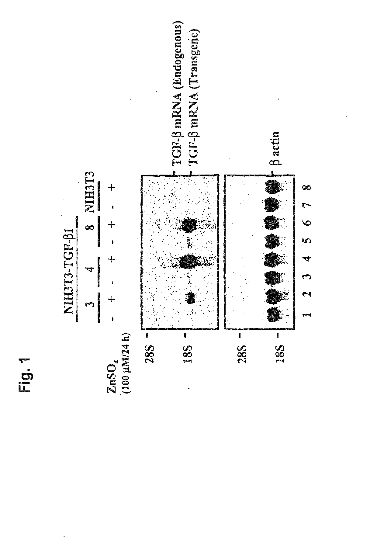
[0138]To generate the metallothionein expression construct (pM), the metallothionein I promoter (−660 / +63) was generated by polymerase chain amplification using genomic DNA using Xba I and Bam HI restriction sites built into the oligonucleotides used for amplification. The amplified fragment was subcloned into Xba I-Bam HI sites of pBluescript (Stratagene, La Jolla, Calif.). The plasmid pmTβ1 was generated by subcloning a 1.2-kb Bgl II fragment containing the TGF-β1 coding sequence and a growth hormone poly A site at the 3′ end into the Bam HI-Sal I sites of pM.

[0139]Cell Culture and Transfections—The TGF-β cDNA was transfected into fibroblasts (NIH 3T3-TGF-β1) or human foreskin fibroblast / TGF-β1. They were cultured in Dulbecco's Modified Eagle's Medium (GIBCO-BRL, Rockville, Md.) with 10% concentration of fetal bovine serum. The TGF-β1 cDNA sequence was added into the pmTβ1 vector with a metallothionein gene promoter. A neomycin resistance gene sequen...

example ii

[0144]Stable cell line—Transfection was carried out by using the calcium phosphate coprecipitation method (FIG. 1). About 80% of the surviving colonies expressed the transgene mRNA. These selected TGF-β1-producing cells were incubated in a zinc sulfate solution. When the cells were cultured in 100 μM zinc sulfate solution, they produced mRNA. The TGF-β secretion rate was about 32 ng / 106 cells / 24 hr.

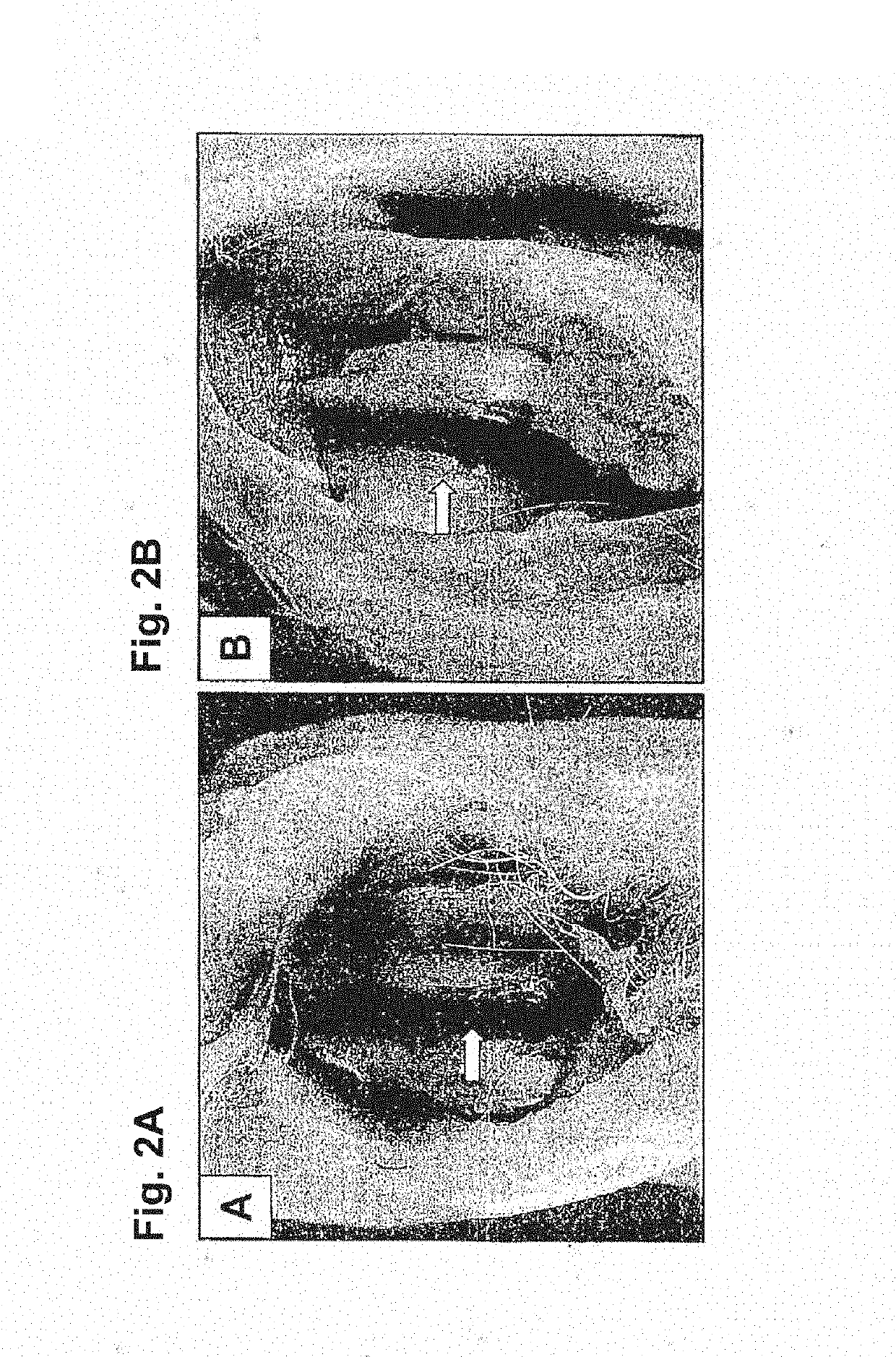
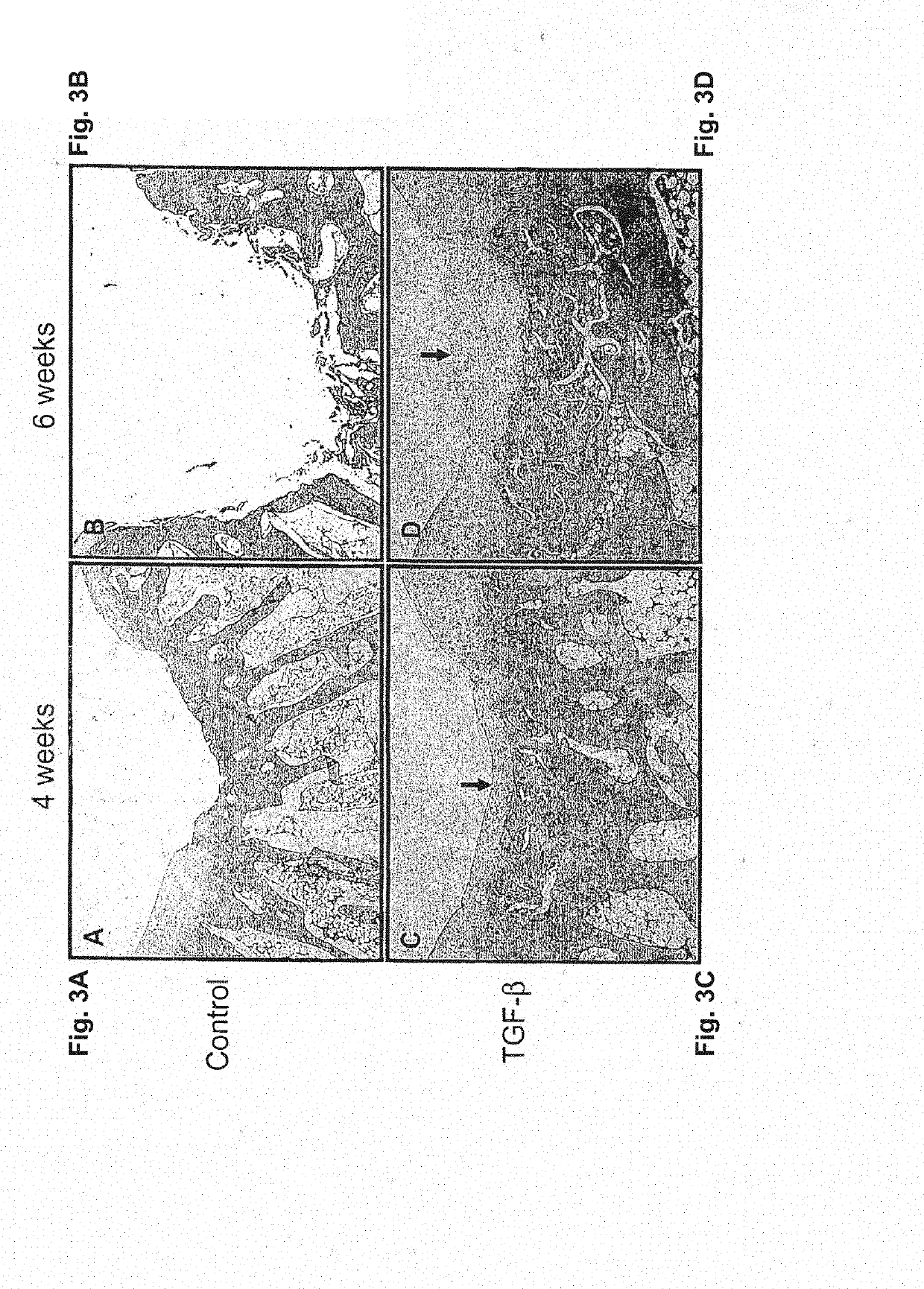
[0145]Regeneration of Rabbit Articular Cartilage Defect—The rabbit achilles tendons were observed to check the viability of NIH 3T3-TGF-β1 cells. At 106 cells / ml concentration, the tendon was grossly thicker than at the other two concentrations of 104 and 105. After making partial and complete cartilage defects, 0.3 ml of 106 cells / ml of the NIH 3T3-TGF-β1 cells were injected into knee joints. The joint was examined 2 to 6 weeks after injection. In partially damaged cartilage, we found newly formed hyaline cartilage; two weeks after injection, hyaline cartilage appeared and six weeks afte...

example iii

[0148]Either control NIH3T3 or NIH3T3-TGF-β1 cells (5-7×10⁵) were irradiated with 6000 rad. and injected into rabbit knee joints. These irradiated cells died completely in 3 weeks in a tissue culture dish. The injection procedure was the same as in the previous protocol with untreated cells. The knee joints were harvested at 3 or 6 weeks post injection. The specimens were fixed in formalin and decalcified with nitric acid. Sections of the specimens were made and embedded with paraffin and then cut into 0.5 μm thickness slices. In FIG. 10, Safranin-O staining (A-D & A′-D′) and Hematoxilin-Eosine staining (E-F & E′-F′) were done in the sections to observe the regenerated cartilage tissue microscopically. (Original magnification: (A, B, A′& B′)×12.5; (C-F & C′-F′)×400).


PUM

PropertyMeasurementUnit
thicknessaaaaaaaaaa
thicknessaaaaaaaaaa
thicknessaaaaaaaaaa

Abstract

The present application is directed to a method of treating osteoarthritis, which includes obtaining a member of a transforming growth factor superfamily of proteins; obtaining a population of cultured mammalian cells that may contain vector encoding a gene, or a population of cultured connective tissue cells that do not contain any vector encoding a gene; and then transferring the protein and the connective tissue cells into an arthritic joint space of a mammalian host, such that the activity of the combination within the joint space results in regenerating connective tissue.

Description

BACKGROUND OF THE INVENTIONField of the Invention[0001]The present invention relates to a method of introducing at least one gene encoding a member of the transforming growth factor β superfamily into at least one mammalian cell for use in regenerating connective tissue in the mammalian host. The present invention also relates to a method of introducing at least one gene product of the transforming growth factor β superfamily and at least one connective tissue cell for use in regenerating connective tissue in the mammalian host. The present invention also relates to a mammalian cell line that harbors a DNA vector molecule containing a gene encoding a member of the transforming growth factor β superfamily.Brief Description of the Related Art[0002]In the orthopedic field, degenerative arthritis or osteoarthritis as well as injuries caused by participation in sports activities is the most frequently encountered condition associated with cartilage damage. As for osteoarthritis, almost e...

Claims


Application Information

Patent Timeline
Patent Type & Authority Applications(United States)
IPC IPC(8): A61K35/32A61K38/18A61K9/00A61P19/02A61K35/35
CPCA61K35/32A61K38/1841A61K9/0019A61P19/02A61K35/35A61K35/00C12N5/00A61L27/00A61K9/0024
Inventor NOH, MOON JONGYI, YOUNGSUKSONG, SUN UKLEE, DUG KEUNLEE, KWAN HEE
Owner KOLON TISSUEGENE INC
Why Eureka
Try Eureka