
Preparation and Applications of 3D Bioprinting Bioinks for Repair of Bone Defects, Based on Cellulose Nanofibrils Hydrogels with Natural or Synthetic Calcium Phosphate Particles

a bioink and cellulose nanofibril technology, applied in the field of 3d bioprinting, bioinks for bioprinting, constructs and tissues prepared by bioprinting, can solve the problems of large defects that cannot be repaired, titanium plates being exposed through the skin, pain in patients, etc., to achieve bone formation and repair large defects.

Active Publication Date: 2019-10-10
CELLINK AB
View PDF0 Cites 14 Cited by
  • Summary
  • Abstract
  • Description
  • Claims
  • Application Information

AI Technical Summary

Benefits of technology

The patent describes a 3D printing technology that creates a strong and stable structure that can be used to repair damaged bone tissue. This technology is designed to be safe and compatible with the body's natural ability to heal. The created structure can also be "vascularized," meaning it can have its own blood flow, which can help it grow and develop more like natural bone tissue. Overall, this technology is leading the way in creating high-quality bones using 3D printing technology.

Problems solved by technology

Bone has in contrast to cartilage, a very good healing capability but larger defects cannot be repaired without transplantation of autologous graft or support material.
These solutions also have many disadvantages such as titanium plates being exposed through the skin.
Furthermore, the current solutions are painful for the patients and they are also very expensive and time consuming for doctors and patients.
One of the challenges when using TCP inorganic particles is to fuse them into 3D structure or made them injectable.
Processability of TCP remains however major challenge.
The major limitation of all these hydrogels is the rheological properties.
But this is not sufficient to provide very high printing fidelity which is defined as thin lines which can stay at the same dimension without sagging.

Method used


Image

  • Preparation and Applications of 3D Bioprinting Bioinks for Repair of Bone Defects, Based on Cellulose Nanofibrils Hydrogels with Natural or Synthetic Calcium Phosphate Particles
  • Preparation and Applications of 3D Bioprinting Bioinks for Repair of Bone Defects, Based on Cellulose Nanofibrils Hydrogels with Natural or Synthetic Calcium Phosphate Particles
  • Preparation and Applications of 3D Bioprinting Bioinks for Repair of Bone Defects, Based on Cellulose Nanofibrils Hydrogels with Natural or Synthetic Calcium Phosphate Particles

Examples


example 1

Preparation and Evaluation of Bioinks

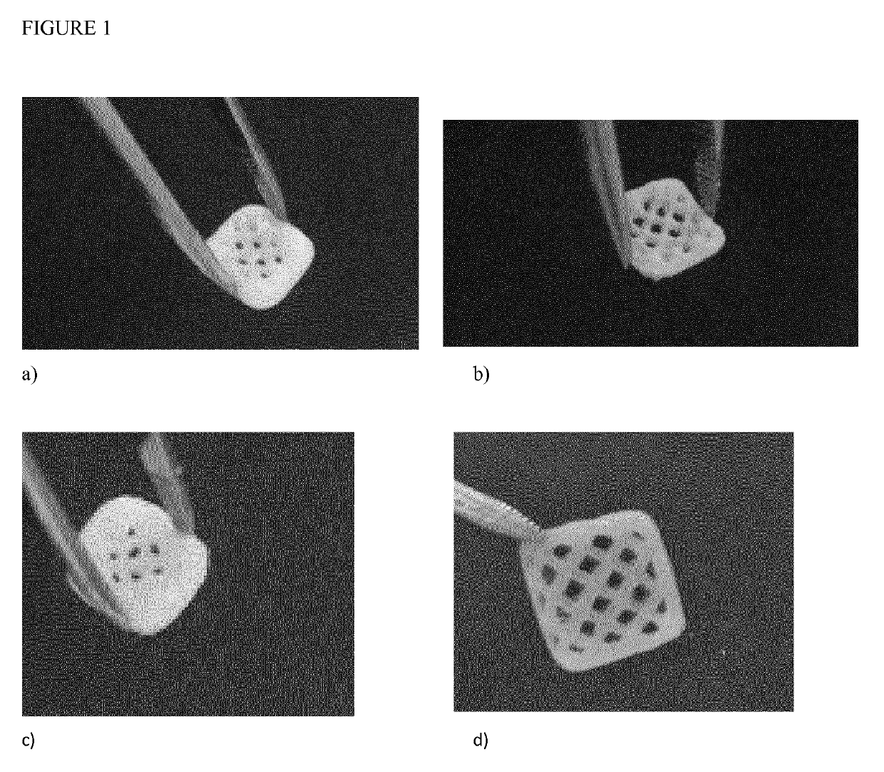
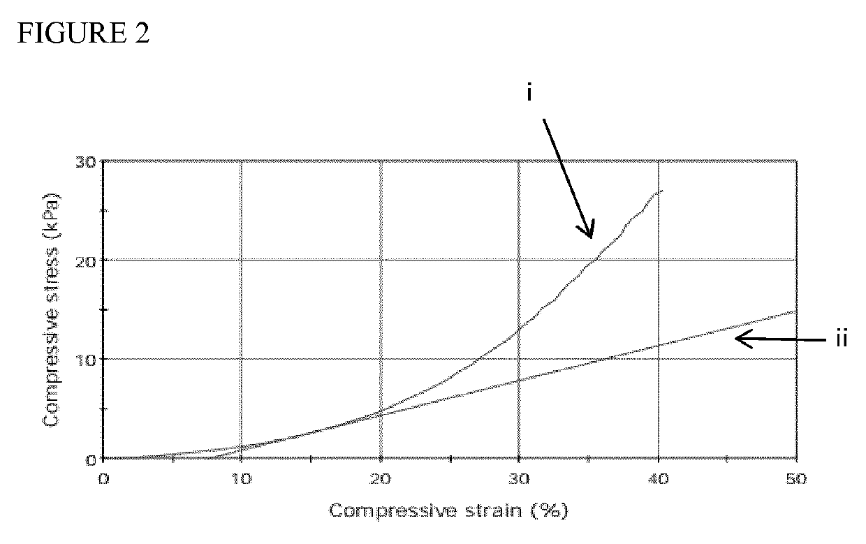
[0064]Granules of β-tricalcium phosphate (β-TCP) 1.4-2.8 mm particle size were grinded in a mechanical grinder to a powder and finally homogenized using a hydraulic press. Other Calcium phosphates from different sources were compared. The grinding process was designed to provide the size of the particles to be below 200 microns as determined by sieving process. Nanocellulose fibril hydrogel was prepared by homogenization of Bacterial Cellulose pellicle which was purified according to protocols published elsewhere [8]. Cellulose nanofibrils isolated from wood have also been evaluated. In order to provide good crosslinking, between 10-20% of alginate based on dry weight of cellulose nanofibrils was added. A typical bioink without mineral phase contains between 2-3% dry weight cellulose nanofibrils and alginate mixture. Different mixing devices were tested. FIG. 1 shows a mixing device which is constructed by combining two cartridges. Mixing was ach...

example 2

3D Bioprinting of Implantable Constructs

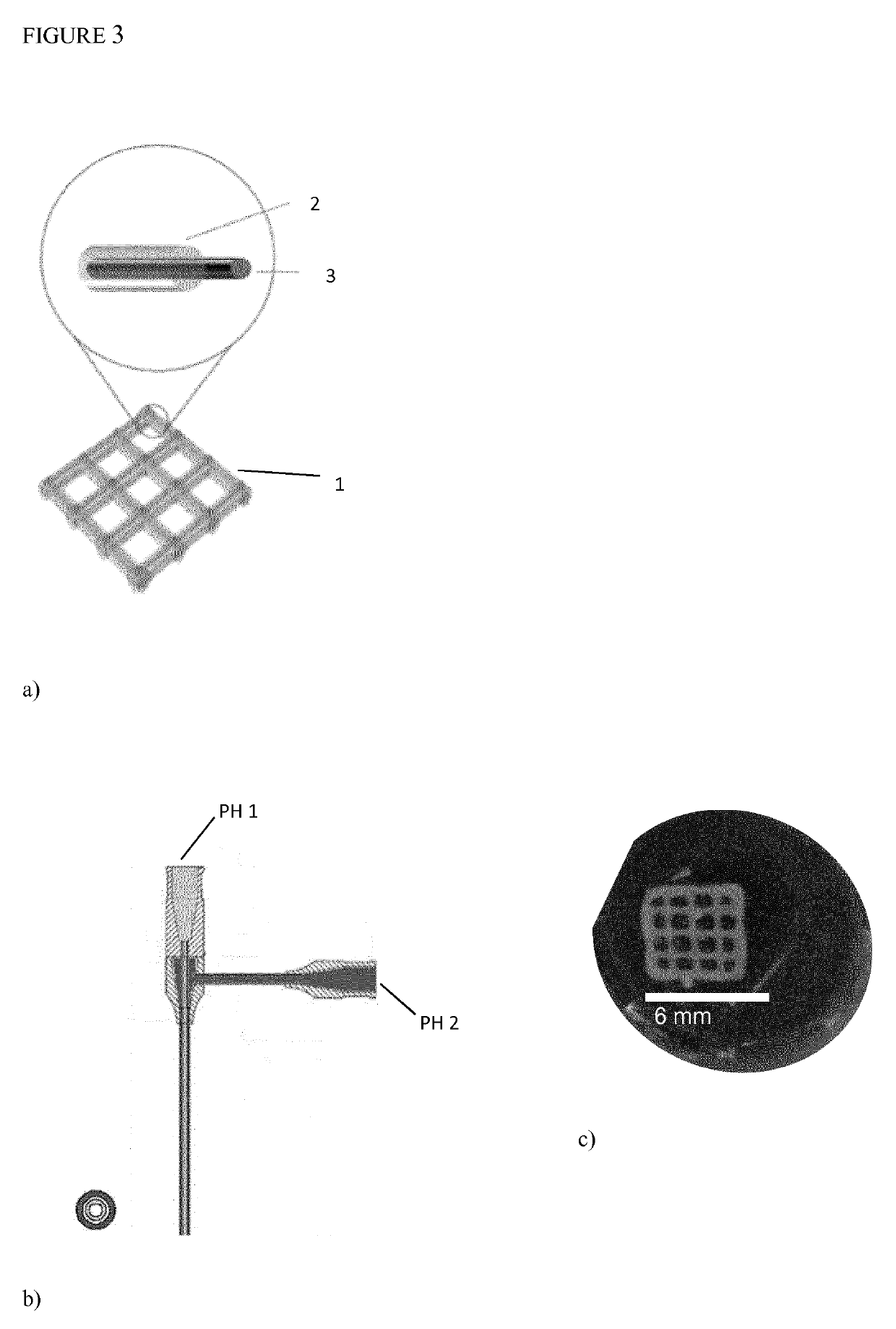
[0065]Very high viscosity of Calcium filled bioinks resulted in much higher pressure necessary to get printed constructs. In order to use bioinks with cells the core shell structures (1) were designed as shown in FIG. 3a. One possible design is to use Calcium filled bioink as a core and cell filled nanocellulose bioink as a shell. In FIG. 3a, the structure 1 comprises an outer shell 2 and an inner core 3. The outer shell may contain nanocellulose bioink and mesenchymal stem cells, and the inner core 3 may contain 20% β-TCP / nanocellulose bioink. Opposite can be also printed. In order to be able to print such structures coaxial needle is installed, see FIG. 3b. The bioink of the inner core is introduced at PH 1, and the bioink of the outer shell is introduced at PH 2. Prior to bioprinting nanocellulose bioink in DMEM medium solution was prepared. Confluent human adipose derived mesenchymal cells AD-MSCs were washed with 6 mL DPBS and then incuba...

example 3

3D Printing Using Human Bone Powder as a Source

[0067]Example 1 and 2 is repeated using human bone powder (LifeNet Health (ReadiGraft Demineralized Cortical Particulate with grind size 125-1000 microns)) instead of the other materials of Example 1 and 2. The powder is sieved before use to obtain the desired particle size (less than 400 microns, or less than 200 microns). The human bone powder is used with the same parameters as for the β-TCP-particles. The same, promising results are expected, i.e. excellent printability with high fidelity.


PUM

PropertyMeasurementUnit
Fractionaaaaaaaaaa
Sizeaaaaaaaaaa
Sizeaaaaaaaaaa

Abstract

The present invention relates to preparation of bioink composed of cellulose nanofibril hydrogel with native or synthetic Calcium containing particles. The concentration of the calcium containing particles can be between 1% and 40% w / v. Such bioink can be 3D Bioprinted with or without human or animal cells. Coaxial needle can be used where cellulose nanofibril hydrogel filled with Calcium particles can be used as shell and another hydrogel based bioink mixed with cells can be used as core or opposite. Such 3D Bioprinted constructs exhibit high porosity due to shear thinning properties of cellulose nanofibrils which provides excellent printing fidelity. They also have excellent mechanical properties and are easily handled as large constructs for patient-specific bone cavities which need to be repaired. The porosity promotes vascularization which is crucial for oxygen and nutrient supply. The porosity also makes it possible for further recruitment of cells which accelerate bone healing process. Calcium containing particles can be isolated from autologous bone, allogenic bone or xenogeneic bone but can be also isolated from minerals or be prepared by synthesis. Preferable Calcium containing particles consist of β-tricalcium phosphate which is resorbable or natural bone powder, preferably of human or porcine origin. The particles described in the present invention have particle size smaller than 400 microns, or more preferably smaller than 200 microns, to make it possible to handle in printing nozzle without clogging and to obtain a good resolution. Cellulose nanofibrils can be produced by bacteria orbe isolated from plants. They can be neutral, charged or oxidized to be biodegradable. The bioink can be additionally supplemented by other biopolymers which provide crosslinking. Such biopolymers can be alginates, chitosans, modified hyaluronic acid or modified collagen derived biopolymers.

Description

BACKGROUND OF THE INVENTIONField of the Invention[0001]The present invention relates to the field of 3D bioprinting, bioinks for bioprinting, constructs and tissues prepared by bioprinting, as well as the medical use of such constructs and tissues for treatment in the field of tissue replacement or defect.Description of Related Art[0002]There is an unmet medical need for novel procedures for repair of larger bone defects. Bone has in contrast to cartilage, a very good healing capability but larger defects cannot be repaired without transplantation of autologous graft or support material. One example can be a reconstruction of the mandible due to traffic accidents, or cancer. The most common cancer affecting the mandible is squamous cell carcinoma. According to Cancer Research UK, in 2012 61,400 cases of oral cavity cancer were reported in Europe [1]. 28,260 cases were reported in 2014 in the United States of America [2]. The most common current solution is transplantation of fibula ...

Claims


Application Information

Patent Timeline
IPC IPC(8): A61L27/46A61L27/44A61L27/48A61L27/38A61L27/52C12N5/077B33Y70/00B33Y10/00B33Y80/00
CPCA61L27/3895C12N2506/45B33Y10/00A61L27/446A61L27/3834A61L27/48A61L27/3865A61L2400/12A61L2430/12C12N2506/1346C12N5/0654A61L27/46B33Y70/00A61L27/3821A61L2430/02A61L27/3847B33Y80/00A61L27/52B29C64/106B29C64/209B33Y70/10B29K2105/122B29K2995/0056C08L1/00B29C64/165
Inventor GATENHOLM, PAULMARTINEZ, HECTORSCHETTINO, MICHELAGATENHOLM, ERIK
Owner CELLINK AB
Learn More