
Methods for improved reproductive management of ruminant ungulates

a technology for ungulates and ruminants, applied in the field of methods for improving the management of ruminant ungulates, can solve the problems of affecting the ability to measure interferon-induced analytes, reducing the probability that interferon-induced analytes will degrade, aggregate or unfold before detection,

Pending Publication Date: 2020-01-09
PENN STATE RES FOUND
View PDF0 Cites 0 Cited by
  • Summary
  • Abstract
  • Description
  • Claims
  • Application Information

AI Technical Summary

Benefits of technology

The present invention provides methods for improved reproductive management of female ruminant ungulates, particularly dairy cows. The methods involve detecting the presence or absence of interferon-induced analytes in biological samples, such as leukocytes, to determine if the cow is open or not open. The method involves contacting the sample with a binding agent specific for the interferon-induced analyte and detecting the complex using flow cytometry. The invention also provides methods for identifying specific types of leukocytes and assigning them to a sample well for further analysis. The methods can be performed using a remote tracking and computer environment, with unique identifiers for each sample. Overall, the invention provides more accurate and reliable methods for managing cow reproductive health.

Problems solved by technology

A female ruminant ungulate that is not open should not be artificially inseminated or undergo additional embryo transfer because doing so is unlikely to result in conception and may terminate an existing pregnancy.
Immediately contacting the blood with a fixative can also stabilize interferon-induced analytes therein, which could otherwise become degraded, denatured, and / or aggregated, thereby impairing the ability to measure the interferon-induced analytes.
Furthermore, in situ fixation decreases the probability that the interferon-induced analytes will degrade, aggregate, or unfold prior to their detection.

Method used


Image

  • Methods for improved reproductive management of ruminant ungulates
  • Methods for improved reproductive management of ruminant ungulates
  • Methods for improved reproductive management of ruminant ungulates

Examples


example 1

eparation Via In Vitro Treatment for Analysis

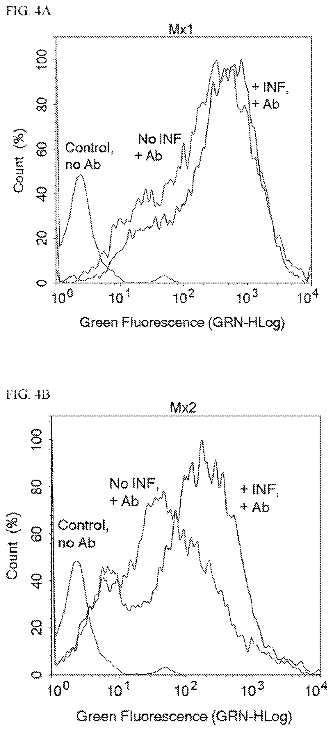
[0165]Bovine blood was drawn and fractionated by centrifugation. The buffy coat fraction was isolated from other blood fractions, and leukocytes of the buffy coat were incubated for 24 hours with or without INF-τ. The leukocytes were then fixed and frozen. Leukocytes were thawed and then contacted with 3 μg / mL of a primary antibody against either Mx1, Mx2, or ISG15 and then contacted with 5 μg / mL of a fluorescently-labeled secondary antibody. All incubations and wash steps were performed in Perm / Wash™ buffer (BD Biosciences). Cells were counted by flow cytometry. FIG. 4A-4C show flow cytometry plots for three antibodies.

[0166]FIG. 4A shows an overlay of three flow cytometry traces in which the x-axis corresponds to the relative fluorescence intensity of a secondary antibody and the y-axis corresponds to relative cell count. The left-most flow cytometry trace corresponds to a control sample that was not contacted with the secondary antibod...

example 2

eparation Via In Vivo Treatment for Analysis

[0169]0.1 mL blood is obtained from a female ruminant ungulate, and the blood is added to a vial containing 4% paraformaldehyde as a fixative (or other fixative such as CytoFix™ (BD Biosciences) or TransFix® (Cytomark)). The fixation reaction is optionally quenched with borohydride depending on the nature of the fixative. The biological sample is then stored for 24 hours at 4° C. The cells of the biological sample are washed and then resuspended in Perm / Wash™ (BD Biosciences) or other detergent such as 1% saponin or 1% Triton X-100. The fixed, permeabilized cells are pelleted and resuspended in blocking buffer. The cells are either pelleted and resuspended in a solution containing a primary antibody (which targets an interferon-induced analyte) and then pelleted and resuspended in a solution containing a secondary antibody (which targets the primary antibody), or the cells are pelleted and resuspended in a solution containing a fluorescent...

example 3

ation of Suitable Blocking Buffers

[0170]A number of blocking buffers were screened according to a protocol similar to Example 1 including 12 blocking buffers provided by SurModics and various blocking buffers containing 4% fetal bovine serum, 1% w / v saponin, and 0-20% goat serum in PBS. The blocking buffers were screened in combination with two different anti-ISG15 antibodies and two different secondary antibodies. Two proprietary blocking buffers obtained from SurModics containing either casein base or milk base were found effective for use with the specific primary anti-ISG15 antibodies and the specific secondary antibodies. 10 proprietary blocking buffers were found ineffective for use with both cow leukocytes and the specific antibodies that were analyzed. Each blocking buffer containing 4% fetal bovine serum, 1% w / v saponin, and 0-20% goat serum in PBS was found effective for use with specific primary anti-ISG15 antibodies and secondary antibodies.


PUM


Abstract

Methods according to aspects of the present invention relate to the determination of whether a female ruminant ungulate is open or not open using flow cytometry to detect an expression level of an interferon-stimulated gene in leukocytes of a biological sample from the female ruminant ungulate.

Description

REFERENCE TO RELATED APPLICATION[0001]This application claims priority from U.S. Provisional Patent Application Ser. No. 62 / 435,432, filed Dec. 16, 2016, the entire content of which is incorporated herein by reference.GOVERNMENT SUPPORT[0002]This invention was made with government support under Hatch Act Project No. PEN04511 awarded by the United States Department of Agriculture. The Government has certain rights in the invention.BACKGROUND OF THE INVENTION[0003]Early determination of pregnancy status is critical for profitable animal production. Pregnancies in cattle have historically been determined by palpation of uterine contents per rectum, but palpation methods cannot determine whether conception occurred until about 30 days after insemination. Palpation is not practicable for other female ruminant ungulates (e.g., sheep, goats, deer, elk and bison). In these species, as well as cattle, detection of the placental protein pregnancy specific protein B (PSPB) by ELISA allows for ...

Claims


Application Information

Patent Timeline
IPC IPC(8): A61D17/00G01N33/68G01N33/50G06K19/06G06K19/07G16H40/67
CPCG16H40/67A61D19/02A61D19/04G01N33/5091G01N33/689G06K19/0723G06K19/06037A61D17/002A61D19/00G01N33/5005
Inventor OTT, TROY L.HAMMERSTEDT, ROY H.
Owner PENN STATE RES FOUND
Learn More