

Rnas for wound healing
a technology of wound healing and rna, which is applied in the direction of peptide/protein ingredients, drug compositions, genetic material ingredients, etc., can solve the problems of acute and chronic wounds, major clinical problems, and prolong the repair process of even minor breaches or ruptures
- Summary
- Abstract
- Description
- Claims
- Application Information
AI Technical Summary
Benefits of technology
Problems solved by technology
Method used
Image

Examples
example 1
ssay—In Vitro Wound Closure
[0652]Experimental Setup
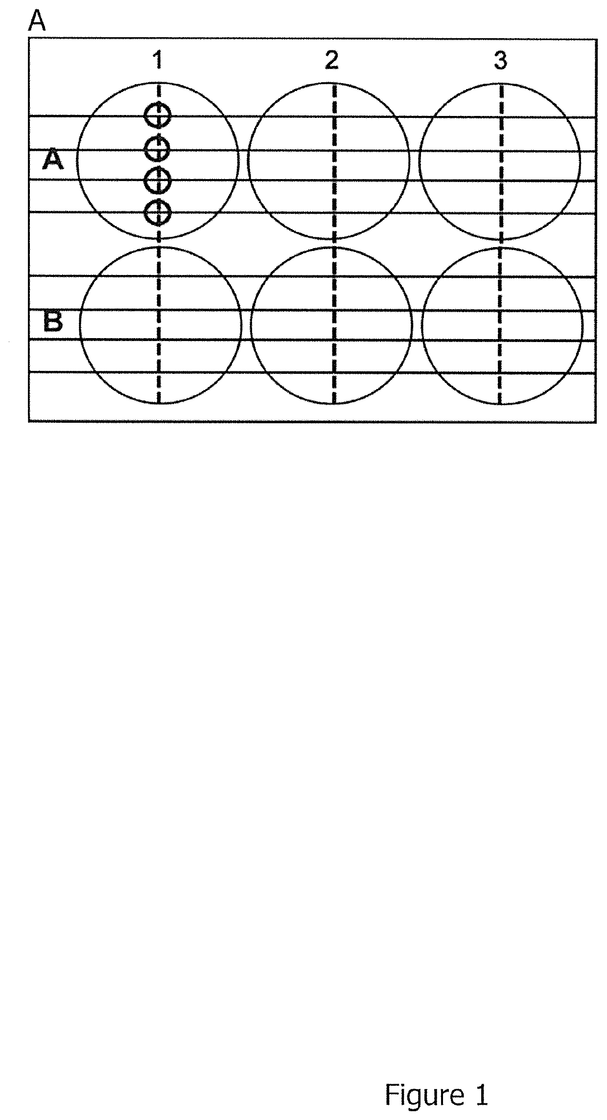
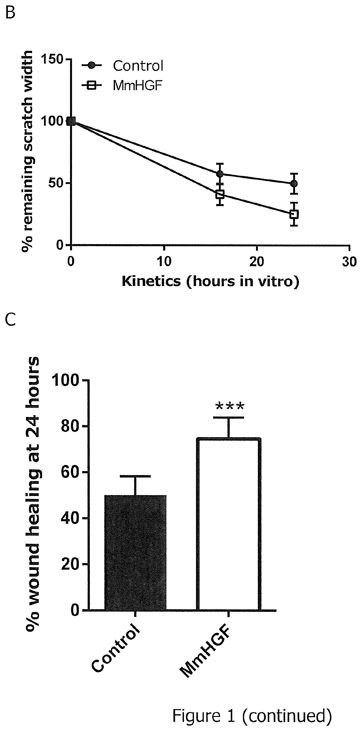
[0653]The 6-well plate was prepared for the scratch assay by drawing five horizontal lines on the bottom of the plate (FIG. 1). 1.000.000 CHO cells (Chinese Hamster Ovary cells) were seeded into the 6-well plate in full culture medium (Ham's F-12+10% Fetal Calf Serum, 1% Penicillin / Streptomycin, 1% L-Glutamine) to obtain a confluent cell monolayer. 24 hours after seeding, cells were transfected with 2 μg mouse Hepatocyte Growth Factor (MmHGF) using Lipofectamine 2000. Transfection complexes remained on the cells for three hours. The transfection medium was changed to full culture medium thereafter.
[0654]8 hours after transfection, the confluent monolayer was wounded by scratching a 100 μl pipette tip perpendicular to the lines drawn on the bottom of the 6-well plate as depicted in FIG. 1A (vertical dotted lines). The serum-containing culture medium and loosened cells were removed by washing twice with PBS (Phosphate Buffered Saline)...
example 2
ound Healing Experiments with Guinea Pigs
[0657]Experimental Setup
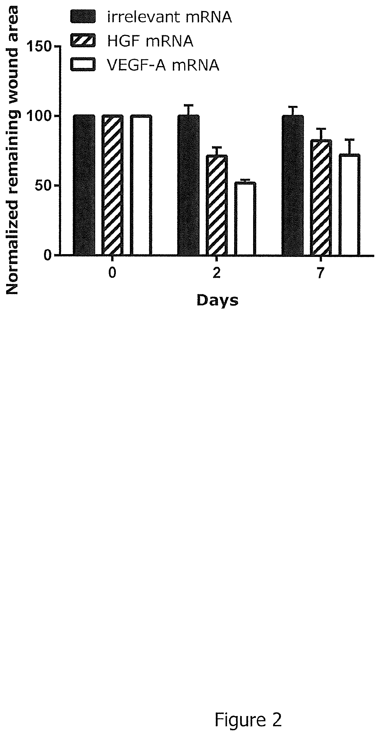
[0658]Five punch biopsies (diameter 6 mm per biopsy) were induced on the back of each guinea pig (2 animals per group, animal model: female hairless guinea pigs from Charles River, strain code 161, animal weight ˜400-450 g). On days 0, 2, 4, 7, and 9 guinea pigs were injected intradermally into the wound edges of four wounds with 4×10 μg mRNA encoding mRNAs described in Example—Table 1; one wound per animal was left untreated for comparison. Injection sites per wound were rotated by 45° on alternating injection days. On days 0, 2, 4, 7, 9 and on the day of termination day 10, wounds were photographed to allow for quantitation of wound closure. Wound area on day 0 at induction of wounds was set to 100%. On day of termination, skin biopsies were extracted, fixed in 4% paraformaldehyde, embedded in paraffin and sectioned for histological analysis. Parameters analysed on histological sections stained with Hematoxylin and E...
example 3
ound Healing Experiments with Diabetic Mice
[0665]Experimental Setup
[0666]Two punch biopsies (diameter 1 cm per biopsy) were induced on the back of each diabetic mouse (db / db mouse strain) (10 animals per group). A silicon ring was adjusted around the wound to prevent wound closure by muscle contraction. On days 0, 3, 6, 9, 12, 15, and 18 mice were injected intradermally into the wound edges of four wounds with 4×10 μg mRNA encoding mRNAs described in Example—Table 2.
Example-Table 2: Study design.# ofTreatment (i.d.) days SEQ db / db0, 3, 6, 9, 12, 15, 18ID NOGroupmiceHarvest Day 21FormulationDesign(RNA)110Buffer onlyRiLa——210MMP1RiLaDesign56323310ColG / HRiLaDesign56325 + 6329
[0667]The efficacy of mRNA treatment is assessed by its ability to enhance wound closure. For evaluation of therapeutic efficacy, 40 μg of RNA (diluted in Ringer Lactate solution) are injected into the animals (n=10) intradermally (i.d.) on days 0, 3, 6, 9, 12, 15, 18 (4×10 μg at four injection sites into the wound...
PUM
| Property | Measurement | Unit |
|---|---|---|
| Molar density | aaaaa | aaaaa |
| Molar density | aaaaa | aaaaa |
| Molar density | aaaaa | aaaaa |
Abstract
Description
Claims
Application Information