
Tissue specific promoters and transgenic mouse for the screening of pharmaceuticals

a tissue specific promoter and mouse technology, applied in the field of tissue specific promoters and transgenic mice, can solve the problems of difficult determination, high risk of repeated damage, and animals treated with chemicals that exhibit a multitude of genetic and metabolic alterations, and achieve the effect of restoring the activity of hinv promoters

Inactive Publication Date: 2001-11-06
CASE WESTERN RESERVE UNIV
View PDF4 Cites 34 Cited by
  • Summary
  • Abstract
  • Description
  • Claims
  • Application Information

AI Technical Summary

Benefits of technology

DNA sequence analysis indicates that the DRR contains binding sites for several transcription factors, in including functionally important Sp1 and AP1 sites (Welter, et al. "Fos-related antigen (Fra-1), junB, and junD activate human involucrin promoter transcription by binding to proximal and distal AP1 sites to mediate phorbol ester effects on promoter activity" J. Biol. Chem. 270:12614-12622, 1995; Banks, et al. " Characterization of human involucrin promoter distal regulatory region transcriptional activator elements-a role for Sp1 and AP1 binding sites" Biochem. J. 331:61-68, 1998; Efimova, et a

Problems solved by technology

The location of the epithelium as the lining of tissue surfaces in the body places it at a particularly high risk for repeated damage from a variety of agents in the environment.
However, one drawback to such a model is that animals treated with chemicals exhibit a multitude of genetic and metabolic alterations.
The multiplicity of genetic and metabolic changes makes it difficult to determine which of this multitude of changes is causally related to the resulting disease state, and hence makes it also difficult, if not impossible, to identify candidate therapeutics which target only relevant genetic and / or metabolic lesions.
The further problems of unpredictability and variability of genetic and metabolic changes in response to chemical treatment make such animals poor models for the evaluation of therapeutics.
Furthermore, because the development of a single cancer phenotype may be caused by more than one genetic alteration, even those cancers for which there is available a transgenic animal model having a defined genetic lesion, such a single transgenic animal model is potentially of limited use in comprehensive screening of therapeutics.

Method used


Image

  • Tissue specific promoters and transgenic mouse for the screening of pharmaceuticals

Examples


example 1

Generation of Transgenic Mice

A. Construction of hINV transgenes.

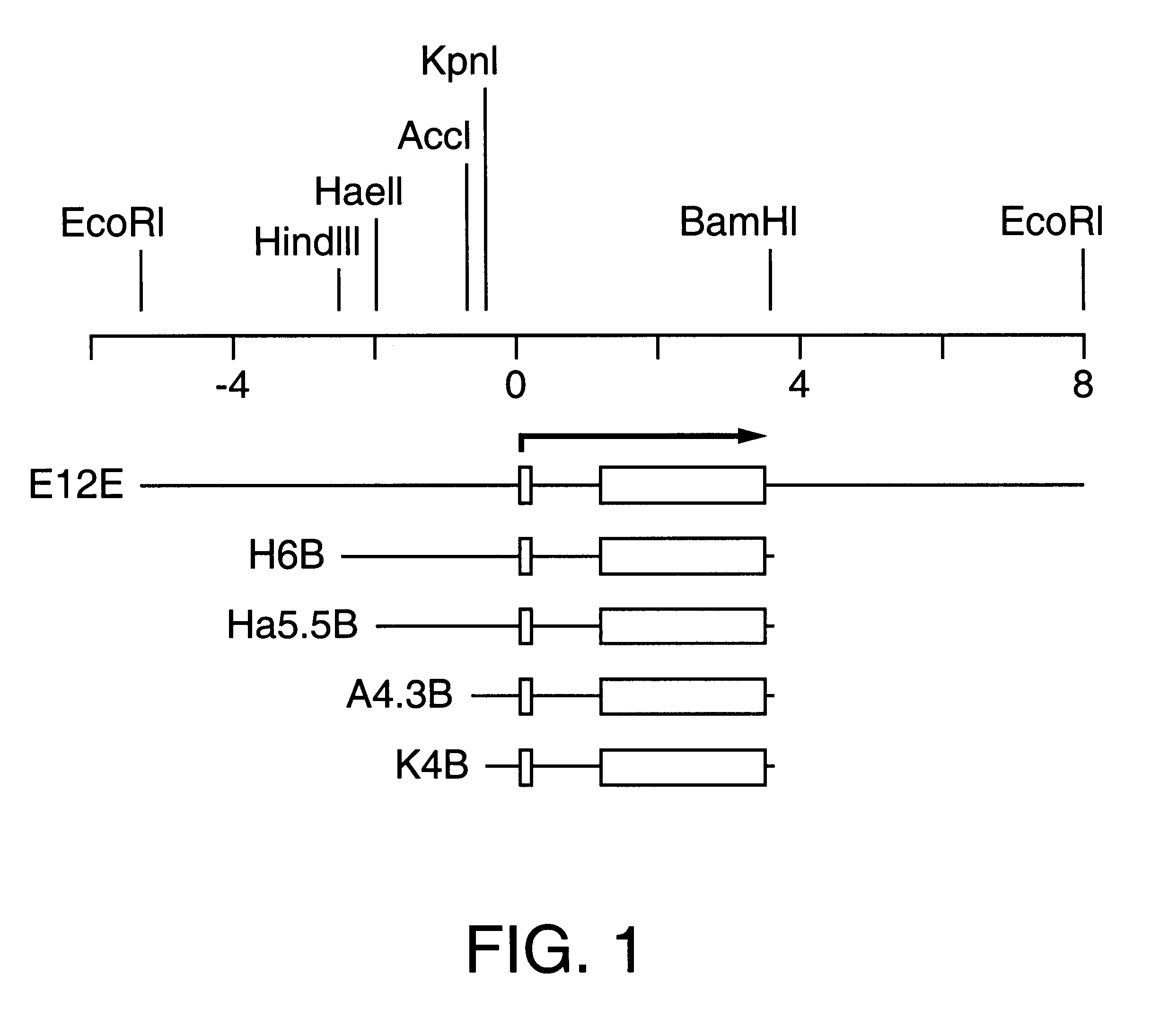
E13E was constructed by EcoR1 digestion of .lambda. phage, Charon 4A .lambda.I-3 (Eckert and Green "Structure and evolution of the human involucrin gene" Cell 46:583-589, 1986). A 13-kb EcoR1 fragment was then subcloned into pBKS (+) to yield pBKS-E13E. The EcoR1 insert from this plasmid is shown in FIG. 3. The H6B transgene is a 6-kb HindIII / BamHI fragment that was derived by restricting Charon 4A .lambda.I-3 with HindIII / BamHI and subcloning the resulting 6-kb fragment into HindIII / BamHI-restricted pSP64 to yield pS64.lambda.I-3 H6B (Eckert and Green "Structure and evolution of the human involucrin gene" Cell 46:583-589, 1986). Promoter deleted transgenes were constructed by taking advantage of unique restriction sites located upstream of the transcription start site. Consequently, the Ha5.5B transgene was generated by digesting pSP64.lambda.I-3 H6B with HaeII / BamHI and isolating the HaeIIBamHI. Likewise, the A4.3B an...

example 2

A 520-bp Segment of the hINV Upstream Regulatory Region is Required for hINV Expression in Epidermis.

A. Detection of hINV protein expression.

To detect expression of the hINV protein in mice, expression of the hINV protein by immunoblot of whole cell extracts was assayed in epidermis and kidney as described proviously (Crish, et al., "Tissue-specific and differentiation-appropriate expression of the human involucrin gene in transgenic mice: an abnormal epidermal phenotype" Differentiation 53:191-200, 1993). Briefly, to detect hINV expression in mouse tissues, total protein extracts were prepared from tissue samples in Laemmli sample buffer, electrophoresed on acrylamide gels, and transferred to nitrocellulose for immunoblot. The blot was incubated with a primary antibody prepared against human involucrin, diluted 1:8000 as described previously (Crish, et al., "Tissue-specific and differentiation-appropriate expression of the human involucrin gene in transgenic mice: an abnormal epide...

example 3

Differentiation Appropriate Expression of the hINV Transgene

We used immunological techniques to evaluate the differentiation-dependence of expression (FIG. 4). hINV was detected in the upper spinous and granular layers in footpad epidermis (EP1, left hand column) in E13E and H6B mice but no expression was detected in the basal layer (arrowheads). Suprabasal expression was also observed in the ectocervical epithelium (EC, right hand column) in these mice. In contrast, no expression was observed in epithelium or epidermis of transgenic strains Ha5.5B or K4B (FIG. 4), and no expression was observed in nontransgenic mice (not shown). FIG. 5 shows transgenic expression in the kidney of K4B mice. In this, and all other lines, expression in kidney was confined to the epithelia lining the distal convoluted tubule in transgenic lines.

The results discussed in Examples 3 and 4 suggest the regulatory elements are localized within the -2473 / -1953 segment. To identify the sequence of these regula...


PUM

PropertyMeasurementUnit
Volumeaaaaaaaaaa
Nucleic acid sequenceaaaaaaaaaa

Abstract

The present invention provides human involucrin (hINV) sequences having tissue specific and cell type specific promoter activity. The sequences provided herein direct expression to suprabasal cells of stratifying epithelia. The invention further provides methods for the production of transgenic animals which contain a hINV promoter sequence which directs the expression of human papillomavirus 16 oncogenes (or other oncogenes). These animals display cervical and epidermal hyperplasias as well as cancer of the trachea, esophagus, colon, epidermis, anus / rectum, lymph nodes, spleen and lung. The animals of the invention provide a useful model for screening potential anti-neoplastic compounds, carcinogens, and co-carcinogens for a number of cancers.

Description

The present invention pertains to the identification and characterization of a nucleic acid sequence of the human involucrin gene which targets expression of any desired nucleic acid sequence to specific tissues and specific cells. In particular, this invention relates to nucleic acid sequences which target expression of nucleic acid sequences to suprabasal cells in stratifying squamous epithelial tissue and to uroepithelial cells. In another aspect, this invention pertains to transgenic animals which exhibit certain cancers and hyperplasias. In yet another aspect, this invention pertains to methods of screening for therapeutics for epithelial neoplasia.Diseases of epithelial cells are the single most common cause of morbidity and mortality of humans. Foremost among these diseases is cancer. Other diseases which are epithelial in origin include, for example, blistering disease (e.g., epidermolytic hyperkeratosis, and Dowling-Meara disease) proliferative disease (e.g., psoriasis, epi...

Claims


Application Information

Patent Timeline
IPC IPC(8): C07H21/02C07H21/00C12N5/00C12N15/00G01N33/00A01K67/027A01K67/033C07H21/04C07K14/82C12N15/63C12N15/85
CPCA01K67/0275C07K14/82C12N15/8509A01K2217/05A01K2227/105A01K2267/0306C12N2830/008
Inventor ECKERT, RICHARD L.CRISH, JAMES F.
Owner CASE WESTERN RESERVE UNIV
Learn More