Method and apparatus for protein manipulation

a protein and apparatus technology, applied in the field of protein manipulation, can solve the problems of low quantitative recovery, difficulty in recovering, and small quantity of these peptides, and achieve the effect of reducing the quantity of starting material, and facilitating the production of large quantities of antibodies

Inactive Publication Date: 2003-11-18
ANDERSON FORSCHUNG GROUP
View PDF30 Cites 117 Cited by
  • Summary
  • Abstract
  • Description
  • Claims
  • Application Information

AI Technical Summary

Benefits of technology

Recovery of denatured proteins may present a problem if recovery is based on the protein being in its native state. Some antibody epitopes, binding to cofactors, associated proteins and substrates may be compromised until the protein is renatured. One advantage of the present invention is the recovery of proteins on beads by means of an interaction that is not specific to a native protein structure (e.g., via biotinylation or hydrophobic attraction), which is operable regardless of whether the protein was denatured or not. Likewise, the bead may provide a scaffolding from which renaturation can begin without concern for whether the denatured protein is soluble, and the immobilized protein is prevented from interacting with other protein molecules until after it is renatured. By omitting a resolubility step, at least one problem inherent to denatured proteins is avoided. Thus, the renaturation aspect of the present invention is broadly applicable to renaturing proteins regardless of source and particularly for inclusion bodies and proteins denatured during handling and purification. After each renaturation, one may test for enzymatic activity or receptor binding and comparing that to the natural form as a measure of the effects of renaturation.
is the recovery of proteins on beads by means of an interaction that is not specific to a native protein structure (e.g., via biotinylation or hydrophobic attraction), which is operable regardless of whether the protein was denatured or not. Likewise, the bead may provide a scaffolding from which renaturation can begin without concern for whether the denatured protein is soluble, and the immobilized protein is prevented from interacting with other protein molecules until after it is renatured. By omitting a resolubility step, at least one problem inherent to denatured proteins is avoided. Thus, the renaturation aspect of the present invention is broadly applicable to renaturing proteins regardless of source and particularly for inclusion bodies and proteins denatured during handling and purification. After each renaturation, one may test for enzymatic activity or receptor binding and comparing that to the natural form as a measure of the effects of renaturation.
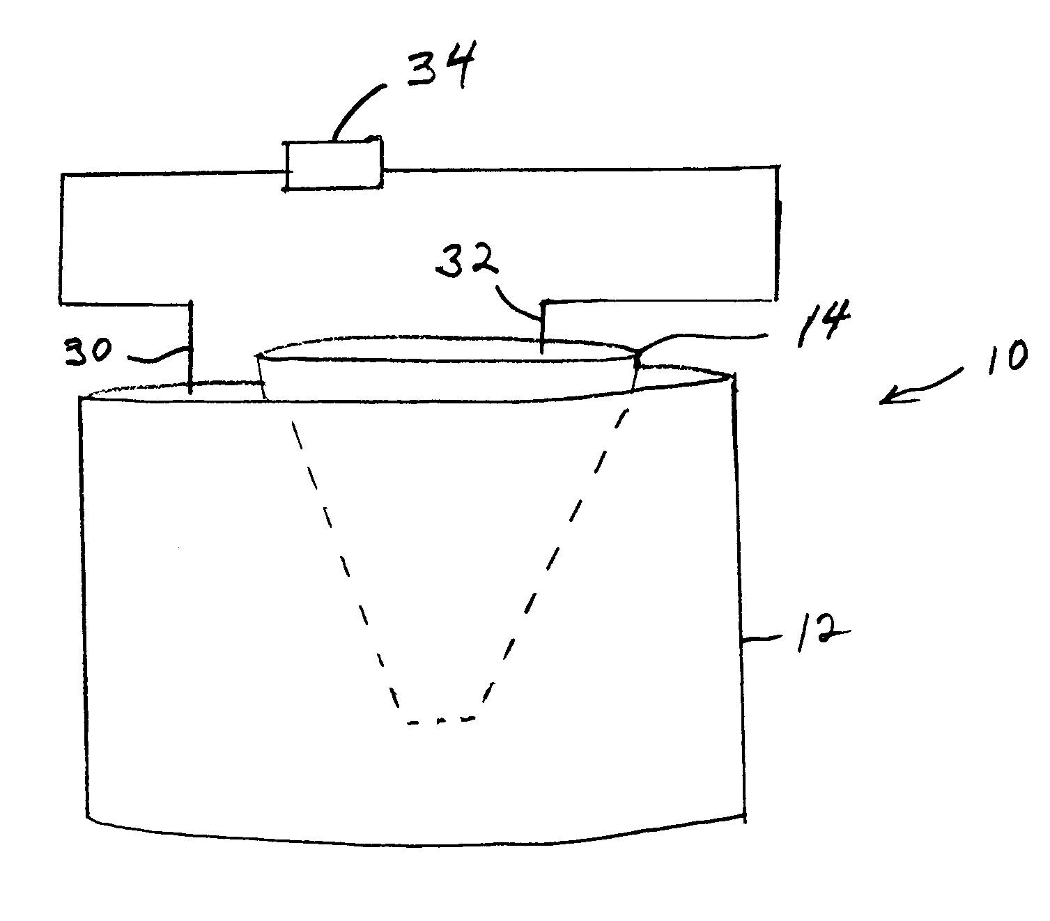
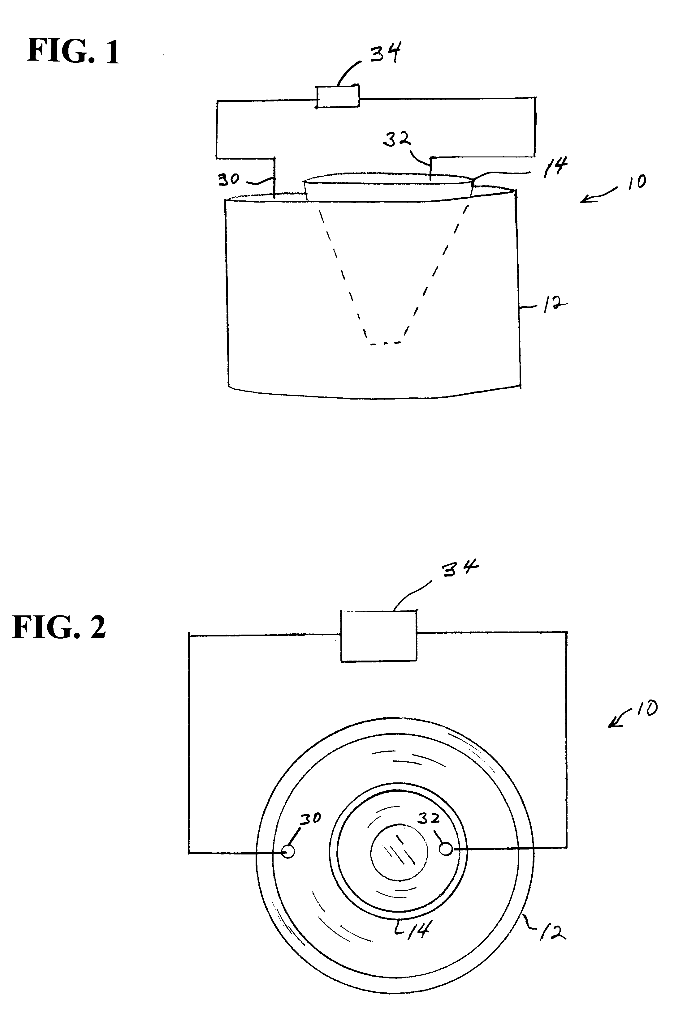
In another embodiment of the electroelution system, the entire 2DE gel, which may be pretreated with trypsin or other peptide treating agent, is placed on a thin layer of coated magnetically responsive beads. More preferred is for a thin layer of coated magnetically responsive beads to be placed on top of the 2DE gel. The entire system is then electrophoresed to electroelute the proteins out of the gel into the layer where they are adsorbed. The region of beads containing each desired protein of interest is then removed from the bead / gel construct preferably by a punch, scoop or the like to remove a small region of beads. A magnetic probe may be used directly once the desired beads are magnetically separated from surrounding beads by a non-magnetic ring, mesh, magnetically pulled through the gel to the opposite side. The location of which beads to remove is determined by scanning the gel and using coordinates from the scan signal to a computer to control the bead removal apparatus. The gel may be scanned from the opposite side from the layer of beads or it may be scanned before the beads are ever added. Supporting the gel on a porous solid phase is recommended for proper registering of the locations of each spot as well as preventing deformation of the gel or shifting of the beads.
Contact between the beads and the gel may be enhanced by mechanically pressing, magnetically driving or centrifugally sedimenting the beads onto or into the gel. If the concentration of the protein in the spot is sufficiently high, driving coated beads into the gel and allowing diffusion and protein binding to occur may be sufficient for qualitative measurements. A mesh can be placed over the gel surface to prevent lateral movement of beads during electroelution and selective bead recovery. Since beads are then throughout the gel, including areas without any protein, proteins in spots are inhibited from diffusing from one to another as they will pass by a coated bead that should adsorb much of the protein. Once removed by cutting or driving them out of contact with the gel, they may be manipulated as any other protein coated beads of the present invention.
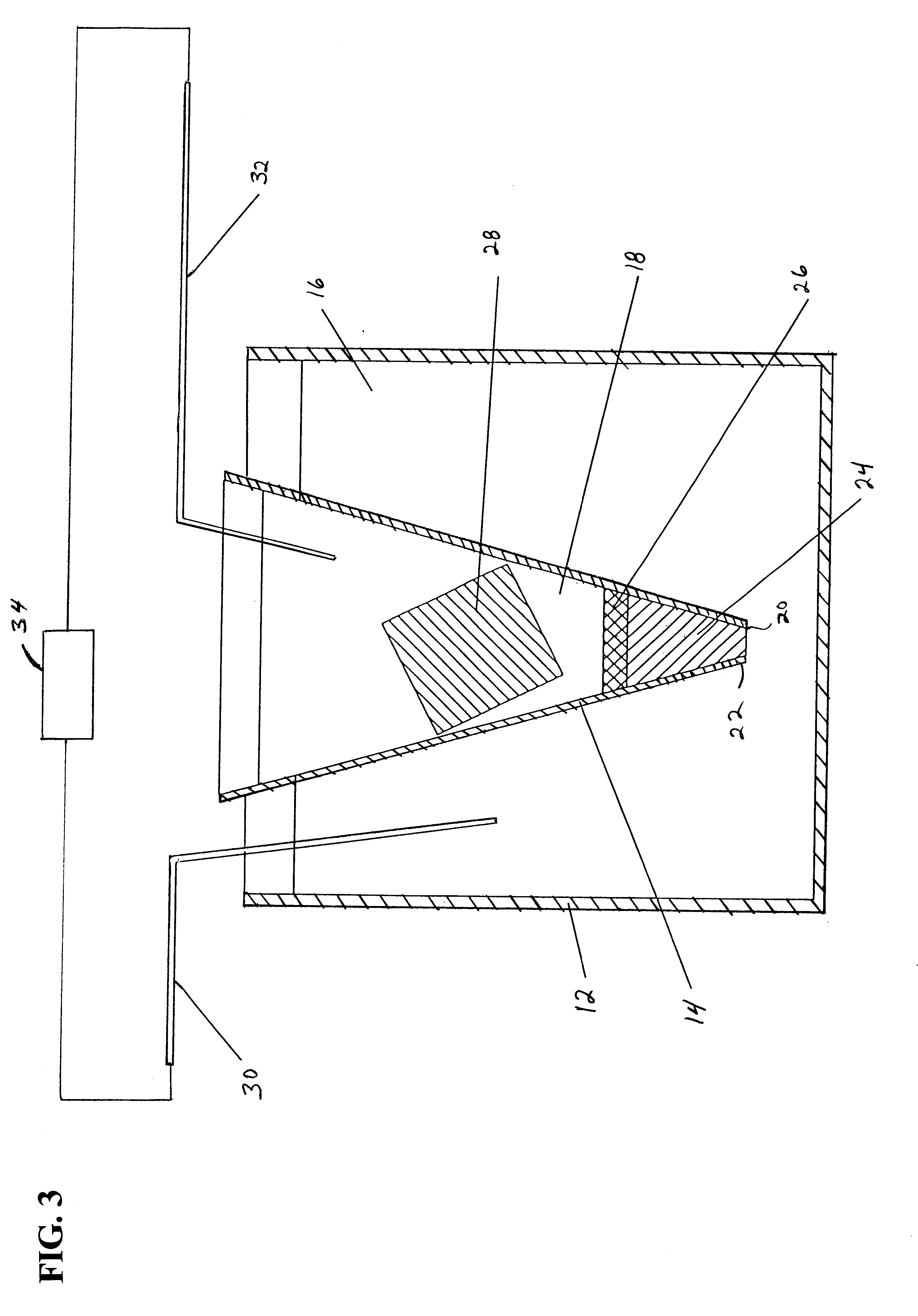
In another embodiment of the invention shown in FIG. 3A, an electroelution apparatus 11 includes vessel 13 for containing a first buffer solution 15. A conduit 17 extends from a sidewall of vessel 13 and includes a substantially U-shaped trap 19 and an open top end 21. A bed of magnetically responsive beads 23 having an affinity coating are placed in trap 19. U-shaped trap 19 defines a first leg 25 in communication with vessel 13 and is filled with first buffer solution 15. A second leg 27 of U-shaped trap 19 is filled with a second buffer solution 29. A gel core 31 with a protein spot and a first electrode 33 are placed in first buffer solution 15 in vessel 13. A second electrode 35 is placed in second buffer solution 29 and an electric current from a power source 37 is applied to electrodes 33 and 35 to electro-elute the proteins from gel core 31 toward second electrode 35 where the proteins or peptides are captured on beads 23. The beads 23 form a bed in U-shaped trap 19 to separate first buffer solution 15 from second buffer solution 29 without a gel plug as in the embodiment of FIGS. 1-3.
In an alternative embodiment, a protein spot is identified on a gel slab and buffer solutions are placed on both sides of the gel slab. In a preferred form of this embodiment, a first vessel having an open end is placed against the gel slab around the protein spot for containing a first buffer solution. A second vessel having an open end is placed against the opposite side of the protein spot for containing a second buffer solution An amount of magnetically responsive beads having an affinity coating is placed in one buffer solution contained in one of the vessels. Electrodes are placed in the respective buffer solutions and an electric current is applied to the electrodes to elute the protein directly from the gel slab and into solution for capture by the beads. The gel slab can be oriented vertically or horizontally and the vessels shaped appropriately to maintain the buffer solutions in contact with the gel.

Problems solved by technology

One of the difficulties of this practice is that the quantity of these peptides is small and they diffuse freely from the gel into the surrounding liquid and interact with the walls of the container.
This recovery step is fraught with difficulties, as the dilute peptides are exposed to plastic and other surfaces upon which they may be captured and thus lost to the investigator.
Such techniques suffer from low quantitative recovery probably due to the imperfect recovery of protein off the barrier membrane.
However, gas bubbles frequently form in the column and block the flow of current.
A drawback of the blotting approach is the difficulty of subsequently removing the blotted proteins from the membrane for subsequent manipulation in a different format.
A common problem with the elution methods is that the protein solution is very dilute, not easily recovered in a small volume and in a form that is difficult to manipulate by robotic methods.

Method used


Image

  • Method and apparatus for protein manipulation
  • Method and apparatus for protein manipulation
  • Method and apparatus for protein manipulation

Examples


example 2

g of Protein

The protein on the beads is renatured by transporting the beads through a series of buffer solutions having a concentration of about 8 M urea, 1% mercaptoethanol plus a buffer used along in increments of 0.5 M urea. Each stage involves an incubation period of 15 minutes at 37.degree. C., followed by withdrawing the beads with the probe and transporting them to the next vessel.

example 3

on of Antibodies and Proteins

The beads of Example 1 ("antigen-coated" beads, the beads of Example 2 may be used also) are next introduced into a well containing a polyclonal antiserum to human haptoglobin, and dispersed to allow binding for 1 minute. Next, the beads are removed via a probe, and washed twice in separate wells containing 200 .mu.l PBSG for 1 minute at 25.degree. C. temperature. Finally, the beads are introduced into a well containing 0.9% NaCl plus HCl pH 2.5 for 1 minute at 25.degree. C. temperature. This treatment releases the bound anti-haptoglobin antibodies into the medium.

Following release, the beads are removed with a probe. The probe is a "KingFisher" (Labsystems OY, Helsinki, Finland) (previously used for mRNA purification) modified by adding a plastic collar. 50 .mu.l of a pH 8 buffer concentrate is then pipetted into the well and mixed, raising the pH to 8. Next 10 .mu.l of a 10% suspension of paramagnetic beads coated with Protein G is introduced into the ...

examples 1 and 3

demonstrate the use of material purified by 2-D gel and immobilized on paramagnetic beads to isolate specific antibody from a polyclonal antiserum. This antibody is subsequently used to capture native antigen from serum, also determining what other subunits (in this case the heavy chains) are combined with the 2-DE gel isolated light chains in the native state.


PUM

PropertyMeasurementUnit
dissociation constantaaaaaaaaaa
pHaaaaaaaaaa

Abstract

A method and apparatus for extracting, identifying, and manipulating proteins or peptides from a solution uses paramagnetic beads having a coating with an affinity for the target component. In one embodiment, paramagnetic beads coated with C18 are used to adsorb proteins and peptides. The beads can be used to purify, immobilize and assay antibodies. By cycling the beads, many times greater molar amount of binding partner may be separated from a solution. A magnetic probe is used to capture the beads and transfer the beads to selected processing stages.

Description

The present invention relates to methods and apparatus for the manipulation of proteins and peptides by reversibly binding proteins and peptides to beads having a coating capable of binding with native proteins and peptides. More particularly, the invention is directed to methods and apparatus for capturing native proteins and peptides on magnetically responsive beads.Magnetically responsive beads or particles of small sizes are well known and have been used for a number of separation and diagnostic purposes. Small magnetic beads have been used to bind to cells and other particles in order to manipulate and separate them. See, for example, U.S. Pat. Nos. 4,230,685, 3,970,518, 5,508,164, 5,567,326 and 4,018,886.Magnetic beads have been prepared from a variety of polymers with metal particles therein, including those prepared by gelatin, colloidal metal and other agents. An example of this type of bead is disclosed in U.S. Pat. No. 4,582,622. Chemical coupling of proteins to reactive ...

Claims


Application Information

Patent Timeline
Patent Type & Authority Patents(United States)
IPC IPC(8): B03C1/02B03C1/28C07K1/36C07K1/00C07K1/22C07K1/24G01N33/553B01D43/00B01D57/00B03C1/00C07K17/08C07K17/14
CPCB03C1/286C07K1/22C07K1/24C07K1/36Y10S436/807Y10T436/25375Y10S436/806Y10S977/914Y10T436/25Y10T428/298Y10S436/805B03C1/0332B03C1/284B03C2201/18B03C2201/26B03C2201/28
Inventor ANDERSON, N. LEIGH
Owner ANDERSON FORSCHUNG GROUP
Why Eureka
Try Eureka