
Biofunctional hydroxyapatite coatings and microspheres for in-situ drug encapsulation

a technology of hydroxyapatite coating and in-situ drug encapsulation, which is applied in the direction of prosthesis, inorganic non-active ingredients, metabolic disorders, etc., can solve the problems of insufficient heat dissipation, inability to process fast setting functional gradient cement microspheres and coatings with in-situ drug encapsulation, and increase in the temperature of the cemented area

Inactive Publication Date: 2004-05-04
THE UNIV OF BRITISH COLUMBIA
View PDF7 Cites 104 Cited by
  • Summary
  • Abstract
  • Description
  • Claims
  • Application Information

AI Technical Summary

Benefits of technology

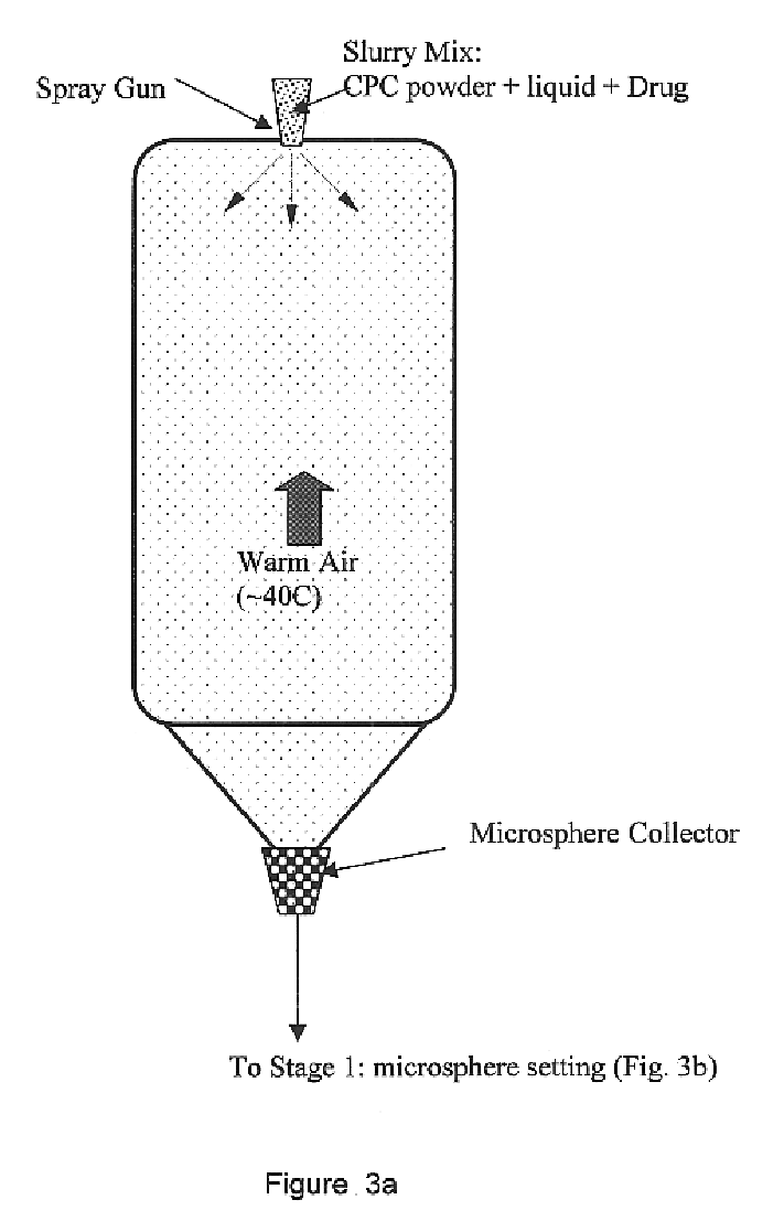
The resulting cement exhibits an apatitic phase (HA) with either an acicular grain or plate-like grain morphology, depending upon processing parameters. A nano-structured cement can also be synthesized and is used particularly for encapsulating drugs, antibiotics, proteins, and enzymes. The apatite phase develops within 1-6 h of incubation within the CPC system disclosed. Fast formation of the apatite phase is advantageous for an early biological response with the surrounding host tissue. A fast setting (2-40 min) and hardening time (less than 6 hours) benefits enormously clinical applications of the cement. Controllable crystallinity, from amorphous (easily resorbable) to highly crystalline (i.e. stable in physiological environment), of the final apatitic structure allows application-oriented customization of the cement. Fine (nano-to-submicron) starting ingredients allow final microstructure to be well controlled, allowing high strength of the final body. Nano-structure of the novel CPC offers great advantage for encapsulating drugs, as well as for their controlled release. Apatite microspheres can be easily fabricated using the novel CPC formulation, with size ranging from 10-1000 microns, using simple spray-drying equipment. Control of the setting and hardening process of the cement microspheres allows a variety of biomedical or clinical applications, e.g., injection-packing, and in-situ hardening for repair, restoration, augmentation of defective tissues, and many others.
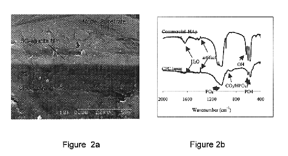
We disclose here a novel approach to achieve a multi-layer, functionally gradient HA coatings or microspheres. That is, several layers of HA coatings are deposited using different techniques, with different functions assigned to any particular layer. In particular, we describe an in-situ drug encapsulation within the functionally gradient HA coatings processed by combining elevated-temperature sol-gel processing with room-temperature precipitation processing. As a result of this protocol, a 10-30 .mu.m thick, nano-to-submicron porous HA matrix, firmly attached to a metallic substrate through a 0.6-0.8 .mu.m thin intermediate HA film is achieved. The thin sol-gel HA film (SG-HA) develops good bond with the underlaying substrate at the 400 C. processing temperature, and acts as an intermediate nucleation / bonding layer between the underlying substrate and the HA thick film matrix. The CPC-HA thick film matrix is deposited at room-temperature on the thin SG-HA film by dip coating in non-aqueous suspension of monocalcium phosphate and calcium hydroxide precursor powders. The drug material is dispersed in the suspension and initially deposited within the pores of the precursor powder film. Subsequently, a porous thick film apatite is formed under physiological environment, through dissolution-precipitation process. The thin SG-HA film acts as a seeding template allowing nucleation and growth of the thick film apatite crystals, and therefore imparts sufficient interfacial strength to the HA matrix. This room-temperature coating process, associated with an in-situ encapsulation of drugs, can be applied onto variety of metallic or non-metallic substrates, providing potential advantages in various biomedical / biochemical applications.

Problems solved by technology

Despite the fact that several techniques were investigated in the past in use of static macro-components of CaP for drug delivery, none of them allows processing of fast setting functionally gradient cement microspheres and coatings with in-situ drug encapsulation.
When setting fast (which is an important requirement in certain applications), this heat may not dissipate fast enough and temperature of cemented area increases, sometimes to a level high enough to damage, e.g. crack, the cement.
More importantly, if the cement is applied in human body, e.g. as part of an implant, this temperature increase may damage the surrounding biological constituents such as cells, proteins, and enzymes when the heat is rapidly dissipated upon setting.

Method used


Image

  • Biofunctional hydroxyapatite coatings and microspheres for in-situ drug encapsulation
  • Biofunctional hydroxyapatite coatings and microspheres for in-situ drug encapsulation
  • Biofunctional hydroxyapatite coatings and microspheres for in-situ drug encapsulation

Examples


example 1

CPC-HA / SG-HA Coatings on Stainless Steel

Stainless steel metallic substrates (316 L) were coated with a 0.6-0.8 .mu.m thin layer of apatite (SG-HA) using the recently developed sol-gel technique. Specifically, the sol-gel process for preparing a crystallized hydroxyapatite, comprises: (a) hydrolysing a phosphor precursor (phosphite) in a water based medium; (b) adding a calcium salt precursor to the medium after the phosphite has been hydrolysed to obtain a calcium phosphate gel such as a hydroxyapatite gel; (c) depositing the gel on the substrate through dip coating, and (c) calcining the film to obtain crystallized hydroxyapatite, at 400.degree. C. for 20 min. An inorganic colloidal slurry containing calcium phosphate precursor Ca(OH).sub.2 and calcium phosphate salt monocalcium phosphate anhydrate, was ball milled in ethanol. The two starting inorganic ingredients had particle size 0.3-2 .mu.m and 0.5-4 .mu.m, respectively. The initial Ca / P ratio in the slurry was kept at 1.5. As ...

example 2

CPC-HA Coatings on Stainless Steel

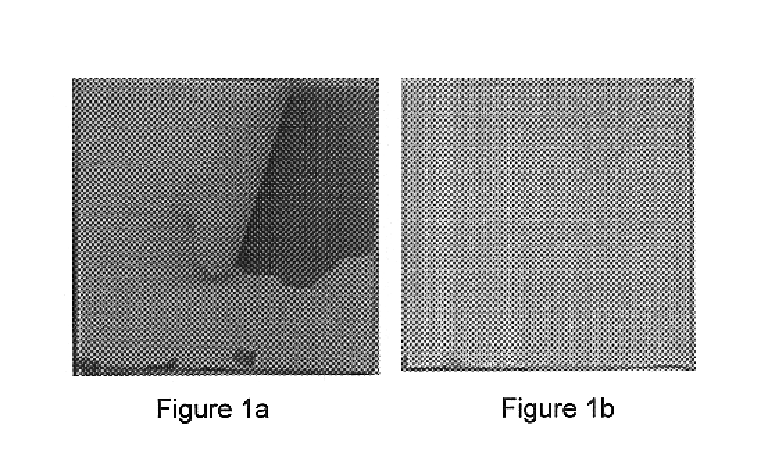
Stainless steel metallic substrates (316 L) free of intermediate SG-HA film were coated with CPC-HA, as described in Example 1. During HA incubation period, the coating spontaneously separated from the substrate, as illustrated in FIG. 1a. Obviously, the CPC-HA coating did not bond to the metallic substrate, i.e. bonding strength was zero MPa. Other properties of the coating, that is phase content and porosity, was similar to that obtained in Example 1.

example 3

BM-HA / SG-HA Coatings on Stainless Steel

Stainless steel metallic substrates (316 L) were coated with a 0.6-0.8 .mu.m thin layer of apatite (SG-HA) as described in Example 1. One group of samples was annealed at 400.degree. C. for 20 min to achieve crystalline SG-HA(C) film and another group at 375.degree. C. for 60 min to achieve amorphous SG-HA(A) film. These films were used as nucleation site for precipitation of BM-HA film. The SG-HA coated samples were immersed into "simulated body fluid" (SBF) of ionic composition (in units of mmol / l) 142 Na.sup.+, 5.0 K.sup.+, 2.5 Ca.sup.2+, 1.5 Mg.sup.2+, 103 Cl.sup.-, 25 HCO.sub.3.sup.31 , 1.4 HPO.sub.4.sup.2-, and 0.5 SO.sub.4.sup.2-. The SBF was buffered at pH 7.4 with tris(hydroxymethyl)-aminomethane and HCl. This in-vitro static deposition (i.e. the SBF was not renewed during the deposition period) at .about.24.degree. C. produced good quality, dense 3-5 .mu.m thick BM-HA film deposits on flat SG-HA substrates, as illustrated in FIG. 6. T...


PUM

PropertyMeasurementUnit
temperatureaaaaaaaaaa
temperaturesaaaaaaaaaa
diameteraaaaaaaaaa

Abstract

This invention relates to novel room-temperature process for obtaining calcium phosphate, in particular hydroxyapatite, coatings and microspheres that encapsulate drugs, proteins, genes, DNA for therapeutical use. The coatings and microspheres are designed to perform a defined biological function related to drug delivery, such as gene therapy through gene delivery. A novel method for encapsulation, and subsequent controlled release of therapeutically active agents from such biofunctional coatings and microspheres is disclosed. Such coatings and microspheres are useful for side-effects free, long-term, targeted, controlled release and delivery of drugs, proteins, DNA, and other therapeutic agents.

Description

This invention relates to novel room-temperature process for obtaining calcium phosphate, in particular hydroxyapatite, microspheres and coatings with encapsulated drugs, proteins, genes, DNA for therapeutical use. The coatings and microspheres are designed to perform a defined biological function related to drug delivery, such as gene therapy through gene delivery. A novel method for encapsulation, and subsequent controlled release of therapeutically active agents from such biofunctional coatings and microspheres is disclosed. Such coatings and microspheres are useful for side effects-free, long-term, targeted, controlled release and delivery of drugs, proteins, DNA, and other therapeutic agents.Rapid progress in the human genome project promises that diseases that could not be treated before can be curable in near future. The expectation is that the trial-and-error era of fighting illnesses by addressing the symptoms is coming to an end. Consequently, the issue of drug and gene re...

Claims


Application Information

Patent Timeline
Patent Type & Authority Patents(United States)
IPC IPC(8): A61K9/16A61K9/00A61L27/00A61L27/32A61L27/12C04B41/67C04B40/00C04B41/60C04B41/50C04B41/45C01B25/00C01B25/32C23C30/00A61K6/02A61K47/02A61K9/20A61K9/28A61K9/50A61K31/711A61K38/00A61K45/00A61K48/00A61P3/04A61P5/00A61P25/28A61P35/00A61P43/00
CPCA61K9/0024A61K9/1611A61K9/1694A61L27/12A61L27/32C01B25/32C04B40/00C04B41/009C04B41/5092C04B41/67C23C30/00C04B41/455C04B28/34C04B2111/00836A61P25/28A61P3/04A61P35/00A61P43/00A61P5/00
Inventor TROCZYNSKI, TOMASZLIU, DEAN-MOYANG, QUANZU
Owner THE UNIV OF BRITISH COLUMBIA
Learn More