Immunological detection of prions

Inactive Publication Date: 2004-07-20
UNIV ZURICH
View PDF1 Cites 79 Cited by
  • Summary
  • Abstract
  • Description
  • Claims
  • Application Information

AI Technical Summary

Benefits of technology

The pharmaceutical preparations have to be administered according to the judgment of the physician in amounts depending on the concentration of the antibodies comprised thereby and the route of administration so that a protective or curative effect is obtained. The amounts and method of administration

Problems solved by technology

In addition, no single nucleic acid capable for coding a protein has been purified until date (Riesner et al., 1993).
However, these in vitro converted PrP.sup.Sc molecules have, to date, not shown to be infectious.
However, a molecular function of PrP.sup.C could not be deduced from these findings.
Remarkably, no diagnostic method suitable for mass screening of infected tissues of cattle has been developed to date.
The drawback of this method is that the inventors claim to detect prion poypeptides that are soluble in vivo, however, it is known since a long time that the disease-associated prion protein PrP.sup.Sc is insoluble in vivo.
However, the inventors do not show in vivo soluble protease-resistant prion polypeptides which would prove their hypothesis about the existence of disease-specific prion polypeptides in CSF.
In addition, to show prion polypeptides in CSF they use an immunoblot (Western blot); this technique is not appropriate to detect naturally oc

Method used


Image

  • Immunological detection of prions
  • Immunological detection of prions
  • Immunological detection of prions

Examples


example 1.1

Method for the Preparation of Tissue Homogenates

One gram of brain, either from the thalamus, medulla or spinal cord, were homogenized with an homogenizer (Omni, USA) in 10 ml 10% sucrose, 20 mM HEPES pH 7.5, 2% sarcosyl and 5 mM EDTA. 10% homogenates were diluted 10 fold, one part of the homogenate was digested with proteinase K at 0, 10 or 100 .mu.g / ml. The probe containing 0 .mu.g / ml served as a control for the specificity of the proteinase K treatment. Probes were then further diluted (in PBS) for the ELIFA test to give a blotting concentration of 0.05% of brain homogenate. To increase the partial protease resistance of bovine PrP.sup.Sc, brain homogenates from BSE-infected and normal cattle was diluted after homogenization in 20, 40 or 80% ethanol / HEPES-sucrose buffer. Suspension of brain homogenates in ethanol was an important step and effectively stabilized the .beta.-sheet structure of the PrP.sup.Sc isoform (Oesch et al., 1994), thereby increasing its protease resistance (Ri...

example 1.2

Western Blotting

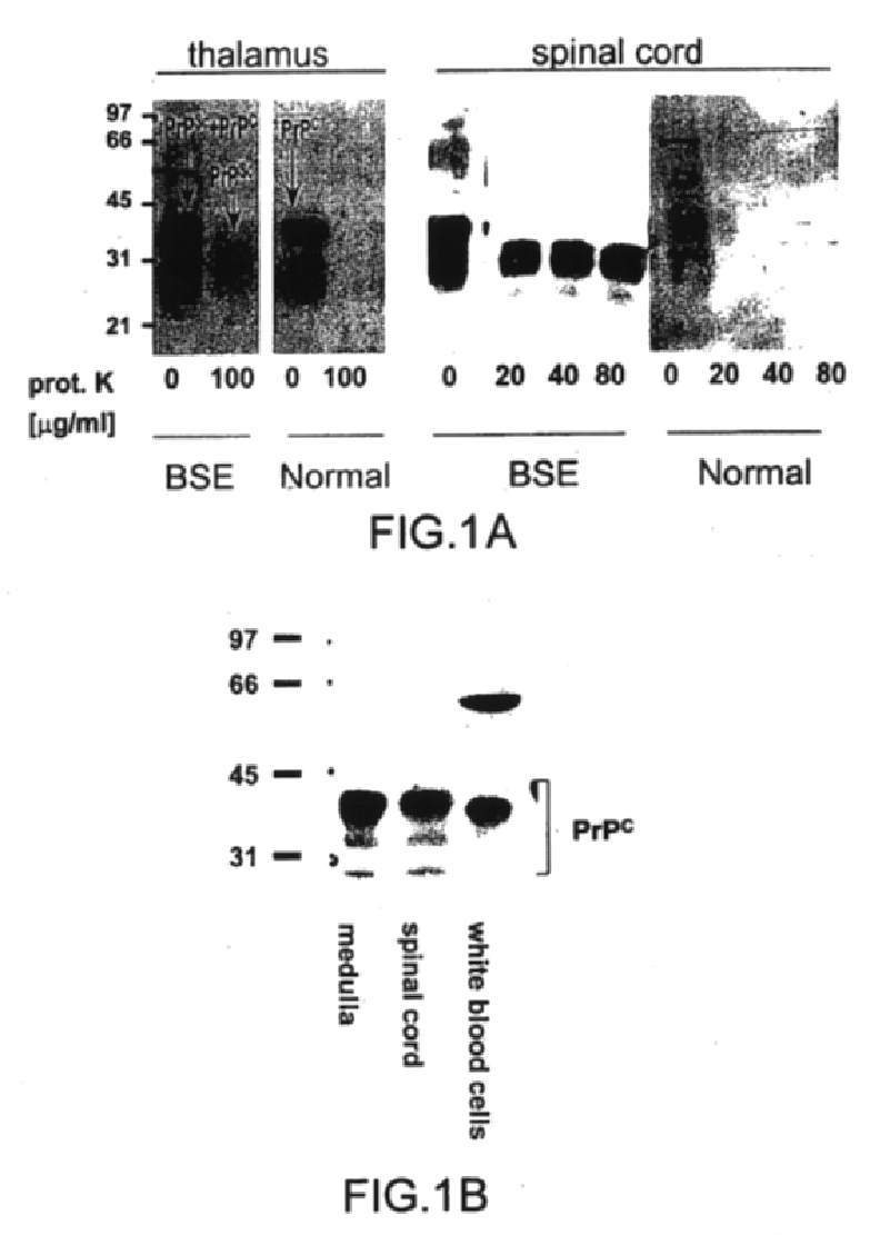
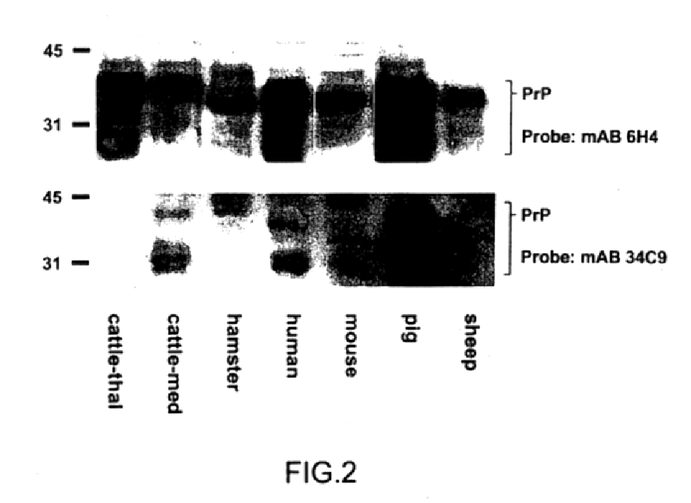
Tissue specimens were homogenized as described in example 1.1, one part protease-digested, the other not (as described above), diluted to 10% and separated by SDS polyacrylamide gel electrophoresis (SDS-PAGE) on 12% gels (Sambrook et al. 1989). Gels were then electroblotted onto 0.45 .mu.m nitrocellulose (NC) membranes, incubated with the respective monoclonal antibodies followed by a secondary anti-mouse IgG antibody coupled to peroxidase. Bound peroxidase activity was detected with a chemiluminescence kit (ECL, Amersham, USA).

Western blots developed with antibodies 6H4 or 34C9 (FIG. 1A) show the characteristic smear of bands for PrP.sup.C and bovine PrP.sup.Sc (33 to 27 kD) in undigested probes while digestion with proteinase K eliminates all of PrP.sup.C, however, leaving a 27 kD band typical of N-terminally truncated PrP.sup.Sc. The smear is due to different glycosylated forms of PrP.

The present antibodies were furthermore able to detect PrP in various tissue ext...

example 1.3

ELIFA (Enzyme Linked Immuno Filtration Assay)

The ELIFA procedure to determine quantitatively the amounts of PrP.sup.C and PrP.sup.Sc in given tissue homogenates has been described for hamster brain homogenates (Oesch et al., 1994). Blotting directly on nitrocellulose has the advantage over the conventional ELISA procedure that the poor solubility of bovine PrP.sup.Sc does not affect its immobilization on the solid phase. Blotting was performed with an ELIFA apparatus (Pierce), i.e. a peristaltic pump created a vacuum below the NC thereby sucking the contents of the wells above onto the NC in a controlled and highly reproducible way. Wells were then washed with PBS. The membrane was removed from the ELIFA apparatus, placed in a plastic tray and then incubated on a rocking table sequentially for the indicated times with the following reagents (inbetween steps, the filters were always washed 3.times. with PBS): 5% BSA / TBST (30 min), avidin (25 .mu.g / ml, 30 min); biotin (2 .mu.g / ml; 30 ...


PUM

PropertyMeasurementUnit
Solubility (mass)aaaaaaaaaa

Abstract

The presented invention relates to monoclonal antibodies useful in sensitive and specific immunological assays for the identification of prions in various tissues and body fluids, the production of such monoclonal antibodies by means of immunization of PrP<0/0 >mice by means of a new recombinant fragment of PrP and the use of the antibodies, e.g. for therapeutic and preventive treatments of humans and animals suffering from prion diseases.

Description

The present invention relates to monoclonal antibodies reacting with certain epitopes of recombinant bovine prion protein, native and denatured normal or disease-specific prion proteins in soluble or insoluble state, stable hybridoma cell lines producing these monoclonal antibodies, recombinant expression vectors for the expression of recombinant bovine prion protein, purified recombinant bovine prion protein, a test kit for the diagnosis of prion diseases, diagnostic methods for the immunological detection of prion diseases, pharmaceutical preparations for the prevention and therapy of prion diseases, a method for clearing biological material from infectious prion proteins, and methods for the production of these materials.Abbreviations used hereinbefore and hereinafter are the following:Prion diseases are transmissible neurodegenerative diseases of the central nervous system (for review see Prusiner, 1991). They can be transmitted, inherited or occur sporadically and are observed ...

Claims


Application Information

Patent Timeline
IPC IPC(8): C07K14/435C07K14/47C07K16/18A61K38/00A61K39/395C12N1/21C12N5/10C12N5/20C12N15/06C12N15/70G01N33/577G01N33/68
CPCC07K14/47C07K16/2872A61K38/00C07K2317/34
Inventor KORTH, CARSTENSTIERLI, BEATSTREGT, PETEROESCH, BRUNOMOSER, MARKUS
Owner UNIV ZURICH
Why Eureka
Try Eureka