
Time-resolved fluorescence assay for the detection of multimeric forms of A-beta 1-40

a fluorescence assay and multimeric technology, applied in the field of methods, can solve the problems of complex method of detecting peptide complexes, abnormal physiological functions at the plaque site, toxicity and death of neuronal cells, etc., and achieve the effects of reducing the amount of reagents required, reducing the cost of the assay per sample, and reducing the cost of the assay

Inactive Publication Date: 2005-01-25
PHARMACIA & UPJOHN CO
View PDF8 Cites 15 Cited by
  • Summary
  • Abstract
  • Description
  • Claims
  • Application Information

AI Technical Summary

Benefits of technology

One advantage of the methods of the present invention is that the rapid method of detecting polypeptide aggregation allows for the rapid assessment of numerous potential modulator compounds. Another advantage of the present invention is that the decreased amount of reagent required per assay reduces cost of the assay per sample.
is that the decreased amount of reagent required per assay reduces cost of the assay per sample.
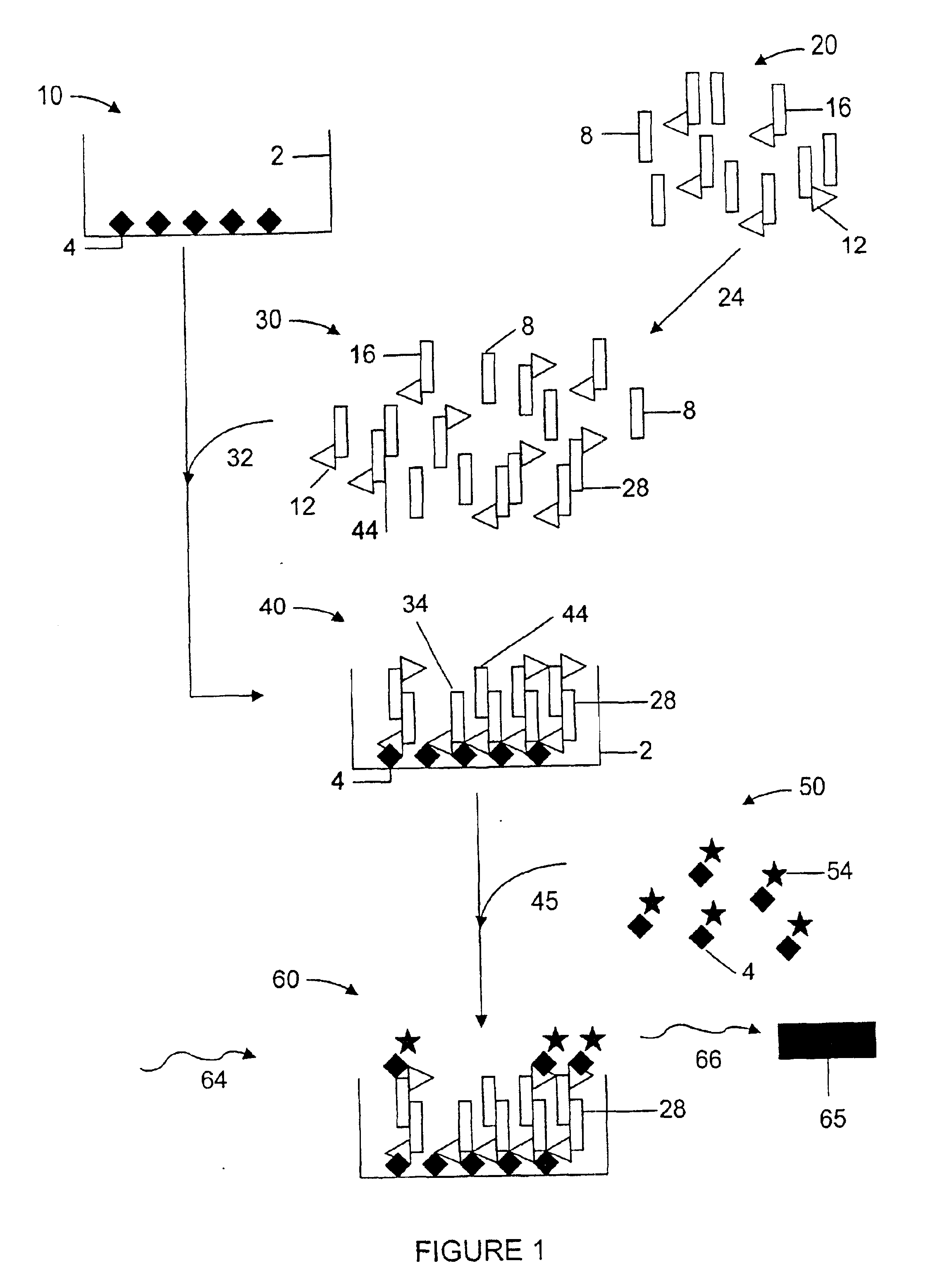
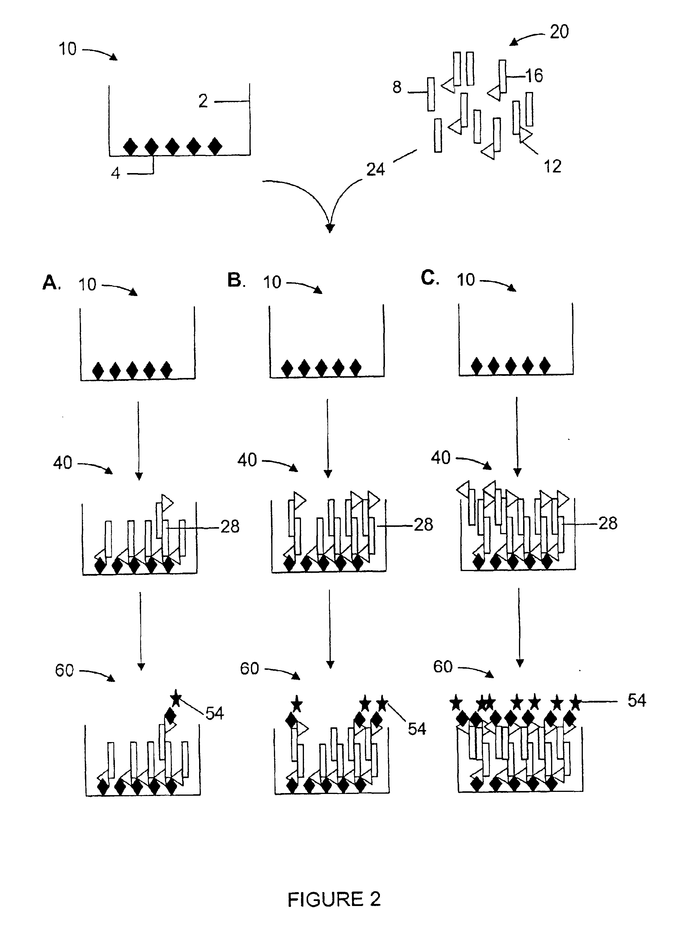
An exemplary method of the invention may be carried out as follows and as schematically illustrated in FIG. 1. As depicted in Panel 10, a 96 well polystyrene dish substrate 2 is coated with a first binding partner 4, such as streptavidin, which is provided by incubating the substrate 2 with a solution containing the first binding partner 4, thereby resulting in the coated plate of Panel 10.
As shown in Panel 20, a composition of polypeptide monomers 8, e.g. β-amyloid 1-40 monomers, is provided, e.g. by making a solution in which some of the monomers 16 are linked to a second binding partner 12, an example of which is biotin, which binds to streptavidin. The monomer composition is first incubated 24 in order to allow the monomers to form polypeptide aggregates 28,44 as shown in Panel 30. The incubation 24 is carried out in the presence or absence of a test compound which may modulate, i.e. inhibit or promote, monomer aggregation.
As shown in Panel 30, after the incubation, the polypeptide composition may comprise a variety of species such as polypeptide monomers 8, biotin-linked polypeptide monomers 16 and polypeptide aggregates 28,44. This incubated polypeptide composition is then contacted 32 with the coated plate depicted in 10 comprising the substrate 2 containing the streptavidin first binding partner 4. As depicted in Panel 40, polypeptide species that include at least one biotin molecule will bind to the coated plate of 10 during the contacting step. Molecules from the polypeptide composition bound to the coated plate from Panel 10 may comprise several species of molecules: single polypeptide monomers linked only to the biotin second binding partner 34 and comprising only one streptavidin binding site; aggregated molecules comprising biotinylated polypeptide monomers in association with monomers lacking the second binding partner 44, and therefore also comprising only one stretpavidin binding site; and aggregated monomers comprising biotinylated polypeptide monomers in association with other biotinylated monomers 28, and thus comprising two or more sites for streptavidin 4 binding. Multiple streptavidin binding sites in an aggregated molecule 28 allows for at least one β-amyloid 1-40-biotin to attach to the substrate coated plate 10, while the unoccupied biotin 12 on an associated β-amyloid 1-40-biotin molecule allows for detection of aggregated monomers 28 using a streptavidin first binding partner 4 linked to a detectable label 54, such as Europium3+. Monomers 34 that attach to the substrate lack any free biotin.
Polypeptide aggregation is measured by detecting polypeptide aggregates 28 that have bound to the coated plate of 10. The bound aggregated molecules are detected by contacting 45 a detecting agent in 50 (containing the first binding partner 4 linked to a detectable label 54) to the substrate coated plate of 10. As shown in Panel 60, the detecting agent 50 only attaches to substrate-bound, aggregated polypeptides which comprise two or more biotinylated polypeptide monomers 28. Using techniques common in the art, the substrate-bound streptavidin-Eu3+50 is detected by excitation of the Eu3+ label 54 at an appropriate wavelength 64 and subsequent measurement with a detector 65 of Eu3+ fluorescence emitted 66, in units of light absorbance. The degree of Europium3+54 excitation measured reflects the amount of detecting agent 50 bound, and thereby correlates with the amount of monomer aggregation occurring during the incubation step 24. Test compounds which result in altered levels of Eu3+54 detection when compared to control compounds are considered modulators of polypeptide monomer aggregation.

Problems solved by technology

A number of serious diseases affecting the human population can be closely associated with the improper aggregation of polypeptide fragments and characterized by aggregate deposition in lesions or plaques often resulting in abnormal physiological function at the plaque site.
However, the common denominator in all of these mutations is the formation of protein, or senile plaques derived from a cleavage fragment, designated amyloid beta (Aβ), of the APP molecule which is deposited in the brain of affected individuals, and results in toxicity and death of neuronal cells.
While the TRAMS assay may measure the initial steps in protein aggregation, it is a very complex method of detecting peptide complexes which requires equipment not readily available and involves difficult interpretation of the data.
To carry out this protocol, large amounts of β1-40 are needed per assay and each assay requires a large amount of time to complete.
Thus, this type of assay does not have the high throughput ability needed in the pharmaceutical industry, as well as using potentially hazardous reagents to carry out the protocol.
While this method is useful in detecting aggregation of β-amyloid peptides, the requirement for peptide-specific monoclonal antibodies limits this assay to availability of the particular antibody and the specificity and binding affinity of the antibody for the peptide.
This assay, however, does not address whether a test compound affects the prevention of aggregation, or the nucleation event, but rather only provides compounds which modulate continuing polypeptide aggregation.

Method used


Image

  • Time-resolved fluorescence assay for the detection of multimeric forms of A-beta 1-40
  • Time-resolved fluorescence assay for the detection of multimeric forms of A-beta 1-40

Examples


example 1

Time Resolved Fluorescence Assay For β-Amyloid Polypeptide Aggregation

The following example is an embodiment of the invention which demonstrates a method for identifying modulators of β-amyloid polypeptide aggregation. β-amyloid polypeptide monomers in solution in vitro are pre-disposed toward aggregation of the peptide monomers into larger, multimeric molecules. Aggregation of the β-amyloid monomers in vivo is a primary contributor to neuronal damage in Alzheimer's Disease. The present invention allows for a sensitive in vitro analysis of β-amyloid aggregation and the effects of outside compounds or test agents on the extent of β-amyloid peptide aggregate formation.

Beta-amyloid 1-40 (Aβ) and biotin-labeled β-amyloid 1-40 peptides were purchased from Biosource (Camarillo, Calif.). β-amyloid and biotinylated amyloid stock solutions, diluted in 0.1% acetic acid and corrected for 80% peptide content, were diluted to 83 μg / ml and 42 μg / ml, respectively, in reaction buffer (Dulbecco's PB...

example 2

Comparison of TRF Assay to Standard ELISA Potocol

For the β-amyloid aggregation ELISA protocol (Howlett et al., supra), 96 uL diluted β amyloid solution (51 μg / ml) is added to a 96-well polystyrene plate in the presence of and absence of 2 μL of an aggregation inhibitor test compound diluted in DMSO. The Aβ is incubated overnight to allow for aggregation and an aliquot transferred to another 96-well polystyrene plate, precoated with anti-Aβ antibody (6E10, Senetek, Napa, Calif.) overnight, washed, and blocked with Dulbecco's PBS / 1% BSA solution. After 3-4 hours, the plates were washed and a secondary anti-Aβ antibody demonstrating the same specificity as the coating antibody but biotinylated (6E10-bio) was added to the plate. The secondary, biotinylated antibody was bound with 200 μL Eu3+-streptavidin (100 ng / ml) in DELFIA assay buffer, the excess washed off, and the plate developed in DELFIA enhancement solution and read on a TRF plate reader as outlined above.

Analysis of the absorb...

example 3

Improved Rate of Detection of Modulators of β-amyloid Nucleation and Aggregation

Coupling of the TRF-Assay described above with a turbidity, or optical density, assay can vastly improve the ability to detect modulators of β-amyloid nucleation and aggregation. A turbidity assay measures the onset of aggregation, or nucleation, of aggregating proteins. A nucleation event is characterized by a considerable lag time in the appearance of aggregates precipitating out of assay solution and giving off an optical density (OD) reading, followed by a rapid increase in OD due to acceleration of aggregation.

A test compound that has been determined to modulate β-amyloid aggregation in the TRF assay can also be assayed in an optical density assay (Findeis et al, Biochemistry 38: 6791-6800. 1999; and WO98 / 08868) to verify if the compound is involved with nucleation of β-amyloid aggregates, or if the compound only inhibits β-amyloid aggregation after the initial seeding of aggregates has occurred.

For...


PUM

PropertyMeasurementUnit
Aggregationaaaaaaaaaa

Abstract

The present invention provides for a polypeptide aggregation screening assay for the purpose of detecting modulators of polypeptide aggregation, which can provide for new therapies in pathologic states associated with polypeptide aggregation, especially considered is Alzheimer's Disease.

Description

FIELD OF THE INVENTIONThe present invention relates to methods of detection of modulators of polypeptide aggregation observed in many human disease states.BACKGROUND OF THE INVENTIONA number of serious diseases affecting the human population can be closely associated with the improper aggregation of polypeptide fragments and characterized by aggregate deposition in lesions or plaques often resulting in abnormal physiological function at the plaque site. For instance, a stretch of polyglutamine repeats in a particular protein has been proposed as the cause for many neurological disorders including Huntington's disease and spinal bulbar muscular atrophy, while aggregation of other unrelated proteins are cited as the causes of prion disease (PrP aggregation), Parkinson's disease and amyotrophic lateral sclerosis (ALS) (α-synuclein aggregation), dialysis-related amyloidosis (β-2 microglobulin aggregation), corneal dystrophy (kerato-epithelial deposits), and aggregation of islet amyloid ...

Claims


Application Information

Patent Timeline
IPC IPC(8): A61K38/17A61K38/16G01N33/68A61P25/00A61P25/28A61P43/00
CPCA61K38/16G01N33/6896G01N33/68A61K38/1709A61P25/00A61P25/28A61P43/00
Inventor PEACH, MATTHEW L.LABORDE, ALICE L.
Owner PHARMACIA & UPJOHN CO
Learn More