
Method and apparatus to control microbubble destruction during contrast-enhanced ultrasound imaging, and uses therefor

a technology of contrast agent and ultrasound imaging, applied in the field of contrast agent-enhanced ultrasound imaging, can solve the problems of inability to use doppler ultrasound to measure the flow through small vessels, uncontrolled angiogenesis, and limited use of doppler ultrasound to measure the flow in small vessels. , to achieve the effect of increasing the brightness of ultrasound and doppler images, reducing amplitude, and increasing image brightness

Inactive Publication Date: 2005-02-22
THE TRUSTEES OF THE UNIV OF PENNSYLVANIA
View PDF2 Cites 32 Cited by
  • Summary
  • Abstract
  • Description
  • Claims
  • Application Information

AI Technical Summary

Benefits of technology

A particular advantage of the present invention is that the method relies upon controlled use of ultrasound imaging, which is noninvasive. It can be used repeatedly on a patient, or in a specific area or tissue, with no known side effects. Yet, unlike other conventional methods used commonly to measure superficial blood flow, ultrasound can be used to visualize blood vessels in organs seated deep inside the body. The dosage of the contrast-enhancing agent need not be changed from that which is routinely used for ultrasound imaging, and any known agent may be used that comprises gas-filled microbubbles. The importance of the invention lies not in the contrast enhancing agent selected, so long as it provides microbubbles as a tracer, it is based upon the systematic and controlled exposure of the microbubbles to the ultrasound waves.
is that the method relies upon controlled use of ultrasound imaging, which is noninvasive. It can be used repeatedly on a patient, or in a specific area or tissue, with no known side effects. Yet, unlike other conventional methods used commonly to measure superficial blood flow, ultrasound can be used to visualize blood vessels in organs seated deep inside the body. The dosage of the contrast-enhancing agent need not be changed from that which is routinely used for ultrasound imaging, and any known agent may be used that comprises gas-filled microbubbles. The importance of the invention lies not in the contrast enhancing agent selected, so long as it provides microbubbles as a tracer, it is based upon the systematic and controlled exposure of the microbubbles to the ultrasound waves.
Quantitative Framework for Discounting Bubble Decay and Controlling Fluid Flow
FIG. 1 shows the enhancement of ultrasound and Doppler images with time. Two cases are represented in the figure. The top curve shows a true or idealized enhancement of the images, wherein no bubbles are destroyed. The lower curve shows the measured or observed enhancement. As shown, the measured curve is significantly diminished in amplitude (compared to the idealized curve) due to bubble destruction by the ultrasound pulses.
In both cases, after the contrast agent is injected, there is an increase in image brightness (or color level in Doppler images) due to the inflow of microbubble-scatterers to the imaged organ. This change is rapid and during this interval, the effects of microbubble decay are minimal.
Inflow of the contrast agent is followed by clearance (outflow) of the agent from the organ. Clearance of the contrast agent from the organ occurs over a relatively longer time and follows an inverse exponential function. During this period the microbubbles also collapse and diminish the image enhancement as compared with what would have occurred from outflow of the contrast agent alone. Thus, the observed image enhancement is reduced, as illustrated by the lower curve of FIG. 1.

Problems solved by technology

When these tight controls are breached, uncontrolled angiogenesis ensues.
However, it is not possible to use Doppler ultrasound to measure the flow through small or newly developed angiogenic vessels.
This is, in part, due to the weakness of the signal from the blood moving through such tiny vessels.
When these agents are injected, the ultrasound images are enhanced over time, but their use to date for measuring the flow in small vessels has been very limited because the microbubbles are unstable at ambient pressure, causing the bubbles to collapse.
However, it has been observed that the microbubbles that produce enhancement are destroyed by the pulses of ultrasound used for constructing images (Porter et al., Circulation 92:2391-2395 (1995)).
Repeated measurements are possible because the microbubbles are rapidly cleared from circulation following administration, but image enhancement is short-lived.
However, while that approach prolongs the time of enhancement, it does not provide quantitative measurements of blood flow or of any other related parameters.
Nevertheless, the method of Wei et al. has several limitations.
During this interval, there is considerable motion, making it difficult to keep the plane fixed during data acquisition.
Moreover, the method also requires image matching for background subtraction, which in view of the motion can be cumbersome and difficult to implement.
Furthermore, the method fails to take into consideration the attenuation effect of the overlying tissue, or the bubble destruction during the transit of ultrasound through the scan plane.
The problem, however, with the conventional method of image enhancement is that the amount of contrast agent entering an organ not only leaves with the outflow of blood, but it is also removed from circulation due to the destruction of microbubbles.
Because the blood flow perfusion and the extent of vascularity are related to these measurements, the underestimation of area under the curve and the time of transit lead to erroneous measurements.
Therefore, a direct measurement of flow (or any other related parameter) cannot be made from the video-intensity curves unless the effect of bubble destruction is taken into consideration.

Method used


Image

  • Method and apparatus to control microbubble destruction during contrast-enhanced ultrasound imaging, and uses therefor
  • Method and apparatus to control microbubble destruction during contrast-enhanced ultrasound imaging, and uses therefor
  • Method and apparatus to control microbubble destruction during contrast-enhanced ultrasound imaging, and uses therefor

Examples


example 1

Effectiveness of Invention by Both Gray Scale and Power Dopple Imaging

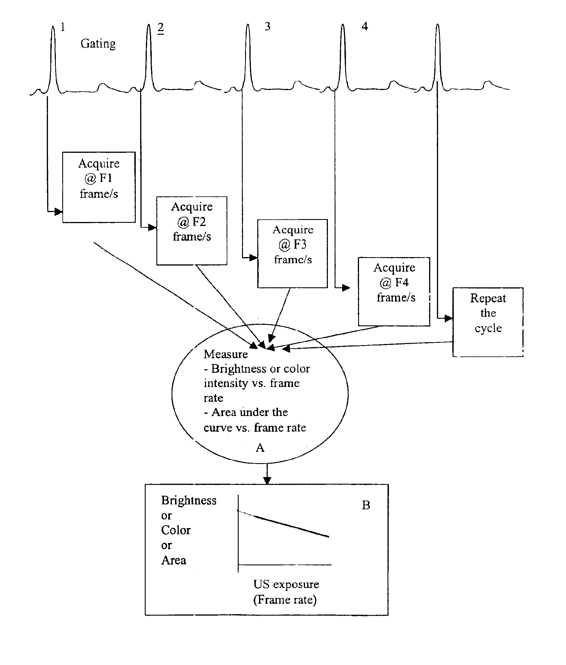
To permit multiple dilution curves to be acquired by the use of only a single contrast injection an apparatus has been developed, along with a novel, gating scheme. Briefly the apparatus comprises a laptop based computer device that allows acquisition of images at a variable rate using a commercial ultrasound scanner. Conceptually this device, which is referred to as a data acquisition controller (DACC), controls the on / off time of the scanner through the EKG port of the scanner.

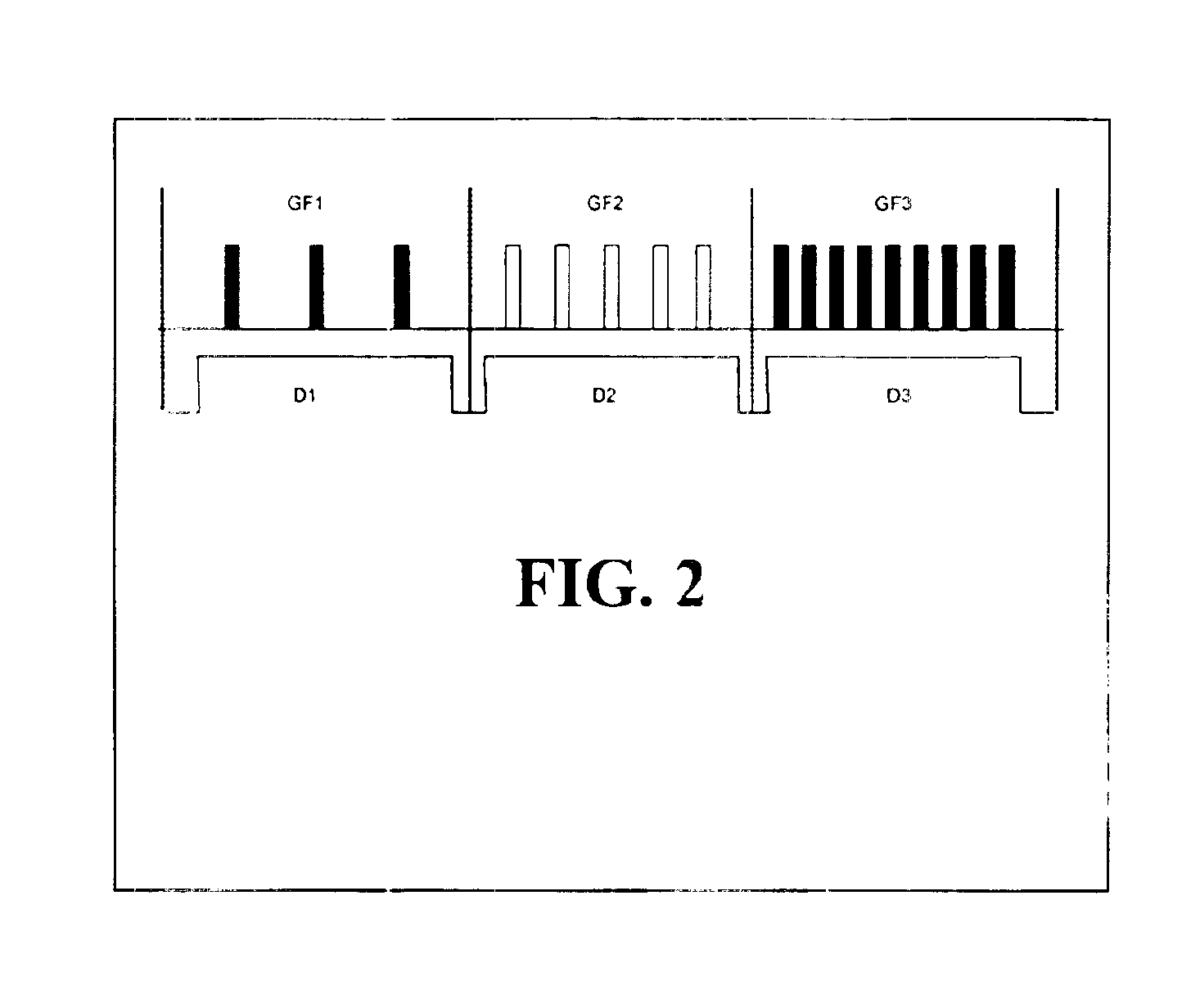
The DACC generates a gating pulse sequence at a variable repetition frequency. With each gating cycle, the ultrasound image is updated with a new frame. After the initial cycle, the scanner is turned “off” until the next trigger cycle, at which point the scanner updates the image. The on / off gating cycle (GF) is varied by changing the pulse repetition rate. The DACC also controls the duration (D) for which each gating frequency is maintain...

example 2

In vivo Studies

While the phantoms provide measurements under controlled conditions, in vivo studies provide proof of the effectiveness of the invention under actual blood flow imaging conditions. To demonstrate that the method can be used to measure blood flow in real situations, measurements were made in mice by injecting 0.1 ml of contrast agent. FIG. 7 shows the change in power Doppler images as the ultrasound contrast transits through the imaged cross-section.

Power Doppler was chosen for imaging because of its higher sensitivity in detecting contrast agent (see, Sehgal et al., J. Ultrasound Med. 14:741-748 (1995)). During the transit of contrast agent, the images were acquired by varying the frame rates to 0.5, 2, and 4 frames per second (fps). The numbers at the bottom left corner of each image in FIG. 7 represent the time in seconds relative to the time of the contrast injection (e.g., 0.1 ml bolus injection of Optison). The first frame (FIG. 7A) shows an image of the tumor be...

example 3

Apparatus

Various configurations of the apparatus are possible. In the 2D mode, the scan system could be a stand-alone system or a module that controls the operation of a conventional scanner. FIG. 9 shows a configuration of a “stand-alone” system. In the preceding examples, the apparatus was used in conjunction with a commercial scanner to generate the reported data.

Unlike conventional imaging systems, the image acquisition in the present apparatus is initiated by an external signal (ETS, Box A) either provided by a user or by provided automatically using physiological signal. Some example of external trigger are listed in Box A and include, but are not limited to ECG, respiration, bolus injection or any other physical event.

The ETS signal initiates the function of the master pulse controller (MPSC). The main purpose of this module, Box B, is to vary the exposure level during imaging. It achieves this by synthesizing a pulse sequence and using it to guide image acquisition. In the s...


PUM


Abstract

Methods and apparatus are provided for controlling fluid flow or perfusion, wherein gas-filled microbubbles are used as ultrasound contrast-enhancing agents, and wherein the method comprises separating the removal of the contrast agent due to flow from the removal of the contrast agent due to bubble destruction for enhanced imaging processes. By varying exposure of the microbubbles to ultrasound, the method and apparatus apply the changes observed in the images to measure flow and vascularity, to improve visualization of blood flow and blood vessels, and to guide delivery of drugs locally to the site of imaging.

Description

FIELD OF THE INVENTIONThe present invention relates to the field of contrast agent-enhanced ultrasound imaging. In particular, the invention relates to controlling the destruction of bubbles by varying exposure to ultrasound and using the changes observed in the images to measure flow and vascularity, improve visualization of blood vessels, and guide delivery of drugs locally to the site of imaging.BACKGROUND OF THE INVENTIONTissues and organs of animals and humans develop characteristic vascular architecture to meet specific nutritional and physiologic requirements. Each vascular system comprises networks of large and small blood vessels, wherein each vessel has a different flow rate and function. Under various physiological and pathological conditions, new micro-vessels are formed from existing vasculature. This process of new vessel formation, called angiogenesis, occurs through tightly regulated processes under normal physiological situations.When these tight controls are breach...

Claims


Application Information

Patent Timeline
IPC IPC(8): A61B8/00
CPCA61B8/543A61B8/481
Inventor SEHGAL, CHANDRA M.
Owner THE TRUSTEES OF THE UNIV OF PENNSYLVANIA
Learn More