Devices and methods for obtaining mammary fluid samples for evaluating breast diseases, including cancer

a technology for breast diseases and biological samples, applied in the field of mammary fluid biological samples obtained and assaying kits, can solve the problems of difficult detection of malignant and benign breast diseases, difficult detection of cancer markers, and difficulty in detecting malignant breast diseases, so as to reduce morbidity and cost, and the effect of high sensitive natur

Inactive Publication Date: 2005-05-03
ATOSSA THERAPEUTICS INC
View PDF6 Cites 116 Cited by
  • Summary
  • Abstract
  • Description
  • Claims
  • Application Information

AI Technical Summary

Benefits of technology

[0011]Methods for detecting and measuring cancer markers have been recently revolutionized by the development of immunological assays, particularly by assays that utilize monoclonal antibody technology. Previously, many cancer markers could only be detected or measured using conventional biochemical assay methods, which generally require large test samples and are therefore unsuitable in most clinical applications. In contrast, modern immunoassay techniques can detect and measure cancer markers in relatively much smaller samples, particularly when monoclonal antibodies that specifically recognize a targeted marker protein are used. Accordingly, it is now routine to assay for the presence or absence, level, or activity of selected cancer markers by immunohistochemically staining breast tissue specimens obtained via conventional biopsy methods. Because of the highly sensitive nature of immunohistochemical staining, these methods have also been successfully employed to detect and measure cancer markers in smaller, needle biopsy specimens which require less invasive sample gathering procedures compared to conventional biopsy specimens. In addition, other immunological methods have been developed and are now well known in the art which allow for detection and measurement of cancer markers in non-cellular samples such as serum and other biological fluids from patients. The use of these alternative sample sources substantially reduces the morbidity and costs of assays compared to procedures employing conventional biopsy samples, which allows for application of cancer marker assays in early screening and low risk monitoring programs where invasive biopsy procedures are not indicated.

Problems solved by technology

All too often, breast cancer is discovered at a stage that is too far advanced, when therapeutic options and survival rates are severely limited.
In addition to the problem of early detection, there remain serious problems in distinguishing between malignant and benign breast disease, in staging known breast cancers, and in differentiating between different types of breast cancers (e.g. estrogen dependent versus non-estrogen dependent tumors).
Previously, many cancer markers could only be detected or measured using conventional biochemical assay methods, which generally require large test samples and are therefore unsuitable in most clinical applications.
For the purpose of breast cancer evaluation, the use of conventional or needle biopsy samples for cancer marker assays is often undesirable, because a primary goal of such assays is to detect the cancer before it progresses to a palpable or mammographically detectable tumor stage.
Prior to this stage, biopsies are generally contraindicated, making early screening and low risk monitoring procedures employing such samples untenable.
Efforts to utilize serum samples for breast cancer marker assays have met with limited success, largely because the targeted markers are either not detectable in serum, or because telltale changes in the levels or activity of the markers cannot be monitored in serum.
In addition, the presence of breast cancer markers in serum probably occurs at the time of micro-metastasis, making serum assays less useful for detecting pre-metastatic disease.
Although the evaluation of mammary fluid has been shown to be a useful method for screening nonpalpable breast cancer in women who experience spontaneous nipple discharge, the rarity of this condition renders the methods of Inaji et al, inapplicable to the majority of women who are candidates for early breast cancer screening.
It is likely that other antibodies used to assay other cancer markers may exhibit even lower sensitivity than the anti-CEA antibodies used by Inaji and coworkers, and may therefore not be adaptable or sensitive enough to be employed even in dry chemical immunoassays of small samples of spontaneously discharged mammary fluid.

Method used


Image

  • Devices and methods for obtaining mammary fluid samples for evaluating breast diseases, including cancer
  • Devices and methods for obtaining mammary fluid samples for evaluating breast diseases, including cancer
  • Devices and methods for obtaining mammary fluid samples for evaluating breast diseases, including cancer

Examples


example 1

Induction of Mammary Fluid Expression by Application of a Novel Breast Pump / Mammary Fluid Sample Collection Device

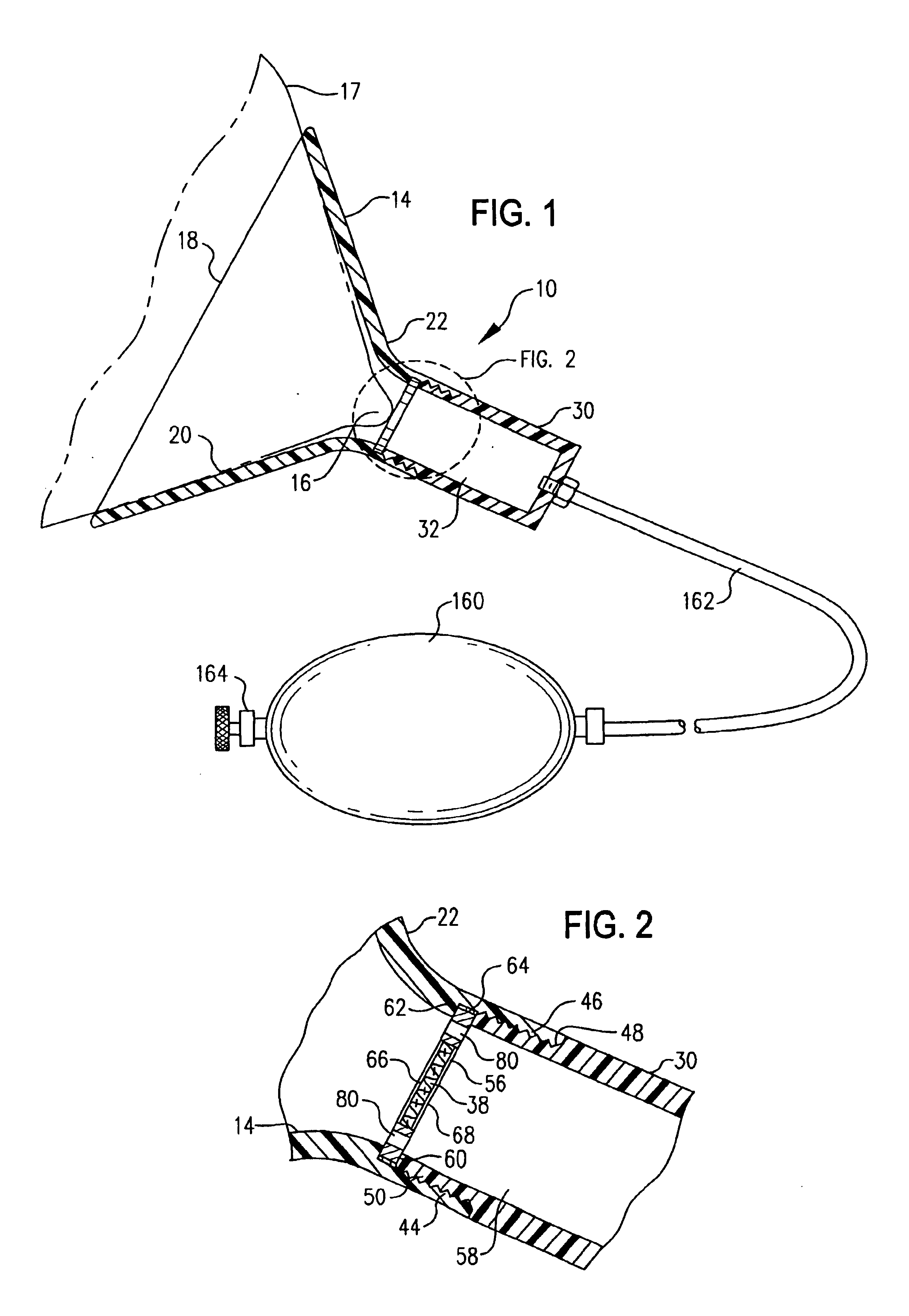
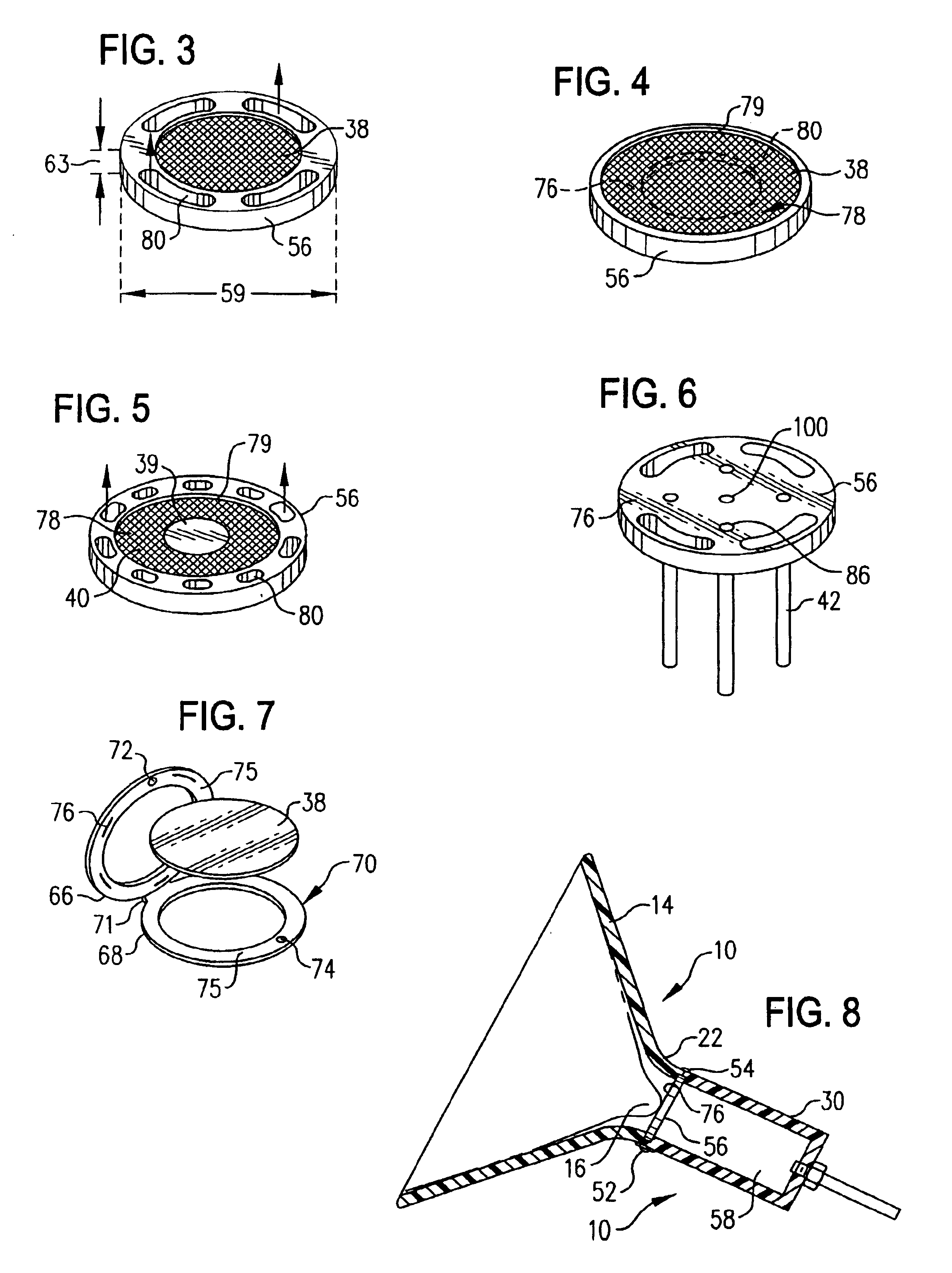
[0216]Within the present example, a hand-held breast pump device 10′ is employed to collect a primary sample of mammary fluid components comprising whole cells and cell fragments for cytological examination. The doctor, technician or patient collects the breast fluid specimen by grasping and operating the hand-held pump as described above to stimulate expression of the mammary fluid and collect a specimen thereof. This operation is a one-handed procedure, leaving the physician or technician free to conduct additional activities with the other, free hand. In this regard, the compact hand-held pump design allows the device to be picked up and manipulated with one hand, to seat the breast engaging element against the breast, apply sufficient vacuum pressure to the breast by manual operation of the vacuum pump to cause a suitable volume of breast fluid to be expressed at or ...

example 2

Cytology in Biological Samples From Mammary Fluid

[0219]This example describes the use of conventional cytological techniques to identify and classify breast diseases from samples obtained as described in Example 1. Following collection of the sample, e.g., in a fluid-retaining reservoir member, the sample may be further processed (e.g., by centrifugation wherein the reservoir member is in the form of a modified cytology vial). Processed samples are then transferred to the central region of a clean glass microscopic slide, and a cover slip is slid over the sample to spread it along the surface of the slide. The slide is allowed to air dry and then is fixed, for example in absolute alcohol, and stained with standard cytological stains, such as methylene blue, hematoxyln and eosin, and other suitable stains.

[0220]The slides are then examined by light microscopy for evidence of atypical growth of cells and clumps of cells, using well known methods, including those described in Diagnosis...

example 3

Stimulation of Mammary Fluid Expression for Sample Collection by Coordinate Administration of Intranasal Oxytocin in Conjunction with Application of a Novel Breast Pump / Mammary Fluid Sample Collection Device

[0221]The foregoing sample collection protocol in Example 1, as well as other sample collection methods within the invention, may be practiced solely by the use of a novel breast pump 10, 10′ of the invention. Alternatively, these sample collection procedures may be practiced in conjunction with the use of oxytocin or oxytocin analogs to facilitate or increase mammary fluid expression induced by operation of the breast pump. As incorporated within the invention, these methods involve application of the breast pump 10, 10′ to the breast, optionally coupled with oxytocin administration in amounts effective to facilitate mammary fluid expression in the patient. After the sample is collected, a bioassay is conducted on the sample to determine the presence and / or amount of a selected ...


PUM

PropertyMeasurementUnit
diameteraaaaaaaaaa
pore sizeaaaaaaaaaa
pore sizeaaaaaaaaaa

Abstract

Biological samples of mammary fluid or components thereof are obtained using a breast pump device coupled with a solid phase sample collection medium, alternatively facilitated by administering oxytocin to the subject. The breast pump device stimulates expression of mammary fluid and provides for collection of diagnostic samples to evaluate breast disease, including cancer. The biological sample may include whole cells or cellular components, purified or bulk proteins, glycoproteins, peptides, nucleotides or other desired constituents comprising a breast disease marker. Methods, kits and adapter devices relating to the breast pump device are also provided. Yet additional methods, devices, accessories, and materials are provided for laboratory handling and processing of breast fluid samples and for related diagnostic methods.

Description

RELATED APPLICATIONS[0001]This application claims the priority benefits of U.S. Provisional Patent Application No. 60 / 248,134, filed by Quay on Nov. 13, 2000 and U.S. Provisional Patent Application No. 60 / 248,136, filed by Quay on Nov. 13, 2000. The disclosures of each of the foregoing priority applications are incorporated herein by reference in their entirety.TECHNICAL FIELD OF INVENTION[0002]The invention relates to methods, devices, and kits for obtaining and assaying biological samples from mammary fluid. More specifically, the invention relates to methods, devices, and kits for obtaining and assaying fluid and cytological samples from the mammary glands of a mammalian subject for evaluating, diagnosing and managing breast disease, including infections, pre-cancerous conditions, cancer susceptibility and cancer.BACKGROUND OF THE INVENTION[0003]Breast cancer is by far the most common form of cancer in women, and is the second leading cause of cancer death in humans. Despite many...

Claims


Application Information

Patent Timeline
Patent Type & Authority Patents(United States)
IPC IPC(8): A61B10/00A61M1/06G01N33/48A61B5/00A61B10/02C12Q1/04
CPCA61B5/14546A61B10/0041A61B10/0045A61M1/066A61M1/06C12Q1/6886A61M2205/075A61B10/0096A61M1/064A61M1/0697
Inventor QUAY, STEVEN C.
Owner ATOSSA THERAPEUTICS INC
Why Eureka
Try Eureka