Measurement of protective genes in allograft rejection

a technology of protective genes and allografts, applied in the field of allograft rejection measurement, can solve the problems of chronic rejection risk factor, unsuitable current monitoring and diagnostic modalities, and common and serious problems, and achieve the effect of accurately detecting allograft rejection and accurately quantifying marker gene expression

Inactive Publication Date: 2005-05-31
BETH ISRAEL DEACONESS MEDICAL CENT INC +1
View PDF3 Cites 44 Cited by
  • Summary
  • Abstract
  • Description
  • Claims
  • Application Information

AI Technical Summary

Benefits of technology

[0027]Thus, as a result of the work described herein, methods are now available to accurately quantitate marker gene expression in biopsy tissue, urine, peripheral blood mononuclear cells and other body fluids, and to correlate the magnitude of expression of these genes with rejection of allografts. Surprisingly, the evaluation of the expression of marker genes in a post-transplant sample, along with the evaluation of expression of an infectious agent gene, also accurately detects allograft rejection.

Problems solved by technology

Acute rejection, despite clinical application of potent immunoregulatory drugs and biologic agents, remains a common and serious post-transplantation complication.
It is also a risk factor for chronic rejection, a relentlessly progressive process.
However, current monitoring and diagnostic modalities may be ill-suited to the diagnosis of acute rejection at an early stage.
Whereas increased serum creatinine levels are currently the best surrogate markers of acute rejection, they lack sensitivity and specificity with respect to predicting rejection.
However, the presence of a modest cellular infiltrate is often not conclusive and can be detected in non-rejecting grafts.
By the time rejection is well-established or is clinically diagnosable, it may be too late to salvage optimal allograft function.
The information yielded by classic biopsy analysis may not provide early indication of an impending rejection episode.
For example, high-dose anti-rejection immunosuppressive treatment is an important contributor to post-transplant morbidity and mortality.

Method used


Image

  • Measurement of protective genes in allograft rejection
  • Measurement of protective genes in allograft rejection
  • Measurement of protective genes in allograft rejection

Examples


example 1

Analysis of Biopsy Samples

Biopsies:

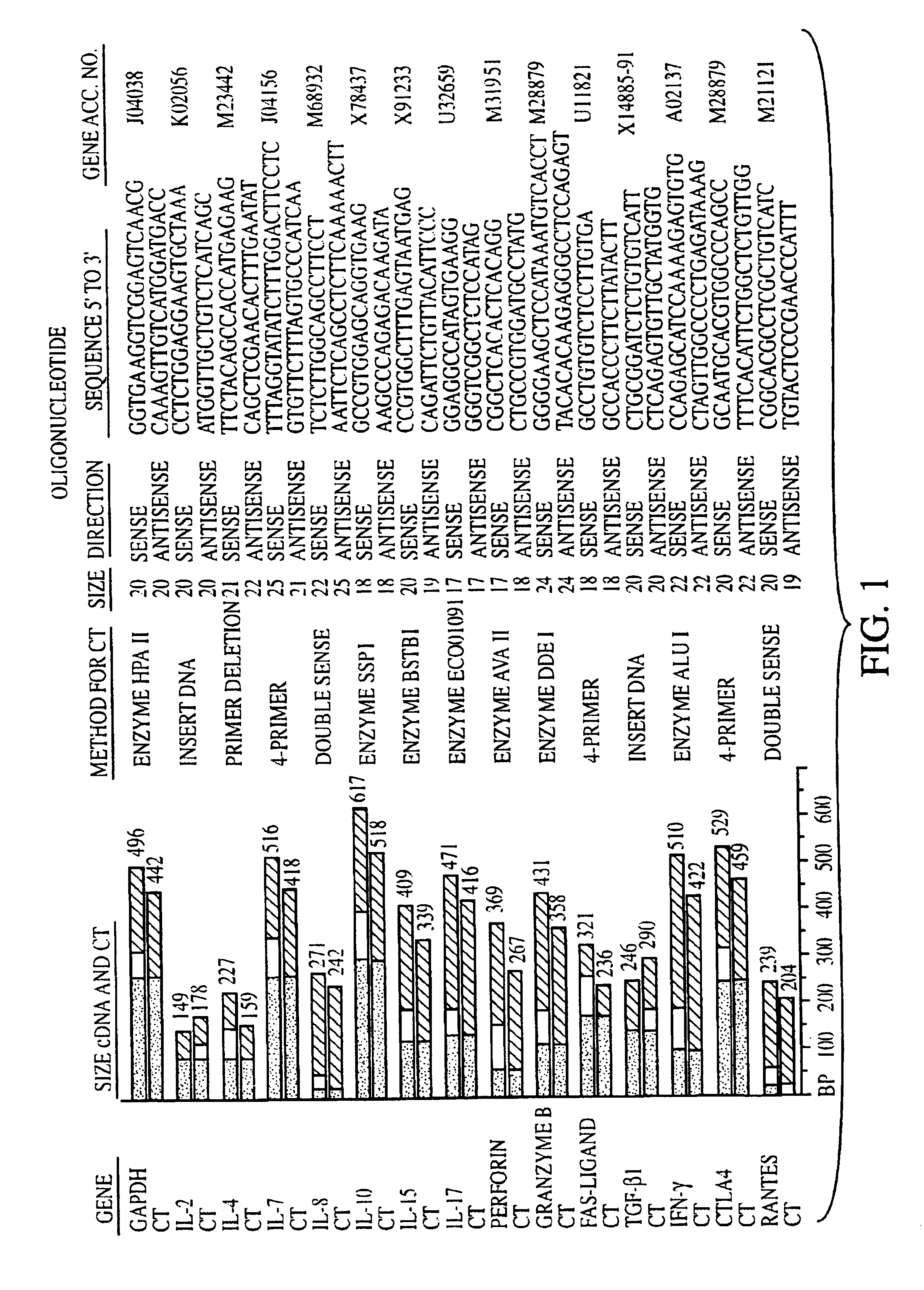
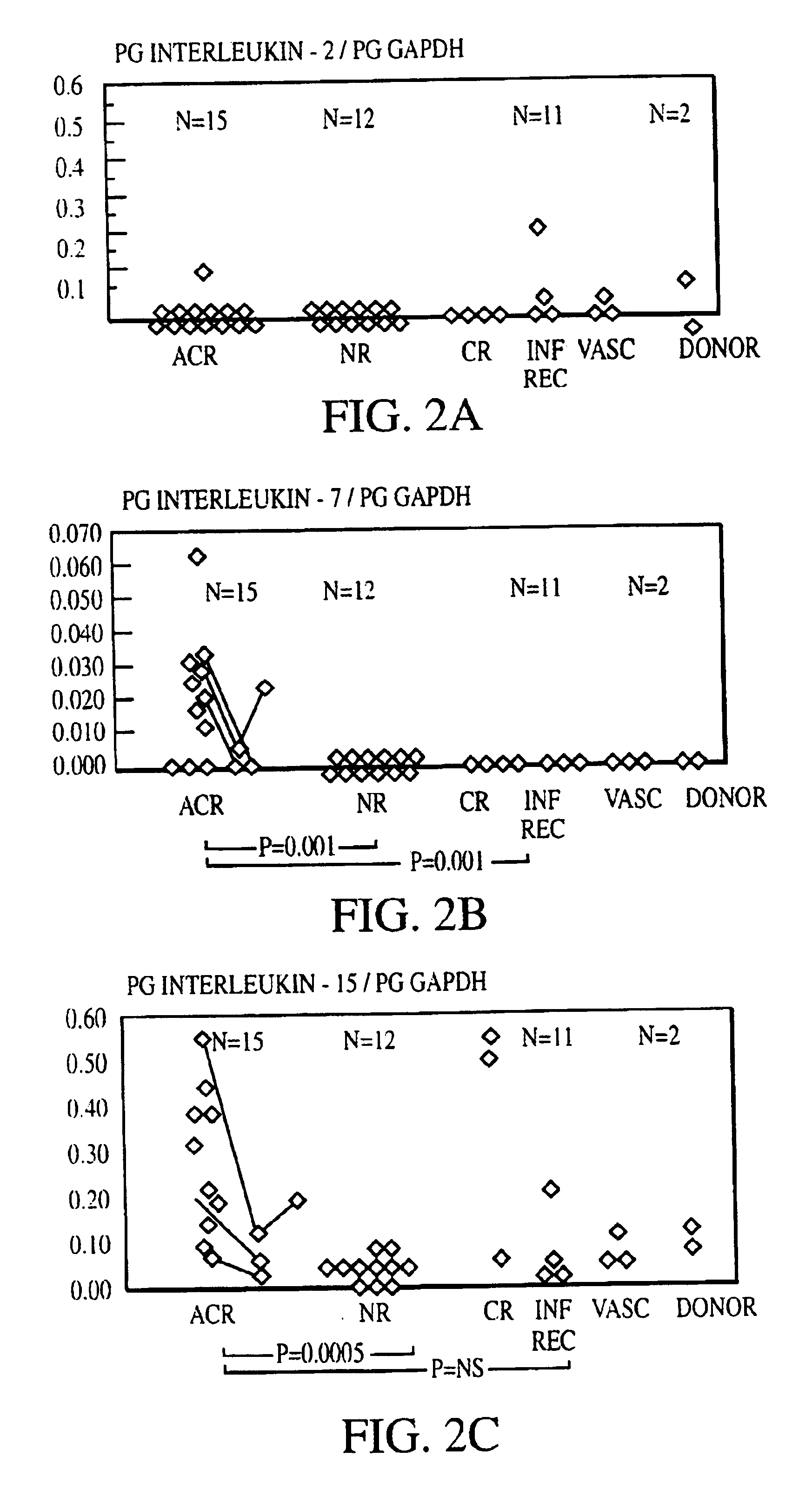
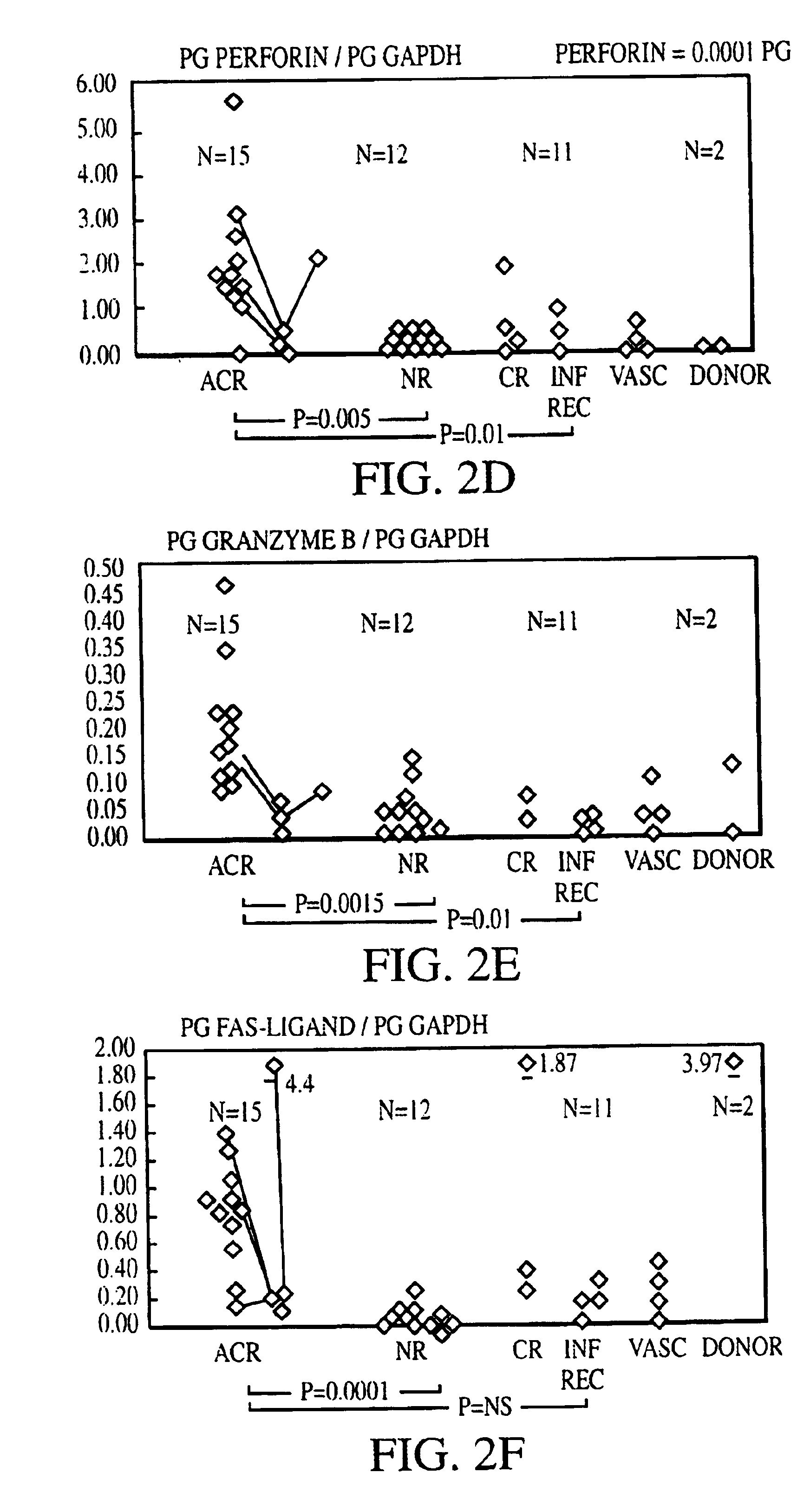
[0133]Sixty kidney transplant biopsies were investigated for gene expression of chemokines (IL-8, RANTES (regulated upon activation, normal T-cell expressed and secreted), T-cell growth factors and other cytokines (IL-2, IL-4, IL-7, IL-10, IL-15, and IL-17), cell surface immunoregulatory proteins (CTLA4), cytotoxic effector molecules (P, GB, FasL), IFN-γ, transforming growth factor (TGF)-1, and the housekeeping protein glyceraldehyde-3-phosphate dehydrogenase (GAPDH). Thirty-eight biopsies were obtained from 34 patients (25 adults and 9 children) to clarify the cause of graft dysfunction, 20 for early post-transplant surveillance and 2 from living related donor kidneys prior to reperfusion. Small portions of biopsy cores ({fraction (1 / 10)}-½) were immediately snap frozen in liquid nitrogen at the bedside and stored at 70° C. The majority of tissue was used for histopathological analysis. Biopsies obtained to evaluate the cause of graft dysfunction ...

example 2

Analysis of PBMCS

[0147]In a study of 16 renal allograft recipients, PBMCs were isolated from whole blood and RNA extracted by a modified QIAGEN™ method. (QIAGEN Rneasy Blood Mini Kits, Cat. No. 74303, 74304 or 74305). The QIAGEN technique involves four steps: 1) a sample is combined with a suitable buffer for isolating RNA in the sample from the remaining components, e.g., 1 part whole blood, is mixed with 5 parts lysing buffer, wherein the blood cells are lysed and RNA released; 2) RNA in the sample is specifically bound to particles or a membrane; 3) the particles or membrane are washed to remove non-RNA components; and 4) the isolated RNA is eluted from the particles / membrane.

[0148]To increase the efficiency of RNA isolation from PBMCs, the second step of the QIAGEN protocol was modified as described in Example 3.

[0149]Gene expression was analyzed by reverse transcription-assisted semi-quantitative PCR in PMBC and in snap frozen transplant core biopsies and was compared to the hi...

example 3

Method for Processing Blood for PCR Analysis

Blood Collection

Supplies:

[0150]2 ml EDTA vacuum tubes (purple top): cat #369651 Vacutainer; Flask with ice.

Procedure:[0151]Label EDTA tubes with Patient ID, date and time.[0152]Draw 2 ml blood into EDTA tube and carefully mix by inversion; transport on ice to the lab to be processed.*

[0153]* For optimal results, blood samples should be processed within a few hours.

White Blood Cell Isolation

Supplies:

[0154]3 cc syringes[0155]15 ml Sterile Conical tubes (Falcon)-Sterile polypropylene tubes (20-200-1000 ul)[0156]RPMI Medium 1640: cat #11875-085 Gibco BRL[0157]EL Buffer: cat #79217 Qiagen[0158]Flask with liquid nitrogen: cat #2123 Lab-Line.[0159]Ethanol (96-100%)-70% ethanol in water[0160]14.5 M-Mercaptoethanol (-ME)[0161]Lab centrifuge with rotor for 15 ml tubes -4C Microcentrifuge with rotor for 2 ml tubes

Instrumentation:[0162]Lab centrifuge with rotor for 15 ml tubes at 4C.

Procedure:[0163]1. Using a 3 cc syringe transfer 1-1.5 ml blood into...


PUM

PropertyMeasurementUnit
areaaaaaaaaaaa
volumeaaaaaaaaaa
volumeaaaaaaaaaa

Abstract

The invention relates to methods of evaluating transplant rejection in a host comprising determining a heightened magnitude of gene expression of genes in rejection-associated gene clusters. The disclosed gene clusters include genes that are substantially co-expressed with cytotoxic lymphocyte pro-apoptotic genes, cytoprotective genes and several other cytokine and immune cell genes.

Description

CLAIM OF PRIORITY[0001]This application claims priority to U.S. Provisional Application 60 / 199,327, filed Apr. 24, 2000; 60 / 238,718, filed Oct. 6, 2000; 60 / 239,635, filed Oct. 12, 2000; and 60 / 240,735, filed Oct. 16, 2000. The contents of these applications are herein incorporated by reference.BACKGROUND OF THE INVENTION[0002]Acute rejection, despite clinical application of potent immunoregulatory drugs and biologic agents, remains a common and serious post-transplantation complication. It is also a risk factor for chronic rejection, a relentlessly progressive process. As the occurrence of acute rejection episodes is the most powerful predictive factor for the later development of chronic rejection in adults and children, many advocate strategies to detect and ablate acute rejection episodes as early as possible. However, current monitoring and diagnostic modalities may be ill-suited to the diagnosis of acute rejection at an early stage.[0003]For example, acute renal allograft rejec...

Claims


Application Information

Patent Timeline
Patent Type & Authority Patents(United States)
IPC IPC(8): C12Q1/68G01N33/68
CPCC12Q1/6883G01N33/6893G01N2800/245C12Q2600/158
Inventor AVIHINGSANON, YINGYOSMA, NALISTROM, TERRY B.SOARES, MIGUEL C.FERRAN, CHRISTIANESUTHANTHIRAN, MANIKKAM
Owner BETH ISRAEL DEACONESS MEDICAL CENT INC
Why Eureka
Try Eureka