Cryosurgical procedures involve
deep tissue freezing which results in tissue destruction due to rupture of cells and or
cell organelles within the tissue.
The application of temperatures of between about −40° C. and 0° C. to such healthy tissues usually causes substantial damage thereto, which damage may result in temporary or permanent impairment of functional organs.
In addition, when the adjacent tissues are present at opposite borders with respect to the freeze treated tissue, such as in the case of
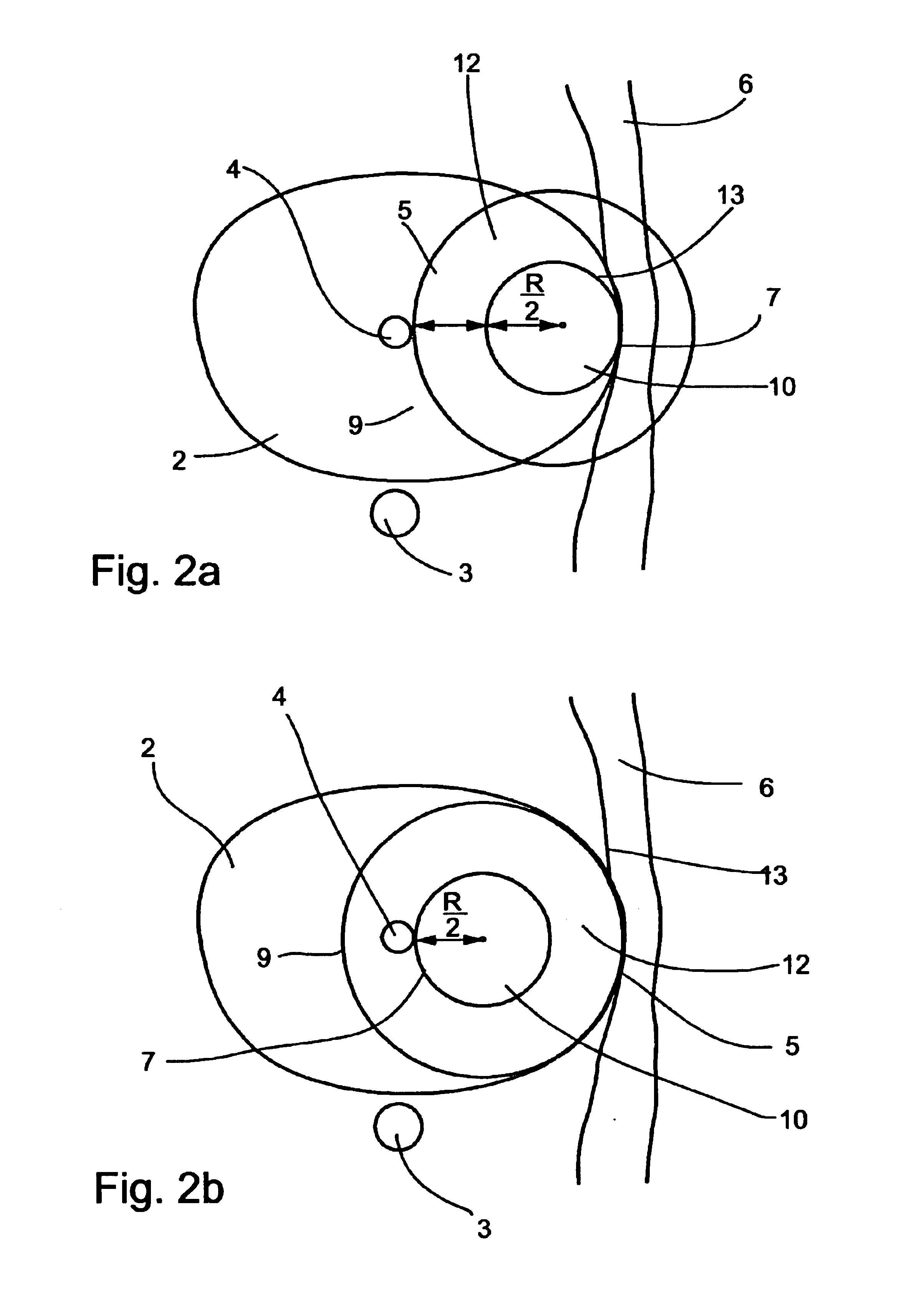
prostate freeze treatments, as is further detailed below, and since the growth of the ice-ball is in substantially similar rate in all directions toward its periphery, if the tip of the cryosurgical device is not precisely centered, the ice-ball reaches one of the borders before it reaches the other border, and
decision making of whether to continue the process of freezing, risking a damage to close healthy tissues, or to halt the process of freezing, risking a non-complete destruction of the treated tissue, must be made.
Treatment of
benign prostate hyperplasia (BPH), while not requiring total destruction of an entire volume of
prostate tissue as does treatment of a
malignancy, nevertheless does run the risk of causing damage to healthy functional tissues and organs adjacent to the
prostate, if care is not taken to limit the scope of destructive freezing to appropriate locations.
Such a modification, however, exposes the patient to the danger of
malignancy because of a possible incomplete destruction of the tumor.
The classical
cryosurgery procedure herein described, therefore, does not provide effective resolution of treatment along the planes perpendicular to the axis of penetration of the cryosurgical probe into the patient's organ.
A further limitation of the classical procedure stems from the fact that anatomical organs such as the prostate usually feature an asymmetric three-dimensional shape.
Consequently, introduction of a cryosurgical probe along a specific path of penetration within the organ may provide
effective treatment to specific regions located at specific depths of penetration but at the same time may severely damage other portions of the organ located at other depths of penetration.
It is, however, a
disadvantage of the HR apparatus and method as taught in U.S. Pat. No. 6,142,991 that the apparatus enables, and the method requires, a high level of diagnostic sophistication in the selection and definition of the particular volume of tissue to be cryoablated.
Real-time imaging capabilities of the HR apparatus provide for imaging of the
target organ at a selected
depth of penetration and thereby assist an operator in deciding where to introduce and utilize a plurality of cryogenic probes, yet the complex three-dimensional geometry of the cryoablation target is poorly rendered by the set of two dimensional images constituting the three dimensional grid as contemplated by the HR method and apparatus.
In this prior art method, little assistance is provided for an operator in understanding the
three dimensional shape and structure of the cryoablation target and the surrounding tissues.
Information vital to the operator may be present in the set of images, yet difficult for the operator to see and appreciate.
In a set of images of this type, the details may be present, yet it may be difficult to appreciate their significance because of the difficulty of seeing them in context.
A three dimensional “grid” composed of a plurality of two dimensional images such as
ultrasound images contain many details, yet do not facilitate the understanding of those details in a three dimensional context.
It is an additional limitation of the HR method and apparatus, and of other prior art systems, that the imaging capabilities contemplated are not well adapted to assist an operator in planning a cryoablation procedure.
In addition to the fact that the imaging facilities there provided are poorly adapted to
visualization of the
three dimensional space by an operator, they are also limited in that the apparatus is poorly adapted to providing images of the target area in advance of the operation, e.g., for planning purposes.
The described HR equipment might, of course, but used to create the described three dimensional mapping of the target area well in advance of a surgical intervention, but no mechanism is provided for facilitating the relating the images so obtained, and any planned procedures based on those images, to a subsequent intervention procedure.
Moreover, the fact that the imaging modality of the HR apparatus is physically connected to parts of the cryosurgery equipment limits its versatility and may in some cases make it awkward to use for creating preparatory images of an intervention site.
It is a further limitation of the HR method that no means are provided for facilitating the relating of images obtained in advance of a surgical intervention to a subsequent intervention.
Yet whereas
ultrasound images of a
target site can be generated in real time during an intervention, and
MRI techniques may also (if somewhat less easily) also be obtained during cryosurgery, other imaging techniques (CT scans, for example) are less well adapted to being produced during the course of an actual cryosurgery intervention.
This information, and this capability for prediction, is underutilized in current cryosurgery practice.
STPS, however, is limited in that it does not provide means for relating a preliminary
three dimensional model of a prostate to the prostate as revealed in real-time during the course of a surgical procedure.
Moreover, the predictive ability of the STPS system is limited to predicting the extent of the freezing produced by a given deployment of a plurality of cryoprobes over a given time.
No assistance is provided to an operator in discerning interactions between the predicted cryoablation and specific structures desired to be protected or to be destroyed.
No assistance is given in predicting long-term effects of a given cryoablation procedure.
This causes patients to experience a frequent urge to urinate because of incomplete emptying of the bladder and a
burning sensation or similar discomfort during
urination.
Left untreated, the obstruction caused by BPH can lead to acute
urinary retention (complete inability to urinate), serious urinary tract infections and permanent bladder and
kidney damage.
Various disadvantages of existing therapies have limited the number of patients suffering from BPH who are actually treated.
It will be appreciated that this procedure results in a substantial damage inflicted upon the
prostatic urethra.
Again, it will be appreciated that these procedures result in a substantial damage inflicted upon the
prostatic urethra.
Heat
ablation techniques, however, burn the tissue, causing irreversible damage to
peripheral tissue due to
protein denaturation, and destruction of nerves and blood vessels.
However
TUNA is less effective than traditional
surgery in reducing symptoms and improving
urine flow.
TUNA also burn the tissue, causing irreversible damage to
peripheral tissue due to
protein denaturation, and destruction of nerves and blood vessels.
Furthermore, as already discussed above,
heat generation causes
secretion of substances from the tissue which may endanger the surrounding area.


