
Monoclonal antibodies to the ClfA protein and method of use in treating or preventing infections

a technology of monoclonal antibodies and clfa protein, which is applied in the direction of antibody medical ingredients, instruments, aerosol delivery, etc., can solve the problems of dramatic alteration of their physiology

Inactive Publication Date: 2005-12-27
INHIBITEX INC
View PDF16 Cites 42 Cited by
  • Summary
  • Abstract
  • Description
  • Claims
  • Application Information

AI Technical Summary

Benefits of technology

"The present invention provides monoclonal antibodies that can bind to the S. aureus ClfA protein and can be used to treat or prevent staphylococcal infections. These antibodies can also be used to prevent adherence of Staphylococcal bacteria by inhibiting or impairing the binding of the ClfA protein to fibrinogen or fibrin. The invention also includes the isolation and use of monoclonal antibodies to the ClfA protein and its binding subdomains, as well as suitable compositions and vaccines based on these antibodies. Overall, the invention provides a valuable tool for the diagnosis, treatment, and prevention of staphylococcal infections."

Problems solved by technology

Once the bacteria have successfully adhered and colonized host tissues, their physiology is dramatically altered and damaging components such as toxins and proteolytic enzymes are secreted.
However, it has still remained a problem to identify and utilize the information concerning MSCRAMM™S from S. aureus such as the ClfA protein to generate effective monoclonal antibodies because of the variability in the binding properties of the different MSCRAMM™s and their role in infectivity and spread of bacterial infections.

Method used


Image

  • Monoclonal antibodies to the ClfA protein and method of use in treating or preventing infections
  • Monoclonal antibodies to the ClfA protein and method of use in treating or preventing infections
  • Monoclonal antibodies to the ClfA protein and method of use in treating or preventing infections

Examples


example 1

Isolation and Sequencing of Clf40 and Clf33

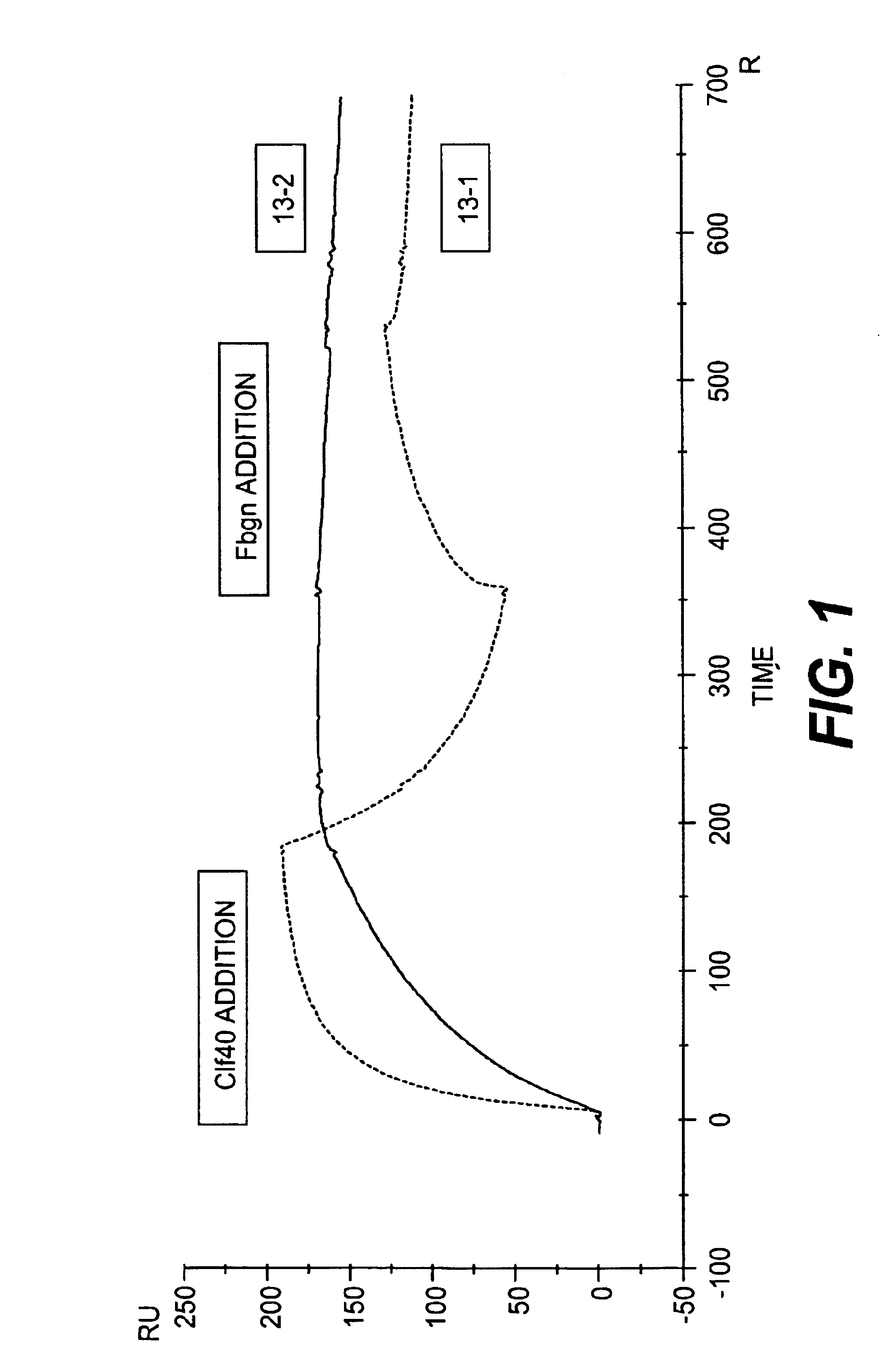
[0064]Using PCR, the A domain of ClfA (Clf40 representing AA 40-559 or Clf33 representing AA 221-550) was amplified from S. aureus Newman genomic DNA and subcloned into the E. coli expression vector PQE-30 (Qiagen), which allows for the expression of a recombinant fusion protein containing six histidine residues. This vector was subsequently transformed into the E. coli strain ATCC 55151, grown in a 15-liter fermentor to an optical density (OD600) of 0.7 and induced with 0.2 mM isopropyl-1-beta-D galactoside (IPTG) for 4 hours. The cells were harvested using an AG Technologies hollow-fiber assembly (pore size of 0.45 μm) and the cell paste frozen at −80° C. Cells were lysed in 1×PBS (10 mL of buffer / 1 g of cell paste) using 2 passes through the French Press @1100 psi. Lysed cells were spun down at 17,000 rpm for 30 minutes to remove cell debris. Supernatant was passed over a 5-mL HiTrap Chelating (Pharmacia) column charged with 0.1M NiCl2. ...

example 2

Monoclonal Antibody Production Using Clf40 and Clf33

[0067]The purified Clf40 or Clf33 protein was used to generate a panel of murine monoclonal antibodies. Briefly, a group of Balb / C mice received a series of subcutaneous immunizations of 50 μg of Clf40 or Clf33 protein in solution or mixed with adjuvant as described below in Table I:

[0068]

TABLE IInjectionDayAmount (μg)RouteAdjuvantPrimary 050SubcutaneousFreund's CompleteBoost #1145(Clf40)IntravenousPBS10(Clf33)

[0069]Three days after the final boost, the spleens were removed, teased into a single cell suspension and the lymphocytes harvested. The lymphocytes were then fused to a SP2 / 0-Ag14 myeloma cell line (ATCC #1581). Cell fusion, subsequent plating and feeding were performed according to the Production of Monoclonal Antibodies protocol from Current Protocols in Immunology (Chapter 2, Unit 2.).

[0070]Any clones that were generated from the fusion were then screened for specific anti-Clf40 antibody production using a standard ELISA...

example 3

Additional Studies of Clf40 and Clf33

[0075]Using PCR, the A domain of ClfA (Clf40 representing AA 40-559, Clf33-N2N3 domain representing AA 221-550 or Clf-N3 domain representing AA370-559) was amplified from S. aureus Newman genomic DNA and subcloned into the E. coli expression vector PQE-30 (Qiagen), which allows for the expression of a recombinant fusion protein containing six histidine residues. This vector was subsequently transformed into the E. coli strain ATCC 55151, grown in a 15-liter fermentor to an optical density (OD600) of 0.7 and induced with 0.2 mM isopropyl-1-beta-D galactoside (IPTG) for 4 hours. The cells were harvested using an AG Technologies hollow-fiber assembly (pore size of 0.45 mm) and the cell paste frozen at −80° C. Cells were lysed in 1×PBS (10 mL of buffer / 1 g of cell paste) using 2 passes through the French Press @1100 psi. Lysed cells were spun down at 17,000 rpm for 30 minutes to remove cell debris. Supernatant was passed over a 5-mL HiTrap Chelating ...


PUM

PropertyMeasurementUnit
optical densityaaaaaaaaaa
pore sizeaaaaaaaaaa
pHaaaaaaaaaa

Abstract

Monoclonal antibodies which can bind to the ClfA protein and which are generated from binding subdomains or active fragments of the ClfA protein from Staphylococcus aureus, including the active fragments proteins from its fibrinogen binding domain such as Clf40 protein, the Clf33 protein, or ClfA N3, are provided which can be useful in the treatment and protection against infection from staphylococcal bacteria such as Staphylococcus aureus. In addition, medical instruments can be treated using the monoclonal antibodies of the invention in order to reduce or eliminate the possibility of their becoming infected or further spreading the infection. In particular, the antibodies of the present invention are advantageous because they can prevent adherence of the bacteria to host cells by impairing or inhibiting the ability of S. aureus ClfA to bind to fibrinogen or fibrin, and thus can be utilized in methods or treating or preventing staphylococcal inventions.

Description

CROSS REFERENCE TO RELATED APPLICATIONS[0001]The present application claims the benefit of U.S. provisional applications Ser. No. 60 / 308,116, filed Jul. 30, 2001, Ser. No. 60 / 298,413, filed Jun. 18, 2001, Ser. No. 60 / 274,611, filed Mar. 12, 2001, and Ser. No. 60 / 264,072, filed Jan. 26, 2001.FIELD OF THE INVENTION[0002]The present invention relates in general to antibodies that have been generated against clumping factor A (or ClfA), a surface localized protein expressed in Staphylococcus aureus and other staphylococcus bacteria, and in particular to monoclonal antibodies against the ClfA protein and its active fragments or proteins from its fibrinogen binding domain such as Clf40, Clf33, or ClfA N3, and their use in inhibiting the binding of the ClfA protein to fibrinogen or fibrin and treating or preventing S. aureus infections.BACKGROUND OF THE INVENTION[0003]The successful colonization of the host is a process required for most microorganisms to cause infections in animals and hu...

Claims


Application Information

Patent Timeline
Patent Type & Authority Patents(United States)
IPC IPC(8): A61K9/12A61K9/72A61K39/395A61P31/04C07K14/31G01N33/53C07K16/12C07K19/00C12N15/09C12P21/08G01N33/532G01N33/577
CPCC07K16/1271A61K2039/505C07K2317/24C07K2317/56C07K2319/00A61P31/04C07K16/12
Inventor PATTI, JOSEPH M.HUTCHINS, JEFF T.DOMANSKI, PAULPATEL, PRATIKSHAHALL, ANDREA
Owner INHIBITEX INC
Learn More