

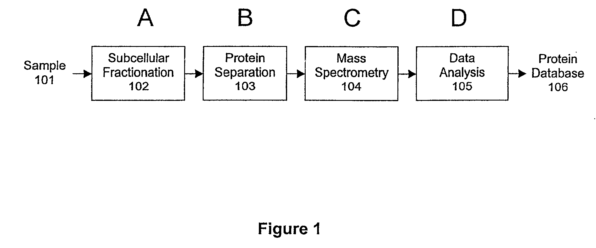
Protein fractionation
a technology of protein fractionation and protein, applied in the field of proteomics, can solve the problem that the rate limitation step of proteomic data collection is generally the protein separation step, and achieve the effect of facilitating an increase in the rate of proteomic analysis
- Summary
- Abstract
- Description
- Claims
- Application Information
AI Technical Summary
Benefits of technology
Problems solved by technology
Method used
Image

Examples
example 1
7.1 Example 1
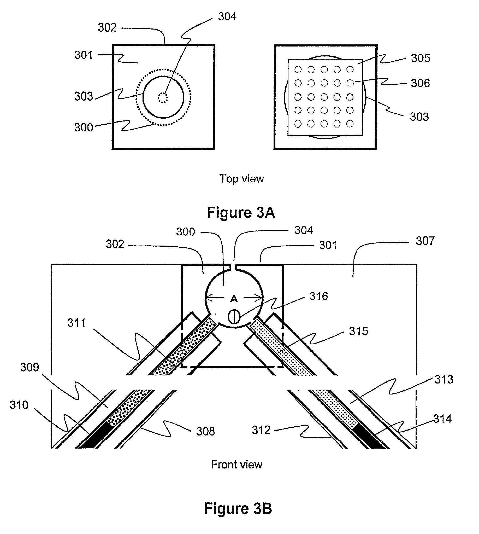
Single Sephadex Bead Protein Analysis of a Complex Mixture
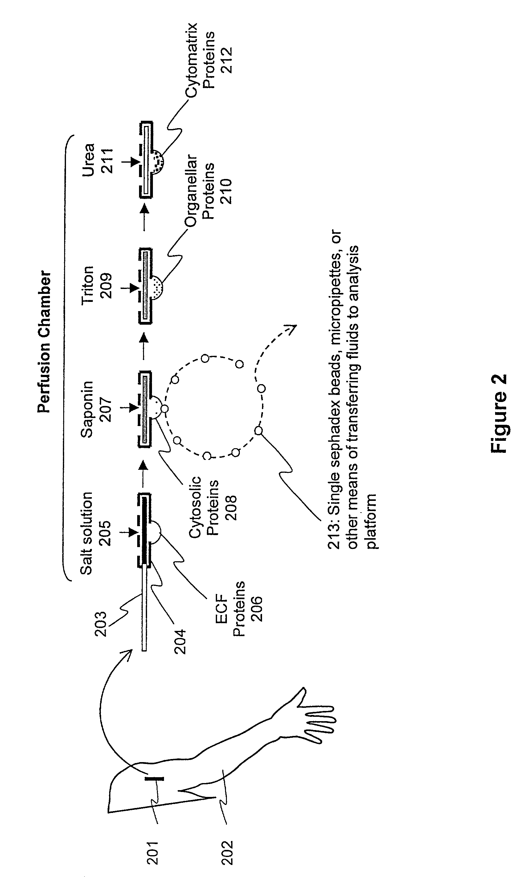
[0126]Summary: A feasibility study using single Sephadex beads for protein analysis was carried out on biopsies of frog skeletal muscle. The results demonstrate that cytosolic proteins can be separated from other muscle proteins using single Sephadex beads. Two exemplar cytosolic proteins, creatine kinase and parvalbumin, were identified alongside purified protein standards, and their concentrations determined, using 1-D SDS PAGE.
[0127]The results support the utility of using single Sephadex beads for capturing relatively undiluted protein samples of intracellular fluid.
[0128]Materials and Methods: Bundles of 10–30 fibers were cut from the isolated semitendinosus muscle of Rana temporaria. The bundles were blotted and placed in a glass-bottom dish filled with mineral oil. The temperature of the oil and bundle was maintained at 4° C. using a thermoelectric device. Fiber segments were isolated by first subdividing ...
example 2
7.2 Example 2
Elemental Mass Spectral Analysis of Vertebrate Muscle Cytosol
[0133]Summary: A feasibility study of elemental mass spectral analysis was carried out on a complex mixture of proteins from biopsies of frog skeletal muscle. Cytosolic fluid samples (50–200 μl) were obtained by placing dry G-200 Sephadex beads on the surface of single skinned muscle fibers under oil, and allowing the beads to directly absorb small volumes of cytosolic fluid. Microsamples (1–5 μl) of the absorbed fluid in the beads were then blotted onto suitable substrates and analyzed by laser mass spectrometry. Distinct spectra from inorganic ionic species and organic compounds (and their pyrolytic fragments) were obtained over a broad range of molecular weight-to-charge ratios (0–2400 amu). The concentration of Na was determined using a method of doping the sample with a known concentration of Co, and comparing the spectral amplitudes of Na and Co with a calibration curve generated using known concentratio...
example 3
7.3 Example 3
Subcellular Fractionation, Single Sephadex Bead Chromatography, and Mass Spectral Analysis of Proteins in Muscle Biopsies.
[0137]Summary: The invention is used to study complex mixtures of proteins from subcellular compartments of biopsy samples obtained from animal models. For example, aspects of the invention yield significant information about the composition of muscle cells. The goal is to determine the proteomes of specific types of muscles in animal models commonly used in muscle research. The proteome of muscle tissue from animals with specific protein alterations is compared with the proteome of muscles from wild type animals.
[0138]As discussed in Section 3 above, proteomics can be broadly defined as the systematic analysis and documentation of all the proteins in a biological sample. This field can be viewed as “a mass-screening approach to molecular biology, which aims to document the overall distribution of proteins in cells, to identify and characterize the i...
PUM
| Property | Measurement | Unit |
|---|---|---|
| mass | aaaaa | aaaaa |
| mass | aaaaa | aaaaa |
| mass | aaaaa | aaaaa |
Abstract
Description
Claims
Application Information