Myeloperoxidase, a risk indicator for cardiovascular disease
a risk indicator and myeloperoxidase technology, applied in the field of myeloperoxidase, can solve the problems of limited ability of the present algorithm to predict a higher probability of developing cvd and markers are not highly specifi
- Summary
- Abstract
- Description
- Claims
- Application Information
AI Technical Summary
Benefits of technology
Problems solved by technology
Method used
Image
Examples
example 1
Levels of MPO Activity and MPO Mass in Blood Samples of Patients with and without Coronary Artery Disease
Methods
[0095]Study Population: Based on logistic regression power calculations (assuming equal size groups), 326 patients were needed to provide 80% power (α=0.05) to detect a statistically significant odds ratio of at least 2.0 for high MPO (upper quartile). Subjects (n=333) were identified from two practices within the Cardiology Department of the Cleveland Clinic Foundation. First, a series of 85 consecutive patients were enrolled from the Preventive Cardiology Clinic. Simultaneously, 125 consecutive patients were enrolled from the catheterization laboratory. Based upon CAD prevalence in this series, a need for 116 additional control subjects was determined. All patients who did not have significant CAD upon catheterization over the preceding 6 months were identified from the catheterization database, and then 140 were randomly selected (based upon area code / telephone number) ...
example 2
Flow Cytometric Analysis of Blood Samples from Subjects with and without CAD
[0109]Blood samples from patients whose leukocytes have above normal or below normal levels of MPO were analyzed by flow cytometry. Whole blood from each patient was injected into a hematology analyzer that identifies leukocytes based upon in situ cytochemical peroxidase staining (the Advia 120 from Bayer). In the instrument, whole blood is first lysed and the intact WBCs heated / fixed with formaldehyde. Peroxidase substrates (hydrogen peroxide and a chromophore) are then incubated with the leukocytes, and the resultant stained cells examined by flow cytometry (20 sec overall time between injection of sample and cytogram obtained). The results are shown in FIG. 11. The clusters of cells shown in different colors refer to: 1) Purple—neutrophils; 2) Green—monocytes; 3) Dark Blue—Lymphocytes; 4) Yellow—eosinophils; 5) Turquoise—large unstained cells; 6) White—RBC Ghosts / noise. Based upon these data, the total wh...
example 3
Dityrosine Levels in Blood from Human Subjects with and without CAD
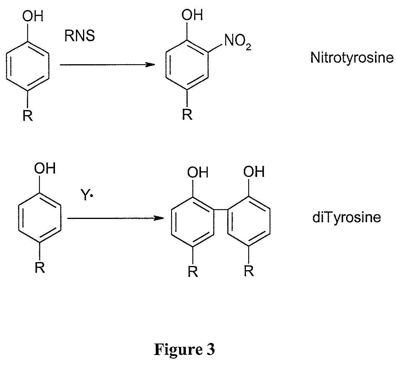
[0113]The levels of protein-bound dityrosine were measured in blood samples from 112 individuals with CAD and from 128 apparently healthy control subjects. The levels were measured by HPLC with on-line fluorescence detection and were quantified using an external calibration curve generated with synthetic dityrosine. Results were normalized to the content of the precursor amino acid, tyrosine, which was simultaneously quantified by HPLC with on-line diode array detection. The results demonstrated that subjects with CAD had higher levels (50% increased, P<0.001 for comparison of CAD vs. healthy subjects) of dityrosine in their serum than that observed in serum from healthy age and sex-matched subjects.
PUM
| Property | Measurement | Unit |
|---|---|---|
| pH | aaaaa | aaaaa |
| time | aaaaa | aaaaa |
| pH | aaaaa | aaaaa |
Abstract
Description
Claims
Application Information
 - R&D
- Intellectual Property
- Life Sciences
- Materials
- Tech Scout
- Unparalleled Data Quality
- Higher Quality Content
- 60% Fewer Hallucinations
Browse by: Latest US Patents, China's latest patents, Technical Efficacy Thesaurus, Application Domain, Technology Topic, Popular Technical Reports.
© 2025 PatSnap. All rights reserved.Legal|Privacy policy|Modern Slavery Act Transparency Statement|Sitemap|About US| Contact US: help@patsnap.com


