Device and system for enhanced SPECT, PET, and Compton scatter imaging in nuclear medicine

Active Publication Date: 2007-11-06
MINNESOTA IMAGING & ENG
View PDF9 Cites 91 Cited by
  • Summary
  • Abstract
  • Description
  • Claims
  • Application Information

AI Technical Summary

Benefits of technology

[0006]In accordance with the present invention, a radiation detection apparatus is provided for radiographic imaging and material composition analysis in which the apparatus can dynamically configure its array geometry and radiation detector parameters for a specific imaging task or it can use an existing radiation detection geometry and settings. This invention is particularly suited for x-ray and gamma ray imaging in nuclear medicine, including scintimammography and nuclear cardiology, and x-ray radiography, specifically, x-ray mammography. There are several advantages inherent to this invention. Superior detectors in cost-effective formats can be utilized and detectors with different properties, including materials, resolution, response time and noise characteristics, can be used within an array. One or more radiation detectors are incorporated into a detector module and one or more modules make up a detector module array. The detector modules transmit detected photon image data and relevant module parameters to a computer

Problems solved by technology

Less-versatile designs reduce the mechanical complexity of the d

Method used


Image

  • Device and system for enhanced SPECT, PET, and Compton scatter imaging in nuclear medicine
  • Device and system for enhanced SPECT, PET, and Compton scatter imaging in nuclear medicine
  • Device and system for enhanced SPECT, PET, and Compton scatter imaging in nuclear medicine

Examples


Embodiment Construction

General Detector Array

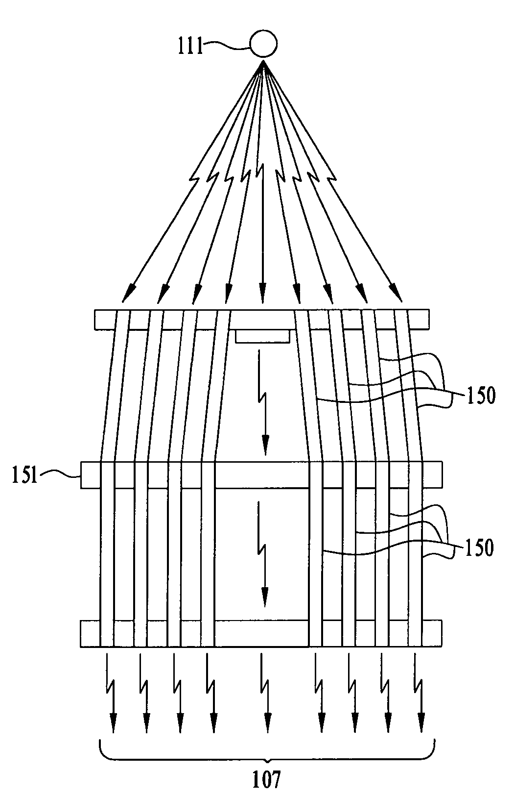
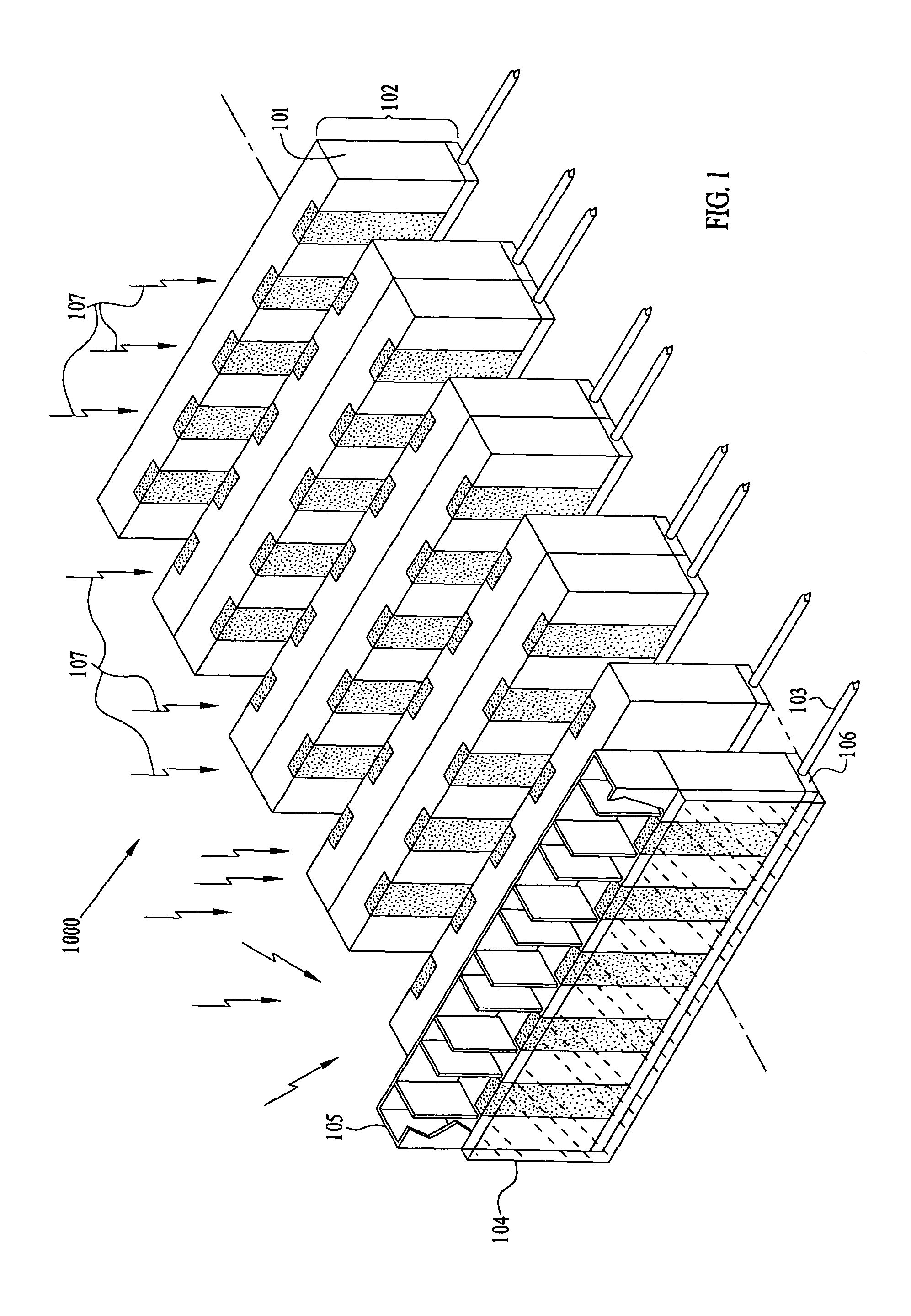
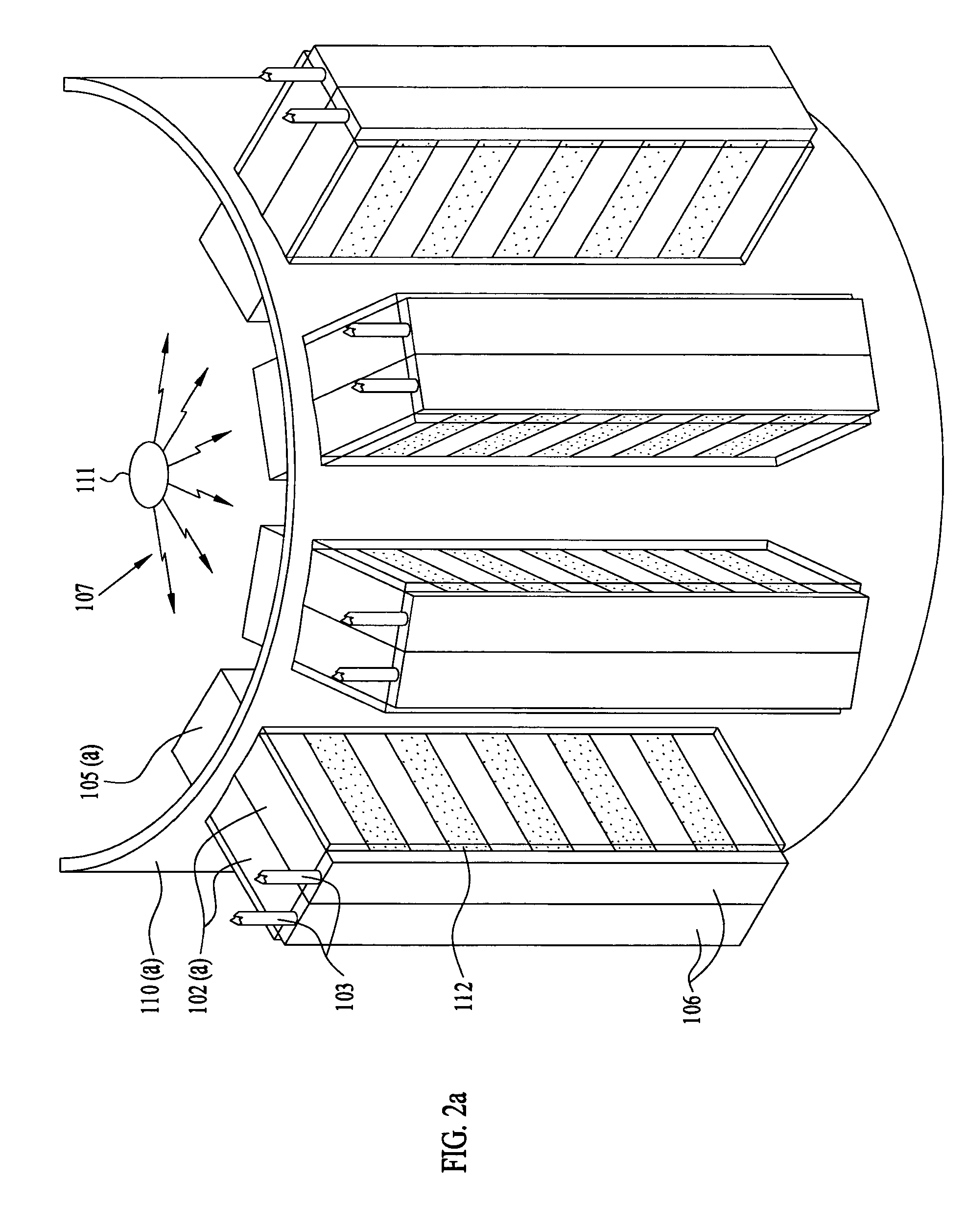
[0040]In one embodiment of the present invention, as illustrated in FIG. 1, a detector array 1000 preferably incorporates separate, discrete detector modules 102, illustrated here as edge-on or strip / micro-strip detectors, configured in a planar geometry to optimize the detection of incident radiation 107. The detector array 1000 may be utilized as part of a gamma camera. Currently, gamma cameras are not based on detector arrays such as detector array 1000 that incorporates discrete modules 102.

[0041]Detector modules 102 utilize one or more detectors 101, typically array detectors, which can have different properties. Note that several of the modules 102 include more than one detector. Additionally, linear array or small, 2-D array semiconductor detectors may be incorporated into the detector modules 102. Each module 102 also includes a base 106 and a communications link 103.

[0042]The base 106 preferably contains detector electronics, power management component...


PUM


Abstract

A method and apparatus for detecting radiation including x-ray, gamma ray, and particle radiation for radiographic imaging, and nuclear medicine and x-ray mammography in particular, and material composition analysis are described. A detection system employs fixed or configurable arrays of one or more detector modules comprising detector arrays that may be electronically manipulated through a computer system. The detection system, by providing the ability for electronic manipulation, permits adaptive imaging. Detector array configurations include familiar geometries, including slit, slot, plane, open box, and ring configurations, and customized configurations, including wearable detector arrays, that are customized to the shape of the patient. Conventional, such as attenuating, rigid geometry, and unconventional collimators, such as x-ray optic, configurable, Compton scatter modules, can be selectively employed with detector modules and radiation sources. Novel Compton gamma camera designs can be implemented. Edge-on detector resolution may be enhanced by measuring the interaction location along the height of the aperture. Edge-on detectors may be irradiated from the side. The components of the imaging chain can be calibrated or corrected using processes of the invention. X-ray mammography and scintimammography are enhanced by utilizing sectional compression and related imaging techniques.

Description

FIELD OF THE INVENTION[0001]This invention relates to an improved system for radiographic imaging and material analysis and more specifically for enhanced Compton scatter, PET, and SPECT nuclear medicine imaging.BACKGROUND OF THE INVENTION[0002]Two general imaging problems in radiology involve the determination of a radiation source distribution and / or the effect of a filter, in this case a patient, on the radiation source distribution. Consider the field of nuclear medicine where the radiation source or other radionuclide distribution emits photons or positrons, Image data acquisition in nuclear medicine presents several challenges in addition to constraints imposed by finite acquisition times and patient exposure restrictions. Most photon energies that are of interest in nuclear medicine are higher than the typical photon energies employed in diagnostic x-ray radiography. In particular, Positron Emission Tomography (PET) involves the detection of pairs of very high energy photons ...

Claims


Application Information

Patent Timeline
IPC IPC(8): G01T1/161G01T1/24
CPCG01T1/243G01T1/249G01T1/244G01T1/2008
Inventor NELSON, ROBERT SIGURDNELSON, WILLIAM BERT
Owner MINNESOTA IMAGING & ENG
Why Eureka
Try Eureka